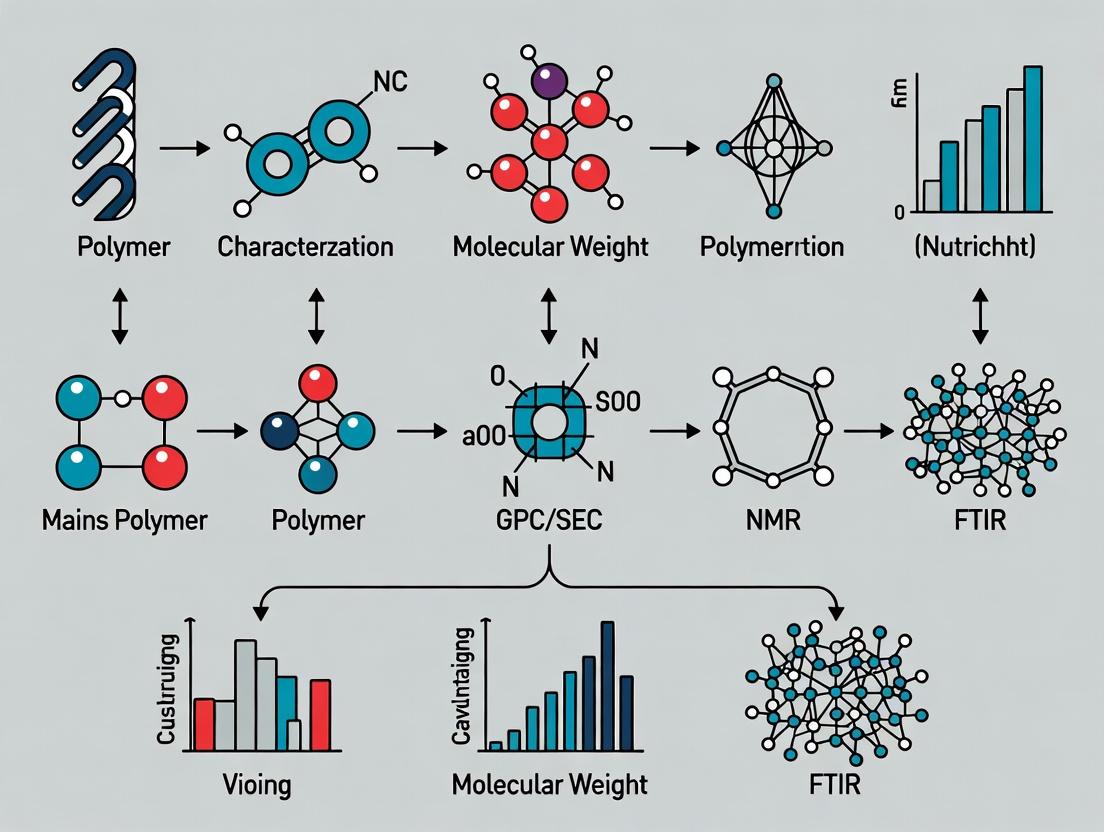
Polymer Characterization in Drug Development: Essential Methods, Applications, and Best Practices for Researchers
This comprehensive guide explores the critical polymer characterization techniques essential for modern drug development.
Polymer Characterization in Drug Development: Essential Methods, Applications, and Best Practices for Researchers
Abstract
This comprehensive guide explores the critical polymer characterization techniques essential for modern drug development. Aimed at researchers and scientists, it covers foundational principles, advanced methodological applications, troubleshooting strategies, and comparative validation frameworks. The article details how techniques like SEC, NMR, DSC, and light scattering are applied to analyze molecular weight, structure, thermal properties, and morphology of polymeric drug carriers, excipients, and delivery systems. It provides practical insights for optimizing characterization workflows and ensuring robust, regulatory-compliant data to advance polymeric biomaterials from lab to clinic.
Core Principles of Polymer Analysis: Understanding Size, Structure, and Properties
This application note, as part of a broader thesis on polymer characterization methods, provides detailed protocols and current data for determining six fundamental parameters critical to understanding polymer structure-property relationships. These parameters—Molecular Weight Averages (Mw, Mn), Polydispersity Index (PDI), Glass Transition Temperature (Tg), Melting Temperature (Tm), and Degree of Polymerization (DP)—are indispensable for researchers, scientists, and drug development professionals working with polymeric materials, excipients, or drug delivery systems.
Parameter Definitions and Significance
- Number-Average Molecular Weight (Mₙ): The total weight of all polymer molecules divided by the total number of molecules. It is sensitive to the presence of low-molecular-weight species.
- Weight-Average Molecular Weight (Mₚ): An average where molecules are weighted according to their mass. It is more sensitive to the presence of high-molecular-weight species.
- Polydispersity Index (PDI or Đ): The ratio Mₚ/Mₙ. It describes the breadth of the molecular weight distribution. A PDI of 1.0 indicates a perfectly monodisperse polymer.
- Glass Transition Temperature (Tg): The temperature range where a polymer transitions from a hard, glassy state to a soft, rubbery state. It is a key determinant of mechanical properties and stability.
- Melting Temperature (Tm): The temperature at which the crystalline domains of a semi-crystalline polymer melt, transitioning from a solid to a viscous liquid.
- Degree of Polymerization (DP or Xₙ): The number of monomeric units (repeat units) in a polymer chain. It is directly related to Mₙ by the formula: DP = Mₙ / M₀, where M₀ is the molecular weight of the repeat unit.
Quantitative Data Table for Common Polymers
The following table compiles typical values for key parameters of polymers frequently used in pharmaceutical and materials research.
| Polymer Name | Typical Mₙ (kDa) | Typical Mₚ (kDa) | Typical PDI | Tg (°C) | Tm (°C) | Application Context |
|---|---|---|---|---|---|---|
| Poly(lactic-co-glycolic acid) (PLGA) | 10 - 100 | 15 - 150 | 1.5 - 2.5 | 45 - 55 | Amorphous¹ | Controlled drug delivery, microparticles |
| Polyethylene glycol (PEG) | 1 - 40 | 1.05 - 44 | 1.01 - 1.1 | -65 to -50 | 55 - 65 | Bioconjugation, solubilization |
| Polystyrene (PS) | 50 - 500 | 55 - 750 | 1.05 - 2.5 | ~100 | ~240 (isotactic) | Standards, model polymer, cell cultureware |
| Polycaprolactone (PCL) | 20 - 80 | 25 - 100 | 1.2 - 1.8 | -60 | 58 - 65 | Long-term implantable devices, tissue engineering |
| Poly(N-isopropylacrylamide) (PNIPAM) | 10 - 100 | 12 - 150 | 1.1 - 2.0 | ~130 | -² | Thermo-responsive gels, smart materials |
| Hydroxypropyl methylcellulose (HPMC) | 10 - 1500³ | - | 2.0 - 10.0³ | 170 - 180 | Decomposes | Oral tablet matrix, controlled release |
¹PLGA is typically amorphous; small crystallinity may appear in high %GA forms. ²PNIPAM is amorphous. ³For polymers like HPMC, viscosity-average molecular weight is often reported instead.
Experimental Protocols
Protocol: Determination of Mₙ, Mₚ, and PDI by Gel Permeation Chromatography/Size Exclusion Chromatography (GPC/SEC)
Principle: Polymers are separated based on their hydrodynamic volume in a column packed with porous beads. Larger molecules elute first.
Materials: See The Scientist's Toolkit section.
Method:
- Sample Preparation: Dissolve the polymer sample in the appropriate, filtered (0.22 µm) eluent (e.g., THF, DMF with salts, water) at a concentration of 1-3 mg/mL. Stir for 6-12 hours at room temperature. Filter through a 0.45 µm (or 0.22 µm for aqueous) PTFE syringe filter.
- System Preparation: Prime the GPC/SEC system with the eluent at the recommended flow rate (typically 0.5-1.0 mL/min). Ensure stable baseline and column oven temperature (e.g., 35°C or 40°C).
- Calibration: Inject a series of narrow-polydispersity polymer standards (e.g., polystyrene, PEG, pullulan) of known molecular weight. Construct a calibration curve of log(M) vs. retention time/volume.
- Sample Injection: Inject 50-100 µL of the prepared sample solution using the autosampler or manual injection valve.
- Data Acquisition & Analysis: Monitor the signal from the concentration detector (RI, UV). Use the calibration curve and software (e.g., Empower, Astra, Cirrus) to calculate Mₙ, Mₚ, and PDI. For absolute molecular weights, use a multi-angle light scattering (MALS) detector in-line.
Protocol: Determination of Tg and Tm by Differential Scanning Calorimetry (DSC)
Principle: Measures the difference in heat flow between a sample and a reference as a function of temperature, detecting endothermic (melting) and glass transition events.
Method:
- Sample Preparation: Precisely weigh 3-10 mg of polymer into a tared, crimpable aluminum DSC pan. For volatile samples, use a hermetic pan with a sealing lid. Ensure an identical, empty pan is used as a reference.
- Instrument Calibration: Calibrate the DSC for temperature and enthalpy using high-purity standards (e.g., indium, tin, zinc).
- Method Programming:
- Equilibrate at -50°C (or well below the expected Tg).
- Ramp at 10°C/min to a temperature well above the expected Tm or thermal degradation point (e.g., 250°C).
- Hold isothermally for 2-5 minutes to erase thermal history.
- Cool at 10°C/min back to the starting temperature.
- Perform a second heating ramp at 10°C/min (this run provides the most reliable Tg/Tm data).
- Data Analysis: Analyze the second heating curve. Identify the Tg as the midpoint of the step change in heat capacity. Identify the Tm as the peak temperature of the endothermic melting transition. Report the associated enthalpy (∆Hf) from the integrated peak area.
Visualizations
Polymer Parameter Determination Workflow
GPC and DSC Analysis Pathways
Relationship Between Molecular Weight Parameters
Polymer Molecular Weight Relationships
The Scientist's Toolkit: Essential Research Reagent Solutions
| Item/Reagent | Primary Function in Characterization |
|---|---|
| GPC/SEC Columns (e.g., Styragel, PLgel, TSKgel) | Porous stationary phase for size-based separation of polymer chains by hydrodynamic volume. |
| HPLC-Grade Solvents (THF, DMF, Water + Salts) | Eluents for dissolving the polymer and carrying it through the GPC/SEC column. Must be filtered and degassed. |
| Narrow Polymer Standards (PS, PEG, PMMA, Pullulan) | Calibrants for constructing the molecular weight vs. retention time curve in GPC/SEC. |
| DSC Calibration Standards (Indium, Tin, Zinc) | High-purity metals with precisely known melting points and enthalpies for temperature and heat flow calibration of the DSC. |
| Hermetic DSC Pans and Lids | Sealed aluminum crucibles that prevent solvent loss during heating, essential for accurate Tg measurement. |
| 0.22 & 0.45 µm PTFE Syringe Filters | For removing dust and micro-gel particles from polymer solutions prior to GPC/SEC injection, preventing column damage. |
| Multi-Angle Light Scattering (MALS) Detector | An absolute detector used in-line with GPC/SEC to determine molecular weight without relying on column calibration. |
| Refractive Index (RI) Detector | A universal concentration detector for GPC/SEC, measuring the change in refractive index of the eluting solution. |
The Role of Polymer Characterization in Drug Delivery System Design
Within the broader research thesis on polymer characterization methods and techniques, this application note delineates the critical role of physicochemical and biological characterization in rational polymer-based drug delivery system (DDS) design. Effective characterization bridges polymer synthesis and in vivo performance, dictulating release kinetics, biodistribution, stability, and therapeutic efficacy.
Key Polymer Properties & Characterization Table
Table 1: Core Polymer Properties and Their Impact on DDS Performance
| Property Category | Specific Parameter | Characterization Technique | Quantitative Impact on DDS (Typical Target Ranges) | Influence on Drug Delivery | ||
|---|---|---|---|---|---|---|
| Molecular Weight & Distribution | Weight-Avg MW (M_w), Polydispersity Index (PDI) | Size Exclusion Chromatography (SEC/GPC) | M_w: 10-200 kDa; PDI: <1.5 for controlled release | Modulates drug release rate, nanoparticle stability, polymer degradation kinetics, and viscosity. | ||
| Chemical Structure & Composition | Monomer ratio, End-group functionality, Block length | NMR Spectroscopy, FTIR, Mass Spectrometry | e.g., Lactide:Glycolide (PLGA) = 50:50 to 85:15 | Determines degradation rate, hydrophilicity/hydrophobicity balance, drug-polymer interactions, and biocompatibility. | ||
| Thermal Properties | Glass Transition Temp (Tg), Melting Temp (Tm) | Differential Scanning Calorimetry (DSC) | Tg > 37°C for solid matrix at body temp; Tm for crystalline polymers. | Affects physical state, mechanical strength, drug stability within matrix, and release mechanism (diffusion vs. erosion). | ||
| Morphology & Solid State | Crystallinity, Phase Separation | X-ray Diffraction (XRD), DSC, Microscopy | Crystallinity: 0-70% for PLGA-based systems | Influences drug loading capacity (amorphous regions favor it), degradation profile, and release kinetics. | ||
| Surface & Bulk Hydrophilicity | Contact Angle, Water Uptake | Goniometry, Gravimetric Analysis | Contact Angle: 20°-80° for tunable protein adsorption | Governs biofouling, cellular interactions, protein adsorption, and rate of hydrolytic degradation. | ||
| Particle/Self-Assembly Properties | Size (Hydrodynamic Diameter), Polydispersity, Zeta Potential | Dynamic Light Scattering (DLS), Electrophoretic Light Scattering | Size: 50-200 nm (systemic delivery); PDI <0.2; Zeta Potential: | ±10-30 | mV | Dictulates in vivo biodistribution, circulation time, cellular uptake pathways, and colloidal stability. |
| Degradation & Drug Release | Mass Loss, Release Kinetics (e.g., k), Erosion Profile | In vitro Degradation Study, USP Dissolution Apparatus | Release rate constant k tailored to therapy (e.g., zero-order targeted). | Directly determines therapeutic dosing profile, duration of action, and need for repeated administration. |
Experimental Protocols
Protocol 3.1: Comprehensive Characterization of PLGA Nanoparticles for Encapsulation
Objective: To synthesize and characterize poly(lactic-co-glycolic acid) (PLGA) nanoparticles for controlled drug release, determining key physicochemical parameters.
Materials: PLGA (50:50, M_w ~30kDa), drug candidate (e.g., hydrophobic small molecule), polyvinyl alcohol (PVA, emulsifier), dichloromethane (DCM, organic solvent), deionized water.
Procedure:
- Nanoparticle Synthesis (Single Emulsion): a. Dissolve 50 mg PLGA and 5 mg drug in 2 mL DCM (organic phase). b. Prepare 20 mL of 1-3% w/v aqueous PVA solution (aqueous phase). c. Emulsify the organic phase into the aqueous phase using a probe sonicator (70% amplitude, 60s on ice). d. Stir the emulsion overnight at room temperature to evaporate DCM. e. Centrifuge the nanoparticle suspension at 20,000 x g for 20 min, wash twice with DI water, and re-suspend in buffer for characterization.
Size and Zeta Potential Analysis (DLS): a. Dilute 50 μL of nanoparticle suspension in 1 mL of 1 mM KCl solution (for zeta) or filtered DI water (for size). b. Transfer to appropriate cuvette (disposable for size, folded capillary for zeta). c. Measure hydrodynamic diameter and PDI via DLS at a 173° backscatter angle. Report intensity-weighted distribution. d. Measure zeta potential via electrophoretic light scattering. Report average of 3 runs with >10 sub-runs each.
Drug Loading and Encapsulation Efficiency (HPLC): a. Digest 1 mg of nanoparticles in 1 mL of DMSO with vortexing. b. Dilute appropriately with mobile phase and analyze drug content via validated HPLC method (e.g., C18 column, UV detection). c. Calculate: Drug Loading (DL)% = (Mass of drug in nanoparticles / Total mass of nanoparticles) x 100. Encapsulation Efficiency (EE)% = (Actual DL / Theoretical DL) x 100.
Protocol 3.2:In vitroDegradation and Release Kinetics Study
Objective: To monitor polymer mass loss and drug release profile under physiologically simulated conditions.
Materials: Weighed polymer films or nanoparticles, phosphate-buffered saline (PBS, pH 7.4), sodium azide (0.02% w/v, preservative), orbital shaker incubator, vacuum oven.
Procedure:
- Sample Preparation: Precisely weigh (W₀) polymer films/nanoparticle pellets (n=5 per time point).
- Incubation: Place each sample in a vial with 5 mL PBS (with azide). Incubate at 37°C under gentle agitation (50 rpm).
- Mass Loss Monitoring: a. At predetermined time points (e.g., days 1, 3, 7, 14, 28), remove a sample set. b. Rinse samples with DI water and lyophilize or dry in a vacuum oven to constant weight (Wₜ). c. Calculate: Mass Remaining % = (Wₜ / W₀) x 100.
- Drug Release Monitoring (Parallel Setup): a. For release, place samples in release medium (PBS, pH 7.4, with 0.5% w/v Tween 80 for sink condition). b. At each time point, centrifuge (for nanoparticles) or sample supernatant, and replace with fresh pre-warmed medium. c. Analyze drug concentration in supernatant via HPLC/UV-Vis. d. Calculate cumulative drug release %.
Visualization: Characterization Workflow
Polymer DDS Design-Characterization Feedback Loop
The Scientist's Toolkit: Research Reagent Solutions
Table 2: Essential Materials for Polymer Characterization in DDS Research
| Reagent/Material | Supplier Examples | Critical Function in Characterization |
|---|---|---|
| Polymer Standards (e.g., PEG, PS) | Agilent, Waters, Polymer Laboratories | Calibration of SEC/GPC columns for accurate molecular weight and PDI determination. |
| Deuterated Solvents (CDCl₃, D₂O) | Merck, Cambridge Isotope Laboratories | Solvent for NMR spectroscopy, enabling analysis of polymer composition and end-group structure. |
| PBS Tablets/Powder | Sigma-Aldrich, Thermo Fisher | Preparation of physiologically relevant buffers for in vitro degradation and drug release studies. |
| HPLC-Grade Solvents (ACN, MeOH) | Honeywell, Fisher Chemical | Mobile phase components for chromatographic analysis of drug content, purity, and release kinetics. |
| PVA (Polyvinyl Alcohol) | Sigma-Aldrich, Polysciences | A common emulsifier/stabilizer for preparing polymeric nanoparticles; grade and degree of hydrolysis affect size. |
| Dialysis Membranes (MWCO) | Spectrum Labs, Repligen | Isolation of nanoparticles or controlled release study by allowing diffusion of small molecules/buffer exchange. |
| Cell Culture Media & Assay Kits | Gibco, ATCC, Promega, Abcam | Assessing in vitro biocompatibility and cytotoxicity (e.g., MTT, LDH assays) of polymer carriers. |
| Stains & Dyes (Nile Red, Coumarin-6) | Invitrogen, Sigma-Aldrich | Fluorescent probes for labeling polymers or drugs to visualize cellular uptake and biodistribution. |
Chromatographic Methods
Chromatographic techniques separate polymer components based on their differential distribution between a mobile and a stationary phase. This is critical for determining molecular weight distributions, chemical composition, and purity.
Size Exclusion Chromatography (SEC/GPC)
Application Note: SEC is the primary technique for determining the molecular weight distribution of polymers. It separates molecules based on their hydrodynamic volume as they elute through a column packed with porous beads. For drug delivery systems, it is essential for characterizing polymeric carriers (e.g., PLGA, PEG) to ensure batch-to-batch consistency, which directly impacts drug release kinetics and biodistribution.
Protocol: Molecular Weight Distribution Analysis of PLGA
- Materials: PLGA sample (10 mg), HPLC-grade THF (mobile phase), Polystyrene standards (narrow dispersity), SEC column set (e.g., Styragel HR series), Refractive Index (RI) detector.
- Procedure:
- Prepare sample solution at 2 mg/mL in THF. Filter through a 0.2 μm PTFE syringe filter.
- Calibrate the SEC system using a series of narrow dispersity polystyrene standards. Construct a log(Mw) vs. retention time calibration curve.
- Inject 100 μL of the filtered sample. Use an isocratic flow of THF at 1.0 mL/min.
- Analyze the chromatogram (RI signal vs. time). Use SEC software to calculate number-average (Mn), weight-average (Mw) molecular weights, and dispersity (Ð) by comparing sample retention to the calibration curve.
- Precautions: Ensure complete dissolution and filtration to prevent column clogging. Maintain constant temperature to ensure reproducibility.
The Scientist's Toolkit for SEC/GPC
| Reagent/Material | Function |
|---|---|
| HPLC-Grade Tetrahydrofuran (THF) | Common mobile phase for polymers soluble in organic solvents. |
| Narrow Dispersity Polystyrene Standards | Calibrants for creating the molecular weight vs. retention time curve. |
| Styragel or Similar SEC Columns | Columns with defined pore sizes for separation by hydrodynamic volume. |
| Refractive Index (RI) Detector | Universal concentration detector for polymers without strong chromophores. |
| 0.2 μm PTFE Syringe Filter | Removes particulate matter to protect columns and ensure smooth flow. |
Diagram: SEC/GPC Workflow and Data Analysis
High-Performance Liquid Chromatography (HPLC)
Application Note: Reversed-Phase HPLC is vital for analyzing polymer additives, residual monomers, and degradation products in pharmaceutical polymers. It offers high sensitivity and resolution for quantifying low-molecular-weight species that may affect drug product safety.
Quantitative Data Table: Chromatographic Methods
| Technique | Key Measured Parameters | Typical Polymer Applications | Resolution/Power |
|---|---|---|---|
| Size Exclusion Chromatography (SEC/GPC) | Mn, Mw, Ð (Molecular Weight Distribution) | PLGA, PEG, Polystyrene, Polyacrylates | Separates by size in solution. |
| Reversed-Phase HPLC | Concentration of additives, monomers, degradants | Analysis of impurities in excipient batches | Separates by hydrophobicity. |
| Two-Dimensional LC (2D-LC) | Chemical composition distribution x MWD | Copolymers (e.g., block, graft), complex mixtures | Highest resolution for complex polymers. |
Spectroscopic Methods
These techniques probe the interaction of electromagnetic radiation with matter to elucidate chemical structure, composition, and interactions.
Fourier-Transform Infrared Spectroscopy (FTIR)
Application Note: FTIR identifies functional groups and chemical bonds via absorption of infrared light. It is indispensable for verifying polymer structure, monitoring curing reactions (e.g., in hydrogels), and detecting surface modifications on drug carrier nanoparticles.
Protocol: Surface Analysis of PEGylated Nanoparticles by ATR-FTIR
- Materials: PEGylated PLGA nanoparticles (lyophilized), Bare PLGA nanoparticles (control), FTIR spectrometer with ATR accessory (diamond crystal).
- Procedure:
- Clean the ATR crystal with isopropanol and background spectrum.
- Place a small amount (~2 mg) of lyophilized nanoparticle powder directly onto the crystal. Apply uniform pressure with the anvil.
- Acquire spectrum from 4000 to 600 cm⁻¹ with 4 cm⁻¹ resolution, 32 scans.
- Compare spectra of PEGylated vs. bare nanoparticles. Identify characteristic PEG peaks: C-O-C stretch at ~1100 cm⁻¹ and -OH stretch at ~3400 cm⁻¹.
- Precautions: Ensure samples are completely dry to avoid interference from water bands (~1640, 3300 cm⁻¹).
Nuclear Magnetic Resonance Spectroscopy (NMR)
Application Note: NMR, particularly ¹H and ¹³C, provides definitive information on polymer microstructure, comonomer sequence, tacticity, and end-group analysis. It is the gold standard for quantifying copolymer composition (e.g., % lactide vs. glycolide in PLGA).
The Scientist's Toolkit for Polymer Spectroscopy
| Reagent/Material | Function |
|---|---|
| Deuterated Solvent (e.g., CDCl₃, DMSO-d₆) | Provides a signal-free environment for NMR analysis without interfering H atoms. |
| Potassium Bromide (KBr) | Used to prepare transparent pellets for transmission FTIR analysis of solid polymers. |
| ATR Crystal (Diamond/Ge) | Enables direct, non-destructive FTIR measurement of solid samples without preparation. |
| Internal Standard (e.g., TMS for NMR) | Provides a reference peak for chemical shift calibration in NMR spectra. |
Diagram: Polymer Characterization by FTIR & NMR
Quantitative Data Table: Spectroscopic & Thermal Methods
| Technique | Key Measured Parameters | Typical Polymer Applications | Information Depth |
|---|---|---|---|
| FTIR / ATR-FTIR | Functional groups, chemical bonds | Polymer ID, degradation, surface chemistry | Surface (~0.5-5 μm for ATR) / Bulk. |
| NMR (¹H, ¹³C) | Composition, microstructure, tacticity | Copolymer analysis, end-group, kinetics | Bulk, quantitative. |
| Differential Scanning Calorimetry (DSC) | Tg, Tm, ΔH, crystallinity % | Amorphous/crystalline phases, stability, purity | Bulk thermal properties. |
| Thermogravimetric Analysis (TGA) | Weight loss %, decomposition onset (Td) | Thermal stability, residual solvent, filler content | Bulk decomposition profile. |
Thermal Analysis Methods
These techniques measure physical and chemical properties of polymers as a function of temperature, critical for understanding processing and application stability.
Differential Scanning Calorimetry (DSC)
Protocol: Determining Glass Transition (Tg) and Crystallinity of a Polymer
- Materials: Polymer sample (5-10 mg), sealed aluminum DSC pans, reference pan.
- Procedure:
- Accurately weigh sample into a pan and hermetically seal it.
- Load sample and reference pan into the DSC chamber.
- Run a heat/cool/heat cycle under N₂ flow (50 mL/min). Common program: Equilibrate at -50°C, heat to 200°C at 10°C/min (1st heat), cool to -50°C at 10°C/min, heat to 200°C at 10°C/min (2nd heat).
- Analyze the 2nd heating curve. Determine Tg as the midpoint of the heat capacity step. For crystalline polymers, calculate percent crystallinity from melting enthalpy (ΔHm): %Cryst = (ΔHm / ΔHm⁰) * 100, where ΔHm⁰ is the enthalpy for a 100% crystalline reference.
- Precautions: Use small sample masses for good thermal contact. The 2nd heat cycle removes thermal history.
Thermogravimetric Analysis (TGA)
Application Note: TGA measures weight change as a function of temperature, quantifying thermal stability, polymer composition (e.g., filler content in nanocomposites), and residual solvent/moisture—key for pre-formulation studies.
Scattering Methods
Scattering techniques use the interaction of X-rays, neutrons, or light with matter to investigate structure from the atomic to the micrometer scale.
X-ray Scattering: SAXS and WAXS
Application Note: Small-Angle X-ray Scattering (SAXS) probes nanostructures (1-100 nm), ideal for studying micelle formation, nanoparticle size/shape, and polymer blend phase separation. Wide-Angle X-ray Scattering (WAXS) analyzes crystalline structure (atomic dimensions, ~0.1 nm), determining crystal polymorphs and orientation in semi-crystalline polymers used in medical devices.
Protocol: SAXS Analysis of Block Copolymer Micelles
- Materials: Aqueous solution of PEG-PLA block copolymer micelles (1% w/v), SAXS instrument (synchrotron or lab-based).
- Procedure:
- Load micelle solution into a capillary flow cell or holder.
- Acquire scattering pattern over the appropriate q-range (q = 4π sinθ / λ). Record for sufficient time for good signal-to-noise.
- Subtract background scattering from solvent/buffer.
- Analyze the scattering curve. For spherical micelles, fit data to a form factor model (e.g., sphere model) to determine core radius (Rcore), overall radius (Rtotal), and aggregation number.
- Precautions: Ensure sample is homogeneous and free of dust. Accurate background subtraction is critical.
The Scientist's Toolkit for Scattering Methods
| Reagent/Material | Function |
|---|---|
| Size Standards (e.g., Au Nanoparticles) | Used to calibrate the q-range and detector distance in SAXS instruments. |
| Quartz Capillary Cells | Hold liquid samples for SAXS/XRD analysis with low background scattering. |
| Kapton or Mica Windows | Low-scattering materials for mounting solid film samples in SAXS/WAXS. |
| Dynamic Light Scattering (DLS) Instrument | Measures hydrodynamic diameter and size distribution of nanoparticles in solution. |
Diagram: Structural Hierarchy Probed by Scattering Techniques
Light Scattering
Application Note: Dynamic Light Scattering (DLS) measures the hydrodynamic diameter and size distribution of nanoparticles and polymer aggregates in solution. Static Light Scattering (SLS), often coupled with SEC, determines absolute molecular weight and radius of gyration (Rg).
Quantitative Data Table: Scattering Methods
| Technique | Key Measured Parameters | Typical Polymer Applications | Size Range Probe |
|---|---|---|---|
| Wide-Angle X-ray Scattering (WAXS) | Crystal structure, d-spacing, crystallinity | Crystalline polymers, polymorphism | Atomic (0.1-1 nm). |
| Small-Angle X-ray Scattering (SAXS) | Particle size/shape, micelle structure, phase separation | Nanocarriers, block copolymers, blends | Nanoscale (1-100 nm). |
| Dynamic Light Scattering (DLS) | Hydrodynamic diameter (Dh), size distribution | Nanoparticles, proteins, micelles in solution | ~1 nm to 10 μm. |
| Multi-Angle Light Scattering (MALS) | Absolute Mw, Rg (with SEC or standalone) | Conformation analysis, branching, aggregation | Varies with Mw. |
Application Notes
The selection of polymer characterization techniques must be driven by the intended function of the material within a drug development context. For a biomaterial serving as a controlled-release matrix, its erosion kinetics and glass transition temperature (Tg) are critical, whereas a polymeric nanoparticle carrier requires precise analysis of size, surface charge, and ligand density. Misalignment between characterization goals and functional requirements leads to incomplete data, failed correlations, and costly developmental delays. The following protocols and data frameworks are designed to ensure this alignment, maximizing the efficiency of the polymer research-to-application pipeline.
Experimental Protocols & Data Presentation
Protocol 1: Determination of Hydrolytic Degradation Kinetics for Erodible Matrices
Objective: To quantify mass loss and molecular weight change of a polyester (e.g., PLGA) as a function of immersion time in physiologically relevant buffer. Materials: Polymer film/sample, Phosphate Buffered Saline (PBS, pH 7.4), orbital shaker incubator (37°C), analytical balance, Size Exclusion Chromatography (SEC) system. Procedure:
- Pre-weigh (Wi) and measure initial molecular weight (Mni) of dry polymer samples (n≥5).
- Immerse samples in PBS (1:100 w/v) in sealed vials. Place vials in an orbital shaker incubator at 37°C, 60 rpm.
- At predetermined time points (e.g., 1, 3, 7, 14, 28 days), remove samples in triplicate.
- Rinse samples with deionized water and lyophilize to constant weight.
- Weigh dry samples (Wd) and calculate mass loss: % Mass Remaining = (Wd / Wi) * 100.
- Analyze dried samples via SEC to determine Mn at each time point. Key Data Output: Degradation profile table (see Table 1).
Protocol 2: Dynamic Light Scattering (DLS) & Zeta Potential Analysis of Polymeric Nanoparticles
Objective: To determine the hydrodynamic diameter, polydispersity index (PDI), and surface charge of drug-loaded polymeric nanoparticles. Materials: Nanoparticle suspension, disposable folded capillary zeta cells, DLS/Zeta potential analyzer, 0.22 µm syringe filter. Procedure:
- Dilute the nanoparticle suspension in appropriate filtered aqueous buffer (e.g., 1 mM KCl for zeta) to achieve a recommended scattering intensity.
- Filter the diluted suspension through a 0.22 µm filter into a clean vial.
- For size/PDI: Load sample into a disposable cuvette, equilibrate at 25°C for 2 min, perform measurement with ≥3 runs.
- For zeta potential: Load sample into a folded capillary cell, equilibrate, measure electrophoretic mobility with automatic voltage selection; convert to zeta potential via Smoluchowski model.
- Report results as mean ± standard deviation from ≥3 independent batches.
Table 1: Representative Characterization Data Alignment
| Material Function | Critical Characterization | Target Metrics | Typical Method |
|---|---|---|---|
| Subcutaneous Implant (Long-term release) | Bulk Erosion, Tg | Mass loss <10% over 30 days; Tg > 37°C | Gravimetric Analysis, DSC |
| IV Nanoparticle (Targeted delivery) | Size, Surface Charge, Ligand Density | Dh: 80-120 nm; PDI <0.2; ζ-Pot: -10 to -30 mV | DLS, NTA, HPLC |
| Mucoadhesive Gel | Rheology, Bioadhesion Strength | Storage Modulus (G') > Loss Modulus (G''); Work of Adhesion > 1 mJ | Rheometry, Texture Analysis |
| Gene Delivery Vector | Polyplex Stability, Binding Efficiency | N/P Ratio > 5; >90% Condensation at 0.5 µg/µL | Gel Retardation Assay, EthBr Exclusion |
Visualizations
Title: Polymer Characterization Selection Workflow
Title: NP Properties Influence Biological Pathways
The Scientist's Toolkit: Research Reagent Solutions
| Item | Function in Characterization |
|---|---|
| Phosphate Buffered Saline (PBS), pH 7.4 | Standard medium for hydrolytic degradation studies and nanoparticle dispersion, simulating physiological pH and ionic strength. |
| Size Exclusion Chromatography (SEC) Columns (e.g., PL aquagel-OH) | Separates polymers by hydrodynamic volume to determine molecular weight distribution and monitor degradation. |
| DLS/Zeta Potential Reference Latex (e.g., 100 nm polystyrene) | Validates instrument performance and measurement parameters for size and surface charge analysis. |
| Differential Scanning Calorimetry (DSC) Calibration Standards (Indium, Zinc) | Provides temperature and enthalpy calibration for accurate determination of Tg, Tm, and crystallinity. |
| Fluorophore-Labeled Ligands (e.g., FITC-PEG-NHS) | Enables quantification of ligand conjugation density on nanoparticle surfaces via fluorescence spectroscopy or HPLC. |
| Ethidium Bromide or SYBR Gold | Intercalating dyes used in gel retardation assays to visualize and quantify polymer-nucleic acid complex (polyplex) formation. |
| Mucin Glycoprotein | Substrate for in vitro testing of polymer bioadhesion strength using texture analyzers or rheometers. |
A Practical Guide to Key Polymer Characterization Techniques and Their Biomedical Uses
Within the comprehensive research on polymer characterization methods, Size-Exclusion Chromatography (SEC), also known as Gel Permeation Chromatography (GPC), stands as a cornerstone technique for the direct determination of molecular weight averages and distribution. This is critical for researchers and drug development professionals, as these parameters dictate polymer properties such as viscosity, solubility, mechanical strength, and drug release profiles from polymeric carriers. This application note details the protocols and considerations for obtaining reliable molecular weight data.
Core Principles and Quantitative Data
SEC separates polymer molecules in solution based on their hydrodynamic volume. Larger molecules elute first as they cannot penetrate the porous matrix of the column packing, while smaller molecules travel longer paths through the pores and elute later. The elution volume is correlated with molecular weight via a calibration curve.
Table 1: Key Molecular Weight Averages Determined by SEC
| Average | Symbol | Definition | Sensitivity To |
|---|---|---|---|
| Number Average | Mₙ | Σ NᵢMᵢ / Σ Nᵢ | Total number of chains |
| Weight Average | Mᵥ | Σ NᵢMᵢ² / Σ NᵢMᵢ | Larger molecules |
| Z-Average | M_z | Σ NᵢMᵢ³ / Σ NᵢMᵢ² | Very large molecules |
| Polydispersity Index | PDI (Đ) | Mᵥ / Mₙ | Breadth of distribution |
Table 2: Common SEC Detection Systems and Their Output
| Detector Type | Primary Measurement | Information Provided | Key Parameter |
|---|---|---|---|
| Refractive Index (RI) | Concentration | Universal concentration detector | dn/dc (specific refractive index increment) |
| UV/Vis Absorbance | Concentration | Concentration for chromophore-containing polymers | Molar absorptivity (ε) |
| Light Scattering (LS) | Scattered Light Intensity | Absolute molecular weight (M), radius of gyration (Rg) | dn/dc, second virial coefficient (A₂) |
| Viscometer | Inherent Viscosity | Intrinsic viscosity ([η]), molecular structure (branching) | Huggins/Kraemer constants |
Detailed Experimental Protocol: Multi-Detector SEC Analysis
Objective: To determine the absolute molecular weight distribution, intrinsic viscosity, and branching information of a synthetic polymer (e.g., polystyrene) in tetrahydrofuran (THF).
Materials & Reagents:
- SEC system with isocratic pump, autosampler, and column oven.
- SEC columns (e.g., 2-3 columns in series with pore sizes spanning 10² to 10⁶ Å).
- Multi-detector array: RI, UV, Multi-Angle Light Scattering (MALS), and Differential Viscometer.
- HPLC-grade solvent (THF, stabilized with BHT).
- Polymer standards for calibration (narrow dispersity polystyrene).
- Sample filters (0.2 or 0.45 μm, PTFE membrane).
- Vials and caps.
Procedure:
System Preparation:
- Purge all solvent lines and prime the pump with fresh, degassed THF.
- Equilibrate the system at a constant flow rate (e.g., 1.0 mL/min) and temperature (e.g., 35°C) for at least 1 hour until a stable detector baseline is achieved.
Column Calibration (Using Narrow Standards):
- Prepare individual solutions of each narrow molecular weight polystyrene standard in THF at a known concentration (~1-2 mg/mL).
- Filter each standard solution through a 0.2 μm syringe filter into an autosampler vial.
- Inject a precise volume (e.g., 100 μL) of each standard sequentially.
- Record the elution volume at the peak maximum for each standard.
- Construct a conventional calibration curve by plotting log(M) versus elution volume.
Sample Analysis:
- Accurately weigh (~5-10 mg) the unknown polymer sample into a vial.
- Add a known mass of THF to achieve a target concentration (typically 1-3 mg/mL). Allow to dissolve fully (may require overnight stirring).
- Filter the sample solution through a 0.2 or 0.45 μm filter into an autosampler vial.
- Inject the sample using the same chromatographic conditions as the calibration.
- Simultaneously collect data from RI, MALS (at multiple angles), and viscometer detectors.
Data Analysis:
- Conventional Calibration Method: Use the calibration curve from Step 2 to convert the sample's elution profile (from RI) to a molecular weight distribution. Calculate Mₙ, Mᵥ, and PDI.
- Absolute Method (Light Scattering): For each data slice, use the Rayleigh ratio (from MALS) and concentration (from RI) to calculate the absolute molecular weight (M) without calibration. Construct a Zimm or Debye plot per slice if needed.
- Intrinsic Viscosity: Use the viscometer and RI detector signals to calculate the intrinsic viscosity [η] for each slice. Plot log([η]) vs. log(M) to obtain the Mark-Houwink parameters (K and a), which inform on polymer conformation.
The Scientist's Toolkit: Essential Research Reagent Solutions
Table 3: Key Reagents and Materials for SEC Analysis
| Item | Function | Critical Notes |
|---|---|---|
| HPLC/SEC-Grade Solvents (THF, DMF, Water w/ salts) | Mobile phase; must dissolve polymer and be compatible with columns. | Must be low in UV absorbance, particulate-free, and often require stabilizers. Degassing is essential. |
| Porous Gel Columns (e.g., Styragel, TSKgel, PLgel) | Stationary phase for size-based separation. | Selected based on pore size range to match polymer's MW. Often used in series. |
| Narrow Dispersity Polymer Standards | For system calibration and validation. | Must match polymer chemistry (e.g., polystyrene, PMMA, PEG) for conventional calibration. |
| Quality Sample Filters (PTFE, Nylon) | Removes particulates that could clog columns. | Membrane material must be chemically compatible with solvent and sample. |
| Refractive Index Increment (dn/dc) Value | Critical constant for light scattering and concentration detection. | Must be known for polymer/solvent/temperature combination. Can be measured offline. |
Visualized Workflows and Relationships
SEC Experimental Workflow
SEC Data Analysis Pathway
This document presents application notes and protocols for Nuclear Magnetic Resonance (NMR) Spectroscopy, framed within a broader thesis on Polymer Characterization Methods and Techniques. NMR spectroscopy is a non-destructive analytical technique that provides atomic-level detail on molecular structure, dynamics, reaction state, and chemical environment. For polymer researchers, scientists, and drug development professionals, it is indispensable for determining monomer composition, sequencing, tacticity, end-group analysis, copolymer microstructure, and confirming polymer-drug conjugate structures.
Key Applications in Polymer and Pharmaceutical Research
Table 1: Quantitative Applications of NMR in Polymer & Drug Development
| Application Area | Measurable Parameter | Typical NMR Nuclei | Data Output |
|---|---|---|---|
| Chemical Structure | Monomer identity, stereochemistry (tacticity), regio-regularity, end-group fidelity | ¹H, ¹³C, ¹⁹F, ³¹P | Chemical shift (δ, ppm), J-coupling constants (Hz) |
| Composition | Copolymer composition ratio, molar mass (via end-group), drug loading in conjugates | ¹H (Quantitative) | Molar ratio from integrated signal areas |
| Sequence Distribution | Dyad/Triad sequences in copolymers (e.g., ethylene/propylene) | ¹³C | Sequence-sensitive chemical shifts |
| Conformational Dynamics | Polymer chain flexibility, glass transition studies, protein-polymer interactions | ¹H (relaxation: T₁, T₂) | Relaxation times, correlation times |
| Diffusion & Size | Hydrodynamic radius, aggregation state of polymer-drug nanoparticles | ¹H (DOSY) | Diffusion coefficient (m²/s) |
Detailed Experimental Protocols
Protocol 3.1: Quantitative ¹H NMR for Copolymer Composition
Objective: To determine the molar ratio of monomers (e.g., MMA:nBA) in a copolymer sample. Materials: See "The Scientist's Toolkit" below. Procedure:
- Sample Preparation: Weigh 5-10 mg of polymer sample into a clean NMR tube. Add 0.6-0.7 mL of deuterated solvent (e.g., CDCl₃). Cap and vortex until fully dissolved.
- Instrument Setup: Load the tube into a 400 MHz or higher NMR spectrometer. Lock, tune, and shim on the deuterium signal of the solvent.
- Parameter Setting: Set probe temperature to 25°C. Use a standard ¹H pulse sequence (zg) with the following critical parameters:
- Pulse Angle (pw): 30° (for quantitative, non-fully relaxed conditions)
- Relaxation Delay (d1): 25 seconds (≥ 5x the longest ¹H T₁, ensures full relaxation)
- Number of Scans (ns): 16-64
- Acquisition Time (aq): 4 seconds
- Data Acquisition & Processing: Run the experiment. Apply exponential window function (lb = 0.3 Hz), Fourier transform, phase, and baseline correct.
- Quantitative Analysis: Identify isolated, non-overlapping signals for each monomer unit. Integrate the peaks. The molar ratio is directly proportional to the ratio of the integrated areas, corrected for the number of protons each signal represents.
Protocol 3.2: ¹H DOSY for Nanoparticle Size Analysis
Objective: To assess the hydrodynamic size and confirm encapsulation of an active pharmaceutical ingredient (API) in a polymeric nanoparticle. Procedure:
- Sample Prep: Prepare a 2-5 mM solution of the nanoparticle formulation in D₂O. Use an internal standard (e.g., TSP, δ = 0 ppm) if absolute concentration is needed.
- Instrument Setup: Load sample and shim carefully. Select a stimulated echo-based DOSY sequence (e.g., ledbpgppr2s).
- Parameter Setting: Key gradient parameters must be calibrated:
- Gradient pulse length (δ): 2-4 ms
- Diffusion delay (Δ): 50-200 ms
- Number of gradient increments (nd): 16-32
- Linear gradient ramp from 2% to 95% of maximum gradient strength.
- Processing: Use inverse Laplace transform or fitting routines within the NMR software (e.g., TopSpin's DOSY processing) to convert decay data into a 2D spectrum with chemical shift vs. diffusion coefficient.
- Interpretation: All signals from a single species will align horizontally. Identical diffusion coefficients for polymer and API signals confirm encapsulation. Use the Stokes-Einstein equation to calculate hydrodynamic radius (Rₕ).
Visualization: Experimental Workflows
Diagram 1: General NMR Experiment Workflow (97 chars)
Diagram 2: NMR Data Analysis Decision Path (95 chars)
The Scientist's Toolkit
Table 2: Essential Research Reagent Solutions for NMR Analysis
| Item | Function & Specification |
|---|---|
| Deuterated Solvents (e.g., CDCl₃, D₂O, DMSO-d₆) | Provides a deuterium lock signal for the spectrometer; minimizes strong ¹H solvent signal. Must be >99.8% D. |
| NMR Reference Standards (e.g., Tetramethylsilane (TMS), 3-(Trimethylsilyl)propionic acid-d₄ sodium salt (TSP)) | Provides a precise chemical shift reference point (0 ppm). TSP is water-soluble. |
| Susceptibility-Matched NMR Tubes (5 mm) | High-quality tubes ensure uniform magnetic field, critical for resolution. |
| Relaxation Agent (e.g., Chromium(III) acetylacetonate - Cr(acac)₃) | Added in trace amounts to reduce long ¹H T₁ times, enabling faster quantitative analysis. |
| Quantitative Analysis Software (e.g., MestReNova, TopSpin) | Used for advanced processing, spectral deconvolution, and accurate integration. |
| Shim Tools (Gradient shimming routines) | Automated protocols to optimize magnetic field homogeneity for each sample. |
Within the broader thesis on polymer characterization methods, thermal analysis, specifically Differential Scanning Calorimetry (DSC), stands as a cornerstone technique for determining key thermodynamic parameters. For researchers and drug development professionals, DSC provides critical, quantitative data on glass transition temperature (Tg), melting temperature (Tm), and percent crystallinity. These parameters are vital for understanding polymer processing, stability, mechanical performance, and, in pharmaceutical contexts, the physical state of amorphous solid dispersions or polymeric excipients, directly impacting drug solubility and shelf-life.
Key Thermal Transitions and Quantitative Data
Table 1: Characteristic Thermal Transitions for Common Polymers & Pharmaceuticals
| Material/Class | Typical Tg (°C) | Typical Tm (°C) | Enthalpy of Fusion ΔHf (J/g) | % Crystallinity* | Key Application Context |
|---|---|---|---|---|---|
| Polyethylene (HDPE) | ~ -120 | 120 - 135 | 293 (100% crystal) | 60-80% | Packaging, medical devices |
| Polyethylene Terephthalate (PET) | 67 - 81 | 245 - 265 | 140 (100% crystal) | 30-50% | Bottles, fibers, films |
| Polystyrene (Atactic) | ~100 | N/A (Amorphous) | N/A | 0% | Laboratory ware, insulation |
| Poly(lactic-co-glycolic acid) (PLGA 50:50) | 40 - 50 | N/A (Amorphous) | N/A | 0% | Biodegradable drug delivery |
| Indomethacin (Amorphous Form) | ~45 | N/A | N/A | 0% | Model poorly soluble drug |
| Polyvinylpyrrolidone (PVP K30) | ~150 | N/A | N/A | 0% | Amorphous solid dispersion polymer |
| Sucrose | ~70 | 185 (Dec.) | N/A | N/A | Lyophilization stabilizer |
*% Crystallinity = (ΔHsample / ΔH100% crystal) x 100
Experimental Protocols
Protocol 1: Standard DSC Method for Tg and Tm Determination
Objective: To determine the glass transition (Tg), melting temperature (Tm), and heat of fusion (ΔHf) of a semicrystalline polymer or pharmaceutical formulation.
Materials & Equipment:
- Differential Scanning Calorimeter (e.g., TA Instruments Q series, Mettler Toledo DSC 3)
- Analytical balance (±0.01 mg)
- Hermetic T-zero aluminum pans and lids
- Encapsulation press
- Nitrogen gas supply (purge gas, 50 mL/min)
- Liquid Nitrogen Cooling System (optional, for sub-ambient Tg)
Procedure:
- Sample Preparation: Precisely weigh 5-10 mg of the sample (polymer or formulation) using an analytical balance.
- Pan Encapsulation: Place the sample in a pre-tared hermetic aluminum pan. Crimp the lid using an encapsulation press to ensure a sealed but pressure-releasable environment.
- Instrument Calibration: Calibrate the DSC for temperature and enthalpy using indium (Tm = 156.6°C, ΔHf = 28.4 J/g) and for heat capacity using a sapphire standard.
- Experimental Setup:
- Place the sealed sample pan in the sample furnace and an empty sealed reference pan in the reference furnace.
- Set the nitrogen purge gas flow to 50 mL/min.
- Program the following temperature method: a. Equilibrate at -30°C (or 50°C below expected Tg). b. First Heat: Ramp at 10°C/min to 200°C (or 30°C above expected Tm). This step erases thermal history. c. Cooling: Ramp at 10°C/min back to -30°C. d. Second Heat: Ramp at 10°C/min again to 200°C. Analyze data from this second heating scan to report Tg and Tm free of processing history.
- Data Analysis:
- Tg: Identify the glass transition as a step change in heat flow (midpoint method).
- Tm: Identify the melting peak. Use tangential integration to determine the onset temperature and peak temperature. Integrate the peak area to obtain ΔHf (J/g).
- % Crystallinity: Calculate using the formula in Table 1, with the appropriate reference ΔHf value for the 100% crystalline polymer.
Protocol 2: Modulated DSC (MDSC) for Complex Transitions
Objective: To separate reversible (heat capacity-related, e.g., Tg, melting) and non-reversible (kinetic, e.g., evaporation, relaxation, crystallization) thermal events in complex materials like amorphous solid dispersions.
Procedure:
- Follow steps 1-3 from Protocol 1.
- Experimental Setup: Use a modulated temperature program. Example parameters:
- Underlying heating rate: 2°C/min
- Modulation amplitude: ±0.5°C
- Modulation period: 60 seconds
- Temperature range: Cover Tg and any other events of interest.
- Data Analysis: The software deconvolutes the signal into Reversing Heat Flow (containing Tg and melting) and Non-Reversing Heat Flow (containing enthalpic relaxation, crystallization, and dehydration).
Visualization of Workflows
Diagram 1: DSC Experiment Decision Workflow
Diagram 2: Data Analysis Pathway for a Semicrystalline Polymer
The Scientist's Toolkit: Essential Reagents & Materials
Table 2: Key Research Reagent Solutions for DSC Analysis
| Item | Function/Benefit | Critical Application Notes |
|---|---|---|
| Hermetic Aluminum Pans & Lids | Provides a sealed, inert environment to prevent sample oxidation, dehydration, or volatile loss during heating. Essential for pharmaceuticals and hygroscopic polymers. | Use pinhole lids for moisture-containing samples. Ensure proper crimping to avoid pan rupture. |
| Calibration Standards (Indium, Zinc, Tin) | High-purity metals with precisely known melting points and enthalpies of fusion. Used for temperature, enthalpy, and heat capacity calibration of the DSC cell. | Always perform calibration before critical experiments. Indium (Tm=156.6°C) is the most common primary standard. |
| Sapphire Disk (Al₂O₃) | Standard reference material with well-characterized heat capacity (Cp). Used for calibrating the Cp signal of the DSC, crucial for accurate Tg measurement. | Required for quantitative heat capacity measurements and precise Tg analysis. |
| Nitrogen Gas (High Purity) | Inert purge gas that flows through the DSC cell to prevent oxidative degradation of samples, ensure stable baseline, and remove volatile contaminants. | Standard flow rate is 50 mL/min. For oxidative stability studies, switch to air or oxygen. |
| Liquid Nitrogen Cooling System (LNCS) | Accessory that enables rapid cooling and sub-ambient temperature operation (e.g., -150°C). Necessary for analyzing polymers with low Tg (e.g., elastomers). | Required for studying crystallization kinetics from the melt and for analyzing materials with transitions below room temperature. |
| Encapsulation Press | Tool to uniformly and reliably crimp hermetic DSC pans, ensuring a consistent seal and optimal thermal contact between the pan and the sensor. | Inconsistent crimping leads to poor thermal contact and artifacts in the DSC curve. |
Within the comprehensive characterization of synthetic and natural polymers for drug delivery, two pivotal techniques stand out: Dynamic Light Scattering (DLS) and Zeta Potential analysis. These methods are indispensable for determining a nanoparticle's hydrodynamic diameter and its surface charge, respectively. For a thesis on polymer characterization, these parameters are critical. They directly inform on the success of polymerization or formulation processes (size), predict in vitro and in vivo behavior (interaction with biological systems), and most importantly, assess long-term colloidal stability—a prerequisite for any viable therapeutic formulation. This document provides detailed application notes and protocols for these techniques.
DLS measures Brownian motion to derive the hydrodynamic size (Z-average and Polydispersity Index, PDI), while Zeta Potential infers electrostatic repulsion from electrophoretic mobility under an applied field. Together, they form the cornerstone of stability assessment.
Table 1: Interpretation Guidelines for DLS and Zeta Potential Data
| Parameter | Typical Range for Stable Formulations | Interpretation in Polymer Characterization | ||||||||||||||||
|---|---|---|---|---|---|---|---|---|---|---|---|---|---|---|---|---|---|---|
| Z-average (d.nm) | Application-specific (e.g., 50-200 nm for IV delivery) | Indicates successful nano-precipitation, polymerization, or self-assembly. Batch-to-batch consistency is key. | ||||||||||||||||
| Polydispersity Index (PDI) | < 0.2: Monodisperse0.2 - 0.7: Moderately polydisperse> 0.7: Very broad distribution | Reflects the homogeneity of the polymer/nanoparticle population. High PDI suggests uncontrolled synthesis or aggregation. | ||||||||||||||||
| Zeta Potential (mV) | > | +30 | or < | -30 | : Excellent stability | +20 | to | +30 | or | -20 | to | -30 | : Good stability | +10 | to | -10 | : Instability zone | Predicts colloidal stability via electrostatic repulsion. Critical for understanding polymer surface chemistry and the efficacy of stabilizers (e.g., PEGylation). |
Table 2: Impact of Common Polymer Formulation Variables on DLS/Zeta Results
| Variable | Expected Impact on Hydrodynamic Size | Expected Impact on Zeta Potential |
|---|---|---|
| Increasing ionic strength | May increase due to shielding and aggregation. | Decreases magnitude (closer to zero) due to double-layer compression. |
| Change in pH | May increase if pH approaches the isoelectric point (IEP). | Sign and magnitude change dramatically if pH crosses the polymer's pKa. |
| Addition of PEG (PEGylation) | Slight increase due to steric layer. | May reduce magnitude, but stability is provided sterically. |
| Polymer concentration | May increase at very high concentrations due to crowding. | Typically minimal direct effect. |
| Storage time | Increase indicates instability and aggregation. | Shift towards zero indicates instability. |
Experimental Protocols
Protocol 3.1: Sample Preparation for DLS & Zeta Potential
Objective: To prepare a representative, debris-free sample suitable for light scattering measurements. Materials: Purified polymer/nanoparticle suspension, appropriate buffer (e.g., 1 mM KCl for zeta), syringe filters (0.22 µm or 0.45 µm, non-protein binding), disposable sizing cuvettes, disposable folded capillary cells (for zeta). Procedure:
- Dilution: Dilute the stock polymer dispersion into the desired buffer. A final concentration yielding a scattering intensity between 200-800 kcps is ideal. Overly concentrated samples cause multiple scattering errors.
- Filtration/Centrifugation: Filter the diluted sample through a syringe filter into a clean vial to remove dust. For large or fragile aggregates, gentle centrifugation (e.g., 500 x g, 2 min) to pellet debris may be preferable.
- Loading: For DLS, pipette ~1 mL into a clean sizing cuvette. For Zeta Potential, load the sample into a folded capillary cell via syringe, ensuring no air bubbles are trapped.
- Equilibration: Allow the loaded cell to thermally equilibrate in the instrument for 2 minutes before measurement.
Protocol 3.2: Standard Operating Procedure for DLS Measurement
Objective: To determine the Z-average diameter and PDI of a polymeric sample. Instrument: Malvern Zetasizer Nano series or equivalent. Procedure:
- Setup: Select the "Size" measurement mode. Set the material RI (typically 1.59 for polymers) and dispersant RI/viscosity (e.g., water at 25°C: RI=1.33, Viscosity=0.8872 cP).
- Temperature: Set equilibration and measurement temperature (e.g., 25.0°C).
- Measurement Parameters: Set number of runs (≥12), run duration (automatic), and measurement angle (173° backscatter for most samples).
- Cell Placement: Insert the cleaned cuvette into the sample chamber.
- Execution: Initiate the measurement. The instrument will perform automatic attenuator selection and positioning.
- Quality Check: Analyze the correlation function decay and the size distribution plot. A single exponential decay and a monomodal distribution are ideal. Report the Z-average and PDI from the cumulants analysis.
Protocol 3.3: Standard Operating Procedure for Zeta Potential Measurement
Objective: To determine the electrophoretic mobility and calculated Zeta Potential of a polymeric sample. Instrument: Malvern Zetasizer Nano series or equivalent. Procedure:
- Setup: Select the "Zeta Potential" measurement mode. Input same material/dispersant parameters as DLS.
- Temperature: Set to 25.0°C.
- Cell Selection: In software, select the correct cell type (e.g., "DTS1070" folded capillary cell).
- Measurement Parameters: Set number of runs (10-100) and voltage selection (automatic).
- Cell Placement: Insert the loaded capillary cell, ensuring electrodes are aligned.
- Execution: Initiate measurement. The instrument measures the particle velocity via Phase Analysis Light Scattering (M3-PALS).
- Analysis: The software reports the mean Zeta Potential (mV) and electrophoretic mobility. The Smoluchowski model is applied by default for aqueous systems. Examine the phase plot for a stable signal.
Visualizations
DLS Measurement Workflow
Zeta Potential Predicts Colloidal Stability
Role of DLS/Zeta in Polymer Characterization Thesis
The Scientist's Toolkit: Research Reagent Solutions
Table 3: Essential Materials for DLS & Zeta Potential Analysis
| Item | Function & Importance |
|---|---|
| Disposable Sizing Cuvettes (e.g., polystyrene, PMMA) | Hold liquid sample for DLS measurement. Must be clean and free of scratches to avoid stray light scattering. |
| Disposable Folded Capillary Cells (e.g., DTS1070) | Specialized cell with electrodes for zeta potential measurement. Disposable to prevent cross-contamination. |
| Syringe Filters (0.22 µm, PES or PVDF membrane) | Crucial for removing dust and particulate contaminants, which are the primary source of artifact signals. |
| Standard Reference Material (e.g., 100 nm polystyrene latex) | Validates instrument performance and protocol. Provides a known size and zeta for quality control. |
| Low Ionic Strength Buffers (e.g., 1 mM KCl, 1-10 mM NaCl) | Essential for accurate zeta potential measurement, as high salt compresses the double layer and masks the true potential. |
| Temperature Control Module | Precise temperature control (e.g., ±0.1°C) is vital as Brownian motion and solvent viscosity are temperature-dependent. |
Within the comprehensive thesis on polymer characterization, elucidating morphology—from the micro to the nanoscale—is paramount. The properties (mechanical, barrier, drug release) of polymers, blends, and composites are dictated by their structural architecture. Scanning Electron Microscopy (SEM), Transmission Electron Microscopy (TEM), and Atomic Force Microscopy (AFM) form a complementary triad for multi-scale, multi-information imaging, essential for advancing research in material science and drug delivery system development.
Application Notes and Comparative Data
Table 1: Core Capabilities and Operational Parameters of Advanced Morphology Tools
| Parameter | Scanning Electron Microscopy (SEM) | Transmission Electron Microscopy (TEM) | Atomic Force Microscopy (AFM) |
|---|---|---|---|
| Primary Information | Surface topology, composition (with EDX) | Internal structure, crystallography, nanoparticle dispersion | 3D surface topography, nanomechanical properties (e.g., modulus, adhesion) |
| Resolution | ~0.5 nm to 5 nm | <0.1 nm to 0.5 nm (atomic scale possible) | ~0.1 nm (vertical), ~1 nm (lateral) |
| Sample Environment | High vacuum (typically) | High vacuum | Ambient air, liquid, vacuum |
| Sample Preparation | Conductive coating (for non-conductors), cryo-fixation | Ultra-thin sectioning (<100 nm), staining (e.g., RuO4, UA), cryo-TEM | Minimal; often requires adhesion to substrate |
| Key Quantitative Outputs | Particle size distribution, porosity %, fractal dimension | Lattice spacing, crystallite size, domain size in blends | Roughness (Ra, Rq), phase imaging, force-distance curves |
| Best for Polymer Studies | Fracture surfaces, composite filler dispersion, pore networks | Block copolymer micelle/core-shell structure, lamellar thickness, nanocrystal dispersion. | Polymer film domains, surface wettability, real-time degradation, single polymer chain imaging. |
Detailed Experimental Protocols
Protocol 1: SEM Imaging of a Polymeric Drug-Eluting Scaffold Surface Objective: To characterize the surface morphology and porosity of a PLGA-based electrospun scaffold.
- Sample Preparation: Mount a ~5x5 mm scaffold on an aluminum stub using double-sided conductive carbon tape.
- Conductive Coating: Sputter-coat the sample with a 10-15 nm layer of gold/palladium using a sputter coater under argon atmosphere to prevent charging.
- Microscope Setup: Load stub into high-vacuum SEM chamber. Set accelerating voltage to 5-10 kV (low voltage to minimize beam damage to polymer).
- Imaging: Use secondary electron (SE) detector. Start at low magnification (e.g., 500X) to locate area of interest, then progressively increase to 10,000X-50,000X to visualize fiber texture and nanopores.
- Analysis: Use integrated software to measure fiber diameter distribution from multiple images (n≥5).
Protocol 2: TEM Sample Preparation for a Block Copolymer Micelle (Drug Carrier) Objective: To visualize the core-shell structure of PCL-b-PEG micelles loaded with an active pharmaceutical ingredient (API).
- Negative Staining (for morphology):
- Dilute the micelle suspension to ~0.1 mg/mL in filtered deionized water.
- Glow-discharge a carbon-coated TEM grid for 30 seconds to render it hydrophilic.
- Place a 5 µL droplet of the suspension on the grid for 60 seconds.
- Wick away excess liquid with filter paper. Immediately add a 5 µL droplet of 2% uranyl acetate (or 1% phosphotungstic acid) solution for 30 seconds.
- Wick away stain, air-dry completely.
- Cryo-TEM (for native state):
- Apply 3 µL of sample to a holey carbon grid. Blot with filter paper to create a thin vitrified film (~100-200 nm thick).
- Rapidly plunge-freeze into liquid ethane cooled by liquid nitrogen.
- Transfer under liquid nitrogen to a cryo-TEM holder.
- Imaging: Insert holder into TEM. For stained samples, use 80-120 kV. For cryo-samples, use cryo-mode at ~-175°C. Use low-dose techniques to minimize radiation damage.
Protocol 3: AFM Nanomechanical Mapping of a Polymer Blend Film Objective: To distinguish phase-separated domains and map their elastic modulus.
- Sample Preparation: Spin-cast a thin film (100-200 nm) of a PS-PMMA blend onto a silicon wafer. Anneal as required to induce phase separation.
- Cantilever Selection: Use a silicon cantilever with a sharp tip (radius <10 nm) and a known spring constant (typically 0.5-5 N/m) calibrated via thermal tune method.
- Imaging Mode: Employ PeakForce Quantitative Nanomechanical Mapping (PF-QNM) or Tapping Mode for topography.
- Data Acquisition: Set a low peak force amplitude (~100-500 pN) to avoid sample deformation. Acquire simultaneous height, adhesion, and DMT modulus channels.
- Analysis: Use analysis software to generate modulus histograms for each domain and overlay modulus maps on topography.
Visualization: Workflows and Relationships
Title: SEM Sample Preparation and Imaging Workflow
Title: Decision Logic for Selecting Morphology Tool
The Scientist's Toolkit: Essential Research Reagent Solutions
Table 2: Key Reagents and Materials for Advanced Morphology Characterization
| Item | Function/Application |
|---|---|
| Conductive Carbon Tape | Adheres non-conductive samples to SEM stubs while providing a conductive path to ground, reducing charging artifacts. |
| Gold/Palladium (Au/Pd) Target | Used in sputter coaters to deposit a thin, uniform conductive metal layer on insulating polymer samples for SEM. |
| Uranyl Acetate (UA) | A common heavy metal negative stain for TEM; accumulates around structures, enhancing contrast of polymer aggregates, micelles, and biological components. |
| Ruthenium Tetroxide (RuO4) Vapor | A selective stain for unsaturated polymers (e.g., PS, PI) in TEM; reacts to provide mass-thickness contrast in multiphase polymer blends. |
| Holey Carbon Film TEM Grids | Grids with a lacey carbon support film, essential for cryo-TEM to hold the vitrified aqueous sample in thin films across the holes. |
| Phosphate Buffered Saline (PBS), Filtered (0.22 µm) | Buffer for preparing and diluting biological or drug-loaded polymeric nanoparticle suspensions for both TEM staining and cryo-TEM. |
| Silicon AFM Cantilevers with Diamond-like Carbon (DLC) Coating | Sharp, wear-resistant tips for nanomechanical mapping (PF-QNM), crucial for accurate modulus measurement on polymers. |
| Polydimethylsiloxane (PDMS) Stamps | Used to create patterned polymer surfaces or as a soft substrate for mechanical testing with AFM. |
| Ultramicrotome with Diamond Knife | Instrument to prepare ultrathin (50-100 nm) sections of bulk polymer samples for high-resolution TEM imaging. |
| Liquid Nitrogen & Liquid Ethane | Cryogens for rapid vitrification of hydrated soft matter (e.g., hydrogels, micelles) to preserve native structure for cryo-TEM and cryo-SEM. |
Within the broader thesis on Polymer characterization methods and techniques research, this case study exemplifies the integrated analytical workflow required to fully characterize a Poly(lactic-co-glycolic acid) (PLGA) nanoparticle (NP) drug delivery system. Moving beyond simple synthesis, this document provides detailed application notes and protocols for determining critical physicochemical parameters that dictate in vitro and in vivo performance, specifically for controlled release applications.
Synthesis & Primary Characterization Protocols
2.1 Protocol: Nanoparticle Synthesis via Double Emulsion (W/O/W) This method is ideal for encapsulating hydrophilic active pharmaceutical ingredients (APIs).
Materials:
- PLGA (50:50 LA:GA, acid-terminated, 10 kDa).
- Dichloromethane (DCM), organic solvent.
- Polyvinyl alcohol (PVA, 1% w/v), aqueous stabilizer solution.
- API (e.g., a model peptide or protein).
- Probe sonicator, magnetic stirrer, rotary evaporator.
Procedure:
- Dissolve 100 mg PLGA and the API in 2 mL DCM (primary water-in-oil emulsion).
- Add 0.5 mL of an aqueous API solution (or PBS for blank NPs) to the PLGA solution.
- Sonicate (70% amplitude, 30 sec, on ice) to form the primary W/O emulsion.
- Immediately pour this emulsion into 20 mL of 1% w/v PVA solution under rapid stirring.
- Sonicate again (50% amplitude, 60 sec) to form the W/O/W double emulsion.
- Stir gently for 4 hours to evaporate the organic solvent.
- Centrifuge at 20,000 x g for 30 min, wash pellet with DI water 3x, and lyophilize.
2.2 Protocol: Size, PDI, and Zeta Potential by Dynamic Light Scattering (DLS) Procedure:
- Re-disperse lyophilized NPs in 1 mM KCl solution to a concentration of 0.1 mg/mL.
- Filter through a 1.2 μm syringe filter into a clean DLS cuvette.
- Equilibrate sample in the instrument at 25°C for 120 sec.
- Perform measurement with backscatter detection (173°).
- Run minimum of 12 sub-runs. Report Z-Average (nm), Polydispersity Index (PDI), and Zeta Potential (mV) as mean ± SD of three independent batches.
2.3 Protocol: Morphology by Transmission Electron Microscopy (TEM) Procedure:
- Dilute NP suspension to 0.01 mg/mL in DI water.
- Apply a 10 μL drop to a carbon-coated copper grid for 60 sec.
- Wick away excess liquid with filter paper.
- Negative stain with 10 μL of 2% uranyl acetate for 30 sec, wick away excess.
- Air-dry completely before imaging at 80-100 kV accelerating voltage.
Quantitative Data from Primary Characterization:
Table 1: Physicochemical Properties of Synthesized PLGA NPs
| Parameter | Analytical Technique | Mean Result (± SD) | Target Specification |
|---|---|---|---|
| Hydrodynamic Diameter | DLS | 185.4 ± 8.7 nm | 150 - 250 nm |
| Polydispersity Index (PDI) | DLS | 0.092 ± 0.021 | < 0.2 |
| Zeta Potential | Electrophoretic Light Scattering | -32.5 ± 2.1 mV | < -20 mV |
| Morphology | TEM | Spherical, smooth surface | Spherical, non-aggregated |
Advanced Characterization for Controlled Release
3.1 Protocol: Determination of Encapsulation Efficiency (EE) and Drug Loading (DL) Procedure:
- Accurately weigh 5 mg of drug-loaded NPs.
- Dissolve completely in 1 mL of DMSO with vigorous vortexing.
- Dilute 100 μL of this solution in 900 μL of release medium (PBS, pH 7.4).
- Quantify API concentration using a pre-validated HPLC-UV or fluorescence method.
- Calculate:
- EE% = (Mass of API in NPs / Total mass of API used in formulation) x 100
- DL% = (Mass of API in NPs / Total mass of NPs) x 100
3.2 Protocol: In Vitro Drug Release Study Procedure:
- Place 10 mg of drug-loaded NPs in a dialysis tube (MWCO 12-14 kDa).
- Immerse the tube in 50 mL of release medium (PBS, pH 7.4, with 0.1% w/v sodium azide) at 37°C under gentle shaking (100 rpm).
- At predetermined time points, withdraw 1 mL of external medium and replace with fresh pre-warmed medium.
- Analyze withdrawn samples for API content.
- Calculate cumulative release as a percentage of total encapsulated drug.
3.3 Protocol: Monitoring Polymer Degradation via Gel Permeation Chromatography (GPC) Procedure:
- During release studies, periodically centrifuge NP suspensions.
- Lyophilize the recovered NPs.
- Dissolve 1 mg of lyophilized solid in 1 mL of THF (HPLC grade).
- Filter through a 0.2 μm PTFE filter.
- Inject into GPC system (PS standards, refractive index detector).
- Track the decrease in weight-average molecular weight (Mw) over time.
Quantitative Data for Controlled Release Performance:
Table 2: Drug Loading and Release Kinetics
| Parameter | Value | Method |
|---|---|---|
| Encapsulation Efficiency (EE%) | 78.3% ± 4.1% | Solvent dissolution/HPLC |
| Drug Loading (DL%) | 9.8% ± 0.5% | Solvent dissolution/HPLC |
| Time for 50% Release (T~50~) | 14.2 days | In vitro release study |
| Mw after 28 days (Release) | 42% of initial Mw | GPC analysis |
Visualization of Workflows and Mechanisms
Title: PLGA Nanoparticle Synthesis via Double Emulsion
Title: PLGA Nanoparticle Drug Release Mechanism
Title: Integrated PLGA NP Characterization Workflow
The Scientist's Toolkit: Research Reagent Solutions
Table 3: Essential Materials for PLGA NP Characterization
| Item | Function / Role | Key Consideration |
|---|---|---|
| PLGA Copolymers | Biodegradable matrix polymer. Determines degradation rate & release kinetics. | Select LA:GA ratio, MW, and end-cap (acid vs. ester) based on desired release profile. |
| Polyvinyl Alcohol (PVA) | Emulsion stabilizer during synthesis. Impacts particle size and surface properties. | Purification grade (e.g., 87-89% hydrolyzed) is critical for reproducible surface chemistry. |
| Dialysis Membranes | Contain NPs while allowing API diffusion for in vitro release studies. | Molecular Weight Cut-Off (MWCO) must be significantly lower than NP size, but higher than API. |
| Dynamic Light Scattering (DLS) Instrument | Measures hydrodynamic size, size distribution (PDI), and zeta potential. | Use appropriate dispersant refractive index and viscosity. Filter all samples. |
| Gel Permeation Chromatography (GPC) System | Monitors the degradation of PLGA by tracking molecular weight loss over time. | Requires appropriate standards (e.g., polystyrene) and solvent (e.g., THF). |
Solving Common Polymer Analysis Challenges: Artifacts, Data Interpretation, and Method Refinement
Within the broader thesis on polymer characterization methods and techniques research, Size Exclusion Chromatography (SEC), also known as Gel Permeation Chromatography (GPC), stands as a cornerstone for determining molecular weight distributions. Its accuracy, however, is critically dependent on avoiding common experimental pitfalls. This application note details protocols to mitigate errors arising from column interactions, improper solvent selection, and calibration inaccuracies, ensuring reliable data for researchers and drug development professionals.
Pitfall: Column-Polymer Interactions
Non-size-exclusion interactions between the analyte and the stationary phase (e.g., adsorption, hydrophobic attraction, ionic interactions) distort elution volumes, leading to inaccurate molecular weight data.
Protocol: Assessing and Minimizing Undesired Interactions
- Objective: To verify the absence of non-SEC interactions for a given polymer/solvent/column set.
- Materials: Polymer sample, candidate mobile phase, SEC columns (e.g., PLgel or TSKgel type), refractive index (RI) detector.
- Method:
- Prepare a series of polymer solutions at a known, low concentration (typically 1-2 mg/mL).
- Inject the sample and record the chromatogram.
- Vary the injection volume (e.g., 20 µL, 50 µL, 100 µL). In an ideal SEC system with no interactions, the elution peak maximum should not shift.
- Calculate the recovery by comparing the integrated peak area to that of a known standard or via mass balance. Recovery <95% indicates significant adsorption.
- Mitigation: If interactions are detected, modify the mobile phase by adjusting pH, ionic strength, or adding a competing agent (e.g., salts for polyelectrolytes, THF for polystyrenes in chloroform). Consider changing column chemistry (e.g., from silica-based to organic gel).
Pitfall: Inappropriate Solvent Choice
Solvent choice affects polymer conformation (via thermodynamic quality), column compatibility, and detection sensitivity.
Protocol: Systematic Solvent Evaluation for a New Polymer
- Objective: To select an optimal SEC solvent that fully dissolves the polymer, matches column compatibility, and provides adequate detector response.
- Materials: Polymer sample, candidate solvents (HPLC grade), columns compatible with organic or aqueous phases, RI and/or UV detectors.
- Method:
- Dissolution Test: Attempt to dissolve 5 mg of polymer in 1 mL of candidate solvent overnight with gentle agitation. Observe for clarity and particulate matter.
- Column Compatibility Check: Ensure solvent viscosity is suitable (<2 cP recommended) and within the column's pH and chemical stability limits.
- Detector Suitability Test: Measure the refractive index increment (dn/dc) of the polymer in the solvent using a differential refractometer, or obtain UV absorbance spectra. Prioritize solvents with high dn/dc (>0.05 mL/g for RI) or strong UV chromophores.
- Data Presentation:
Table 1: Solvent Evaluation for Polylactic Acid (PLA) Characterization
| Solvent | Good Dissolution? | Column Compatible? | dn/dc (mL/g) | Viscosity (cP) | Recommended? |
|---|---|---|---|---|---|
| Tetrahydrofuran (THF) | Yes | Yes (Organic) | 0.054 | 0.48 | Yes |
| Chloroform | Yes | Yes (with caution) | 0.042 | 0.54 | Yes |
| Dimethylformamide (DMF) | Yes | Yes (Organic) | 0.060 | 0.92 | Yes |
| Water | No | N/A | N/A | 0.89 | No |
Pitfall: Calibration Errors
Using a single, incorrect calibration curve is a primary source of molecular weight error.
Protocol: Establishing a Multi-Detector & Universal Calibration
- Objective: To create a calibration that accounts for polymer hydrodynamic volume, providing accurate absolute molecular weights.
- Materials: Narrow dispersity polymer standards (e.g., polystyrene, polymethylmethacrylate), target polymer sample, SEC system with RI, UV, and Multi-Angle Light Scattering (MALS) detectors, viscometer (optional).
- Method A (Conventional Calibration with Broad Correction):
- Run a series of narrow standards covering the molecular weight range.
- Plot log(M) vs. elution volume to create a calibration curve.
- Apply a "Q factor" or "broadening correction" algorithm using software to correct for polydispersity effects.
- Method B (Universal Calibration Using Viscometry):
- Perform Method A.
- Use an online viscometer to measure intrinsic viscosity [η] for each standard and sample slice.
- Plot log(M[η]) vs. elution volume. This curve is "universal" for polymers of the same hydrodynamic volume.
- For the analyte, calculate M from its measured [η] and the universal calibration curve.
- Method C (Absolute Calibration with MALS):
- Connect a MALS detector downstream of the SEC columns.
- The light scattering signal directly measures weight-average molecular weight (Mw) at each elution slice, independent of elution time or calibration standards.
Table 2: Comparison of SEC/GPC Calibration Methods
| Method | Principle | Key Requirement | Accuracy | Suitability |
|---|---|---|---|---|
| Conventional | Elution time of known standards | Narrow standards of identical polymer | Low-Moderate | Routine QC of known polymers |
| Universal Calibration | Hydrodynamic volume (M[η]) | Standards & sample [η] data | High | Unknown polymers, branched chains |
| Light Scattering | Direct Mw measurement | MALS detector, dn/dc value | Highest (Absolute) | Novel polymers, aggregates, conjugates |
The Scientist's Toolkit: SEC/GPC Research Reagent Solutions
Table 3: Essential Materials for Robust SEC/GPC Analysis
| Item | Function & Specification | Example/Catalog Consideration |
|---|---|---|
| SEC Columns | Separation based on hydrodynamic volume. Pore size mix determines range. | PLgel Mixed-B (Agilent), TSKgel SuperMultiporeHZ-M (Tosoh), Acquity APC (Waters). |
| Narrow Dispersity Standards | For system calibration and validation. Must be chemically similar or used for universal calibration. | Polystyrene (PS) in THF, Polyethylene oxide (PEO) in water, Polymethylmethacrylate (PMMA) in DMF. |
| HPLC-Grade Solvents | Mobile phase with low UV absorbance, high purity, stabilized if necessary (e.g., THF with BHT). | THF (stabilized), Chloroform (with ethanol), DMF (with LiBr for polyelectrolytes). |
| In-Line Degasser | Removes dissolved gases to prevent bubbles in detectors and pump. | Essential for RI and light scattering detectors. |
| Syringe Filters | Removes particulate matter to prevent column blockage. | PTFE or Nylon, 0.45 µm or 0.22 µm pore size, solvent compatible. |
| Refractive Index Detector | Universal concentration detector for polymers with known dn/dc. | Optilab, RI-410. |
| Multi-Angle Light Scattering (MALS) Detector | Provides absolute molecular weight and size (Rg) without calibration. | DAWN (Wyatt), Viscotek. |
| Online Viscometer | Measures intrinsic viscosity for universal calibration and branching analysis. | Viscotek, PSS. |
| Data Analysis Software | For processing chromatograms, applying calibrations, and calculating molecular weight averages. | Astra (Wyatt), Empower (Waters), PSS WinGPC. |
Interpreting Complex NMR Spectra for Copolymers and Degradation Products
Within the framework of polymer characterization for advanced material and drug delivery system development, Nuclear Magnetic Resonance (NMR) spectroscopy is an indispensable tool. It provides unparalleled insight into copolymer microstructure, composition, and the structural elucidation of degradation products. This Application Note details advanced protocols for interpreting complex NMR spectra, addressing challenges such as signal overlap, dynamic processes, and low-concentration species.
Key Quantitative Data for Copolymer Characterization
Table 1: Typical Chemical Shift Ranges for Common Polymer Units in Deuterated Chloroform (CDCl₃)
| Polymer Unit / Functional Group | ¹H δ (ppm) | ¹³C δ (ppm) | Key Spectral Feature |
|---|---|---|---|
| Polyethylene (PE) -CH₂- | 1.26 | 30.0 | Strong singlet |
| Polystyrene (PS) -ArH | 6.2-7.2 | 125-146 | Aromatic multiplet |
| Poly(methyl methacrylate) -OCH₃ | 3.60 | 51.5 | Sharp singlet |
| Poly(lactic-co-glycolic acid) -CH (LA) | 5.15-5.25 | 69.0 | Quadruplet (from HH coupling) |
| Poly(lactic-co-glycolic acid) -CH₂ (GA) | 4.65-4.85 | 61.0 | Complex multiplet |
| Poly(ethylene glycol) -OCH₂CH₂O- | 3.65 | 70.5 | Sharp singlet |
| Chain-end hydroxyl (-OH) | 1.5-3.0 (varies) | - | Broad, concentration/temp dependent |
| Degradation Product: Lactic Acid -CH | 4.35 (aq.) | 69.5 | Shifts with pH |
Table 2: NMR Techniques for Specific Characterization Challenges
| Challenge | Recommended NMR Technique | Key Parameter | Information Gained |
|---|---|---|---|
| Microstructure/Tacticity | High-Resolution ¹³C NMR | High Digital Resolution (0.5 Hz/pt) | Triad/Diad sequences, Regioregularity |
| Low-Concentration Degradants | 2D NMR (e.g., HSQC, HMBC) | Long Acquisition Time (>4 hrs) | Connectivity of trace species |
| Dynamic Processes (e.g., hydrolysis) | In-situ NMR Kinetics | Variable Temperature Control | Real-time degradation rates |
| Overcoming Signal Overlap | Diffusion-Ordered (DOSY) NMR | Gradient Strength Variation | Separation by hydrodynamic radius |
Experimental Protocols
Protocol 1: Comprehensive Microstructural Analysis of PLGA Copolymers
Objective: Determine lacticle:glycolide ratio, sequencing, and end-group integrity.
Materials:
- Deuterated solvent (e.g., CDCl₃, DMSO-d₆).
- High-field NMR spectrometer (≥ 400 MHz for ¹H).
- 5 mm NMR tube.
- Purified, dry copolymer sample (~10-20 mg).
Procedure:
- Sample Preparation: Dissolve 15 mg of PLGA in 0.6 mL of CDCl₃. Filter through a basic alumina plug if paramagnetic impurities are suspected.
- ¹H NMR Acquisition: Lock, shim, and tune the probe. Acquire a standard ¹H spectrum (64 scans, 10 sec relaxation delay). Integrate the methane proton signal of the LA unit (δ ~5.2) and the methylene signal of the GA unit (δ ~4.8). Calculate the LA:GA molar ratio.
- Quantitative ¹³C NMR Acquisition: Use an inverse-gated decoupling pulse sequence with a 60-second relaxation delay to suppress Nuclear Overhauser Effect (NOE). Acquire for 8-12 hours to achieve sufficient S/N. Analyze the carbonyl region (δ 165-175 ppm): peaks at ~169.5 and ~170.5 ppm correspond to GA-GA and LA-LA diads, respectively; signals for LA-GA/GA-LA heterodiads appear between them. The relative intensities provide sequence distribution.
- 2D HSQC Analysis: Perform a gradient-enhanced HSQC experiment to correlate ¹H and ¹³C signals. This resolves ambiguities in the crowded ¹H spectrum by spreading signals into a second dimension.
Protocol 2: Identification of Polymer Degradation Products via In-situ NMR Kinetics
Objective: Monitor hydrolytic degradation in real-time and identify soluble oligomeric products.
Materials:
- Phosphate buffered saline (PBS), pD 7.4, in D₂O.
- NMR spectrometer with temperature control and automation software.
- Susceptibility-matched NMR tube (Shigemi tube preferred).
Procedure:
- Initial Setup: Prepare a 5 mg/mL polymer solution in PBS/D₂O buffer. Place in the NMR tube.
- Kinetic Series Programming: Set the probe temperature to 37°C. Program an automated series to collect ¹H NMR spectra (e.g., 16 scans each) at defined time intervals (e.g., every 30 minutes for 48 hours).
- Data Acquisition & Monitoring: Start the series. The software will acquire spectra sequentially. Monitor the decrease in polymer main chain signals and the concurrent appearance of new signals in the aliphatic (δ 1.0-2.5 ppm) and carboxylate regions.
- Product Identification: At the end of the series, acquire a high-sensitivity 2D NMR experiment (e.g., TOCSY or ¹H-¹³C HMBC) on the final degraded mixture. Compare chemical shifts and correlation peaks to known standards (e.g., lactic acid, glycolic acid, short oligomers) for identification.
Visualization of Methodologies
NMR Spectral Analysis Decision Workflow
Polymer Degradation Pathway for NMR Analysis
The Scientist's Toolkit: Essential Reagents & Materials
Table 3: Key Research Reagent Solutions for Polymer NMR
| Item | Function & Specification | Notes |
|---|---|---|
| Deuterated Solvents (CDCl₃, DMSO-d₆, D₂O) | Provide NMR lock signal; dissolve polymer. Must be 99.8% D or higher. | Dry over molecular sieves. Use correct buffer in D₂O for degradation studies. |
| Chemical Shift Reference (TMS, DSS) | Internal standard for ¹H and ¹³C chemical shift calibration (δ = 0 ppm). | TMS for organic solvents; DSS (sodium trimethylsilylpropanesulfonate) for aqueous solutions. |
| Shigemi Tubes | Susceptibility-matched NMR tubes for limited sample volume or aqueous solutions. | Minimizes lineshape distortions, crucial for high-resolution kinetics. |
| Chromatographic Adsorbents (Basic Alumina, Celite) | Remove paramagnetic impurities (e.g., catalyst residues) that cause signal broadening. | Pack a small pipette column for sample filtration prior to NMR. |
| NMR Processing Software (MestReNova, TopSpin) | For spectral processing (FT, phasing, baseline correction), integration, and 2D analysis. | Essential for accurate quantitative analysis and data presentation. |
| Variable Temperature Unit | Precise temperature control of NMR probe for kinetics or studying dynamics. | Required for in-situ degradation studies at physiological (37°C) temperatures. |
Optimizing DSC Run Parameters for Accurate Glass Transition Detection
1. Introduction and Thesis Context Within the comprehensive framework of a thesis on polymer characterization methods and techniques, Differential Scanning Calorimetry (DSC) stands as a cornerstone for thermal analysis. The accurate determination of the glass transition temperature (Tg) is critical for understanding polymer behavior, drug-polymer interactions in solid dispersions, and the stability of amorphous pharmaceutical formulations. This application note provides detailed protocols and data for optimizing DSC run parameters to ensure precise and reproducible Tg detection, a fundamental skill for researchers and drug development professionals.
2. The Scientist's Toolkit: Essential Research Reagents & Materials
| Item | Function in DSC Tg Analysis |
|---|---|
| Hermetic Aluminum Crucibles (Tzero pans/lids) | Ensures an airtight seal to prevent solvent/plasticizer loss during heating, which can artifactually shift Tg. |
| High-Purity Inert Gas (Nitrogen or Argon) | Purge gas to maintain an inert atmosphere, preventing oxidative degradation of samples during runs. |
| Calibration Standards (Indium, Zinc) | Certified pure metals with known melting points and enthalpies for temperature and enthalpy calibration of the DSC cell. |
| Reference Material (Empty, sealed pan) | Provides the baseline heat flow differential against which the sample measurement is compared. |
| Thermal Annealing Oven | For controlled thermal history erasure (physical aging) prior to measurement, ensuring a consistent initial state. |
3. Core Experimental Protocol: Sample Preparation and DSC Measurement
- Sample Preparation: Precisely weigh 5-10 mg of polymer or solid dispersion powder using a microbalance. For films, use a clean punch to obtain a uniform disc. Place the sample in the bottom half of a hermetic aluminum pan. Crimp the lid using a manual or hydraulic press to ensure a complete seal. Verify seal integrity.
- Instrument Calibration: Perform temperature and enthalpy calibration using indium (mp 156.6°C, ΔHf 28.45 J/g) and zinc (mp 419.5°C) at the heating rate intended for use. Perform cell constant calibration.
- Method Programming: Program the following method sequence:
- Equilibration: Hold at a starting temperature (typically Tg - 50°C) for 5 min.
- Heating Scan: Heat to an end temperature (typically Tg + 50°C) at the selected heating rate (β, e.g., 10°C/min).
- Cooling Scan: Cool back to the starting temperature at a controlled rate (e.g., -50°C/min).
- Second Heating Scan: Repeat the heating scan (Step 2) to obtain the thermal history-free Tg.
- Data Analysis: Analyze the second heating scan. Tg is reported as the midpoint of the step change in heat capacity (Cp), determined by the half-height or inflection point method per relevant standards (e.g., ASTM E1356).
4. Optimized Parameter Tables and Data
Table 1: Effect of Heating Rate (β) on Tg Detection for Amorphous Poly(Lactic Acid) (PLLA)
| Heating Rate (°C/min) | Onset Tg (°C) | Midpoint Tg (°C) | Step Change ΔCp (J/g°C) | Signal-to-Noise (S/N) |
|---|---|---|---|---|
| 2 | 58.2 ± 0.3 | 60.1 ± 0.3 | 0.42 ± 0.02 | High |
| 10 | 59.8 ± 0.5 | 62.3 ± 0.4 | 0.43 ± 0.02 | Very High |
| 20 | 61.5 ± 0.7 | 64.0 ± 0.6 | 0.44 ± 0.03 | High |
| 50 | 64.1 ± 1.2 | 66.9 ± 1.0 | 0.45 ± 0.05 | Moderate |
Table 2: Effect of Sample Mass on Tg Detection for an Amorphous Drug (Itraconazole)
| Sample Mass (mg) | Midpoint Tg (°C) | Signal Amplitude (mW) | Thermal Lag (°C)* | Enthalpy of Relaxation (J/g) |
|---|---|---|---|---|
| 3 | 59.5 ± 0.8 | 0.08 | 0.1 | 1.05 |
| 5 | 60.1 ± 0.4 | 0.14 | 0.3 | 1.12 |
| 10 | 60.3 ± 0.3 | 0.27 | 0.8 | 1.08 |
| 20 | 61.0 ± 0.6 | 0.52 | 1.5 | 0.99 |
*Lag calculated at 10°C/min.
5. Summary of Optimal Parameter Recommendations
- Heating Rate: 10°C/min offers the best compromise between sufficient signal strength, acceptable thermal lag, and reasonable experimental time. Use slower rates (2-5°C/min) for extremely weak transitions or high-resolution studies.
- Sample Mass: 5-10 mg is optimal for most polymers and pharmaceuticals. It provides a strong signal while minimizing intra-sample temperature gradients.
- Purge Gas Flow: Maintain a constant 50 mL/min nitrogen purge.
- Pan Type: Always use hermetically sealed pans for Tg detection to prevent mass loss.
- Thermal History: Always analyze the second heating scan after controlled cooling to erase previous thermal history and physical aging effects.
6. Visualizing the Optimization Workflow and Signal Analysis
DSC Tg Optimization and Analysis Workflow
Components of the DSC Tg Signal
Within the broader thesis on advancing polymer characterization, Dynamic Light Scattering (DLS) stands as a cornerstone technique for determining the hydrodynamic size and size distribution of nanoparticles, polymers, and biologics in solution. However, its accuracy is notoriously compromised by specific artifacts: dust/foreign particulates, multiple scattering, and sample polydispersity. These challenges can lead to severe misinterpretation of data, particularly in critical applications like drug formulation development. These Application Notes detail protocols to identify, mitigate, and correct for these artifacts, ensuring robust and reliable DLS analysis.
Key Artifacts and Diagnostic Data
Table 1: Common DLS Artifacts and Their Signatures
| Artifact | Primary Effect on DLS Data | Diagnostic Indicators (Correlogram & PSD) |
|---|---|---|
| Macroscopic Dust / Debris | Overestimation of mean size; false large population. | Correlogram does not reach baseline; large tail in PSD; poor fit quality. |
| Multiple Scattering | Underestimation of true hydrodynamic size. | Apparent size decreases with increasing concentration; inconsistent results. |
| High Polydispersity | Biased intensity-weighted distribution; poor resolution. | Polydispersity Index (PDI) > 0.2; multimodal or very broad PSD. |
| Viscous Sample / Wrong Solvent Params | Systematic error in calculated size. | Consistent offset from expected value; check temperature & viscosity. |
Table 2: Recommended Mitigation Strategies and Their Efficacy
| Mitigation Technique | Target Artifact | Key Parameter Controlled | Typical Efficacy (Size Accuracy) |
|---|---|---|---|
| Ultracentrifugation/Filtration | Dust/Debris | Sample pre-cleaning | High (>90% recovery of true monodisperse signal) |
| Backscatter Detection (173°) | Multiple Scattering | Scattering volume depth | Very High (enables analysis up to ~40% w/v) |
| Attenuation/Neutral Density Filters | Multiple Scattering | Incident laser intensity | High (for moderately turbid samples) |
| Multi-Angle DLS (MADLS) | Polydispersity | Angular sampling | Improves resolution of multimodal distributions |
| SEC-MALS-DLS Online | Polydispersity | Fractionation prior to analysis | Highest (gold standard for polydisperse systems) |
Detailed Experimental Protocols
Protocol 3.1: Sample Preparation for Dust-Free DLS Analysis
Objective: To remove dust and aggregates from samples prior to DLS measurement.
- Clean Equipment: Rinse all vials (preferably disposable) and pipette tips with particle-free, filtered solvent (e.g., 0.02 µm filtered toluene or buffer) 3x.
- Sample Filtration/Ultracentrifugation:
- For proteins/polymers: Filter sample through a 0.02 µm or 0.1 µm syringe filter (e.g., Anotop) compatible with the solvent. Note: For large nanoparticles (>200 nm), use a larger pore size to avoid capture.
- Alternative: Use ultracentrifugation (e.g., 100,000 x g for 1 hour) and carefully extract the top portion of the supernatant.
- Loading: Pipette the cleaned sample directly into a pristine DLS cuvette, avoiding introduction of air bubbles. Cap immediately.
- Verification: Run a short measurement. A clean sample should show a smooth, mono-exponential decay in the correlogram.
Protocol 3.2: Optimizing Measurement for Turbid Samples (Multiple Scattering)
Objective: To obtain accurate size data from concentrated, scattering suspensions.
- Instrument Setup: Utilize a DLS instrument equipped with backscatter (NIBS/173°) detection. Select the backscatter optic position.
- Attenuation Optimization: Use the instrument's automated attenuation (ND filter) selection or manually adjust until the detected photon count rate is within the linear range of the detector (typically 100-500 kcps for many systems).
- Concentration Series Validation: Perform measurements on a dilution series of the sample. The apparent hydrodynamic radius should stabilize at low concentrations. The highest concentration where this stable size is maintained defines the operable range for that sample.
- Data Analysis: Use the viscosity of the continuous phase (not the dispersion) for size calculation.
Protocol 3.3: Deconvoluting Polydisperse Systems via MADLS
Objective: To improve size distribution resolution for multimodal samples.
- Angular Measurement: Using a multi-angle DLS instrument, perform sequential measurements at a minimum of three angles (e.g., 90°, 60°, 120°). Ensure temperature equilibration at each angle.
- Data Collection: Acquire correlograms with sufficient duration to achieve a smooth baseline at each angle. Use the same sample and cell.
- Combined Analysis: Process the correlogram data from all angles simultaneously using a proprietary (e.g., MADLS) or generalized inverse transform algorithm that combines the different
q-vectors (scattering vectors). - Validation: Compare the resulting number-weighted distribution from MADLS to a intensity-weighted distribution from single-angle DLS. The MADLS distribution should show enhanced resolution of distinct populations.
Visualized Workflows and Relationships
Title: DLS Artifact Diagnosis & Mitigation Workflow
Title: SEC-MALS-DLS Online System for Polydisperse Samples
The Scientist's Toolkit: Key Research Reagent Solutions
Table 3: Essential Materials for Robust DLS Analysis
| Item | Function & Rationale | Example/Note |
|---|---|---|
| Anopore / Ultrafine Syringe Filters | Removal of sub-micron dust and aggregates during sample prep. | Whatman Anotop 25 (0.02 µm); material must be solvent-compatible. |
| Disposable, Pre-Cleaned Cuvettes | Eliminates cell cleaning as a source of contamination. | Branded disposable polystyrene or glass cuvettes for specific instruments. |
| Particle-Free Solvents & Buffers | Preparation of samples and blanks without introducing scatterers. | Filter all buffers through 0.02 µm filter; use HPLC-grade solvents. |
| NIST-Traceable Size Standards | Validation of instrument performance and data processing parameters. | Polystyrene or silica nanospheres (e.g., 60 nm, 100 nm) with low PDI. |
| Neutral Density (ND) Filter Sets | Manual attenuation of laser for highly scattering samples. | Used when automatic attenuation is not available or precise control is needed. |
Sample Preparation Best Practices for Reproducible and Reliable Results
Within the comprehensive thesis on polymer characterization methods, sample preparation emerges as the most critical determinant of data integrity. For researchers, scientists, and drug development professionals working with polymeric systems—from excipient polymers in solid dispersions to polymeric nanoparticles for drug delivery—irreproducible preparation can invalidate even the most sophisticated analytical techniques. This document outlines established and emerging best practices to ensure that characterization data from DSC, GPC/SEC, rheology, and spectroscopy truly reflects material properties and not preparation artifacts.
Core Principles of Reproducible Polymer Sample Preparation
The foundational principles for reliable polymer characterization are homogeneity, history erasure (or controlled history), and environmental control. Variability often stems from inconsistent solvent evaporation, inadequate mixing, poor dissolution protocols, or sensitivity to ambient conditions like humidity and oxygen.
Quantitative Impact of Preparation Variables on Data
The following table summarizes key findings from recent literature on how preparation variables affect common characterization outcomes in polymer science.
Table 1: Impact of Sample Preparation Variables on Characterization Data
| Characterization Method | Preparation Variable | Quantitative Effect on Data | Reference Key |
|---|---|---|---|
| Gel Permeation Chromatography (GPC/SEC) | Filter Pore Size (for solvent clarification) | 0.22 µm vs 5.0 µm: Can alter Mn by up to 15% for high Mw aggregates | J. Chromatogr. A, 2023 |
| Differential Scanning Calorimetry (DSC) | Thermal History in Annealing | Annealing time variance of ±10% led to Tg measurement SD of ±1.8°C for PLA | Polymer, 2024 |
| Rheology (Melt) | Sample Loading Geometry & Trimming | Inconsistent trimming caused complex viscosity variance of up to 12% at low shear | Rheol. Acta, 2023 |
| Fourier-Transform Infrared (FTIR) | Film Thickness (from solvent casting) | Thickness CV >10% caused >5% deviation in peak ratio quantitation | Appl. Spectrosc. Rev., 2024 |
| Dynamic Light Scattering (DLS) | Filtration Protocol for Nanoparticles | Use of 0.45 µm vs 0.22 µm PES syringe filter changed PDI from 0.08 to 0.15 | Langmuir, 2023 |
Detailed Experimental Protocols
Protocol 1: Solvent Casting of Thin Films for Thermal & Spectroscopic Analysis
Objective: To produce homogeneous, thickness-controlled, solvent-free polymer films for DSC, TGA, or FTIR. Materials: Polymer sample, appropriate high-purity solvent (e.g., THF, chloroform, DMF), analytical balance, glass vial, magnetic stirrer, Teflon-coated stirring bar, level casting surface (e.g., glass plate, Petri dish), doctor blade or casting knife, vacuum desiccator or oven. Procedure:
- Precise Solution Preparation: Accurately weigh (record to 0.1 mg) polymer to achieve a target concentration (typically 2-5% w/v). Add solvent to the glass vial. Seal and stir at a controlled rate (200-400 rpm) until complete dissolution is visually confirmed (4-24 hours). Note: For crystalline polymers, mild heating (<40°C) may be required.
- Degassing: Briefly sonicate the solution (5-10 min) or let it stand to remove air bubbles.
- Casting: Place the casting substrate on a perfectly level surface. Set the doctor blade gap to the desired film thickness (e.g., 250 µm). Pour the solution steadily in front of the blade and draw the blade across the substrate in a single, smooth, continuous motion.
- Controlled Drying: Cover the cast film loosely with a lid to allow slow, uniform solvent evaporation (Initial: 2 hrs at ambient). Then, transfer the film (on substrate) to a vacuum oven. Dry at a temperature 20°C below the polymer's Tg (or 40°C for amorphous) under full vacuum (<100 mTorr) for a minimum of 24 hours. Critical: Weigh a separate cast aliquot periodically to confirm constant mass, indicating complete solvent removal.
- Sample Extraction: Carefully peel the film from the substrate. Cut samples for analysis using clean punches or cutters. Store in a desiccator until analysis.
Protocol 2: Preparation of Polymer Solutions for GPC/SEC Analysis
Objective: To prepare perfectly dissolved, filtered, and aggregate-free polymer solutions for molecular weight distribution analysis. Materials: Polymer sample, GPC-grade solvent (often THF, DMF, or water with salts), 2 mL clear glass vials with PTFE-lined caps, 0.22 µm or 0.45 µm PTFE syringe filters (non-autosampler vial type), 1 mL or 3 mL glass syringes, analytical balance, low-speed orbital shaker or rotator. Procedure:
- Vial Taring and Weighing: Tare a clean, dry 2 mL vial with cap. Precisely weigh in 1-5 mg of polymer (target: 1-2 mg/mL final concentration). Record mass to 0.01 mg.
- Solvent Addition: Using a calibrated pipette or dispenser, add the appropriate mass or volume of solvent to achieve the target concentration. Cap the vial tightly.
- Dissolution: Place the vial on a low-speed orbital shaker or rotator. Agitate at a speed that creates a gentle vortex. Do not use magnetic stirring (risk of degradation). Allow 6-12 hours for dissolution. For stubborn polymers, allow up to 24-48 hours with intermittent gentle hand shaking. Never apply heat unless the method is explicitly validated for the polymer.
- Clarification and Filtration: Draw the solution into a clean glass syringe. Affix a 0.22 µm PTFE syringe filter. Expel the first 3-5 drops to waste. Gently filter the remaining solution directly into a clean, labeled GPC autosampler vial. Cap immediately.
- Holding Time: Analyze samples within 24 hours of preparation. For aqueous GPC, analyze immediately or within 6 hours to prevent microbial growth or aggregation.
Workflow and Logical Relationship Diagrams
Title: Workflow for Reproducible Polymer Sample Preparation
Title: Polymer Prep Problems, Effects, and Corrective Solutions
The Scientist's Toolkit: Key Research Reagent Solutions
Table 2: Essential Materials for Polymer Sample Preparation
| Item | Function & Importance | Best Practice Selection Guide |
|---|---|---|
| High-Purity, Inhibited Solvents | Ensures complete polymer dissolution without introducing radicals or impurities that could affect stability or analysis (e.g., GPC). | Use HPLC or GPC-grade solvents. For THF, use stabilized with BHT. Check water content for hygroscopic solvents. |
| PTFE Syringe Filters (0.22 µm & 0.45 µm) | Removes undissolved particles and aggregates that can clog instrumentation and skew light scattering or chromatographic data. | Use PTFE for chemical compatibility. 0.22 µm for GPC of high Mw polymers; 0.45 µm for viscous solutions. Pre-rinse with solvent. |
| Glass Microfiber Prefilters | Protects expensive syringe filters and instruments from coarse debris when working with poorly soluble or composite materials. | Place in-line before syringe filter when clarifying suspensions or polymer composites with filler particles. |
| Certified Analytical Balance | Fundamental for preparing solutions of accurate and reproducible concentration, critical for quantitative comparisons. | Use balance with 0.1 mg readability. Calibrate daily. Use anti-static equipment for hydrophobic polymers. |
| Vacuum Oven with Cold Trap | Provides controlled, low-temperature, and efficient removal of residual solvents without oxidative or thermal degradation. | Prefer oven with digital pressure gauge (<100 mTorr achievable). Liquid N2 cold trap protects pump and recovers solvent. |
| Inert Atmosphere Glove Box or Bag | Prevents degradation of moisture- or oxygen-sensitive polymers (e.g., polyesters, polyanhydrides) during preparation and weighing. | Essential for preparing samples for sensitive analyses like melt rheology of biodegradable polymers. Maintain <10 ppm O2/H2O. |
| Doctor Blade/ Film Applicator | Creates polymer films with uniform, reproducible thickness for thermal, mechanical, or spectroscopic testing. | Use adjustable micrometer blades (e.g., 50-500 µm range). Ensure the casting bed is perfectly level. |
| Hermetic Sample Vials | Prevents solvent evaporation or moisture uptake during dissolution and storage, which alters concentration. | Use glass vials with PTFE-lined caps. For long-term storage of solutions, use crimp-top vials with septa. |
Within the broader thesis on advancing polymer characterization methodologies, a significant challenge lies in the accurate analysis of non-ideal polymer samples. This includes highly branched architectures, hydrophobic polymers with limited solubility, and systems available only at trace concentrations. Traditional characterization techniques often fail or provide ambiguous data for these difficult cases. This application note details integrated strategies and protocols to overcome these hurdles, enabling precise structural elucidation, molar mass determination, and compositional analysis.
Table 1: Comparative Efficacy of Characterization Techniques for Difficult Polymers
| Technique | Key Metric | Highly Branched Polymers | Hydrophobic Polymers | Low-Concentration Samples | Notes |
|---|---|---|---|---|---|
| Size Exclusion Chromatography (SEC/MALS) | Branching Factor (g') | 0.5 - 0.9 (measured) | Challenging (solvent mismatch) | Concentration > 0.1 mg/mL | MALS is essential for absolute Mw and branching analysis. |
| Asymmetrical Flow FFF-MALS | Recovery Efficiency | >95% for aggregates | Excellent with apolar carriers | Down to 0.01 mg/mL | Superior for high Mw/aggregates and gentle separation. |
| NMR Spectroscopy (e.g., 1H, 13C) | Signal-to-Noise Ratio | Structural defects detectable | Limited by solubility | Requires >1-5 mg (high-field) | Critical for branching quantification (e.g., <10 mol% LCB). |
| Mass Spectrometry (MALDI-TOF, ESI) | Mass Range Accuracy | < 5 kDa for clarity | Requires optimal matrix | LOD ~ 0.1 pmol | ESI-MS better for polar functional groups; MALDI for hydrophobic. |
| Single Molecule Fluorescence | Count Rate per Molecule | Not primary | Requires labeling | Effective at pM-nM range | For dynamics and sub-population analysis in dilute regimes. |
| Advanced Light Scattering (DLS/SLS) | Hydrodynamic Radius (Rh) | Rh distribution broad | In appropriate solvent | Down to 0.01 mg/mL (SLS) | DLS for size, SLS for absolute Mw at low conc. |
Experimental Protocols
Protocol 1: Branching Analysis via SEC-MALS-QELS (Triple Detection) Objective: Determine absolute molecular weight (Mw), radius of gyration (Rg), and quantify long-chain branching (LCB) frequency.
- Sample Preparation: Dissolve ~2-4 mg of branched polymer (e.g., polyethylene, LDPE) in 1 mL of TCB (1,2,4-Trichlorobenzene) with 0.0125% BHT stabilizer at 150°C for 2 hours with gentle agitation.
- System Configuration: Equip a high-temperature SEC system (150°C) with online MALS (λ=658 nm), quasi-elastic light scattering (QELS), and differential refractive index (dRI) detectors. Use three PLgel Olexis columns in series for optimal separation.
- Calibration: Normalize MALS detectors using pure toluene. Establish dRI concentration calibration with a narrow polystyrene standard of known dn/dc.
- Injection & Run: Inject 200 µL of filtered (0.45 µm PTFE) sample at a flow rate of 1.0 mL/min. The eluent is TCB at 150°C.
- Data Analysis: Use dedicated software (e.g., Astra, Empower) to calculate Mw and Rg at each elution slice. Plot log(Rg) vs. log(Mw) and compare slope to linear standard. Calculate the branching ratio (g' = [Rg²]branched / [Rg²]linear at same Mw). The average number of branches per molecule can be estimated using the Zimm-Stockmayer theory.
Protocol 2: Analyzing Hydrophobic Polymers via AFFF-MALS-dRI Objective: Characterize aggregates and molar mass of hydrophobic polymers (e.g., polyolefins) without column adsorption.
- Carrier Solvent: Prepare a carrier liquid of HPLC-grade THF or CHCl₃ with 0.02% butylated hydroxytoluene (BHT). Degas thoroughly.
- Membrane Selection: Install a compatible, hydrophobic membrane (e.g., PTFE) in the AFFF channel.
- Sample Preparation: Dissolve the hydrophobic polymer at ~0.5 mg/mL in the carrier solvent with overnight stirring. Do not filter if assessing large aggregates.
- Focusing/Injection: Inject 20 µL of sample into the channel. Set a focusing flow rate of 2.0 mL/min for 5 minutes.
- Elution Method: Apply a cross-flow gradient: Start at 3.0 mL/min, hold for 10 min, then exponentially decay to 0 mL/min over 30 min. The channel flow is constant at 1.0 mL/min.
- Detection: Direct eluent to MALS and dRI detectors. The lack of a column eliminates shear degradation and adsorption losses common in SEC.
- Analysis: Software calculates Mw and Rg distributions directly from first principles (light scattering equations) without column calibration.
Protocol 3: Enhancing Sensitivity for Low-Concentration Samples via Pre-concentration SEC Objective: Analyze polymer structure and molar mass from samples with concentration < 0.1 mg/mL.
- Online Pre-concentration: Use an SEC system equipped with an online sample concentrator. This typically consists of a reverse-phase cartridge trap.
- Sample Loading: Dilute the sample in a weak solvent (e.g., water for aqueous SEC). Load a large volume (e.g., 1-5 mL) onto the trap at a high flow rate (2 mL/min). The polymer is retained on the cartridge.
- Elution & Transfer: Switch the valve. A strong, compatible SEC solvent (e.g., DMF, THF) flushes the trapped polymer off the cartridge and directly onto the SEC analytical columns, in a sharp, concentrated band.
- Separation & Detection: Proceed with standard SEC-MALS/dRI or SEC-DAD analysis. The effective on-column concentration is now 10-50x higher, enabling reliable light scattering and dRI signals.
- Quantification: Use a carefully calibrated dRI response. Account for any dilution or focusing factors in the software for accurate concentration determination.
Visualized Workflows & Relationships
Title: Strategy Selection Workflow for Difficult Polymer Analysis
Title: SEC with Triple Detection for Branching Analysis
The Scientist's Toolkit: Research Reagent Solutions
Table 2: Essential Materials for Advanced Polymer Characterization
| Item | Function & Rationale |
|---|---|
| 1,2,4-Trichlorobenzene (TCB) with BHT | High-temperature SEC solvent for polyolefins. BHT prevents oxidative degradation during dissolution and runs. |
| PTFE Syringe Filters (0.1 & 0.45 µm) | For sample filtration prior to injection. PTFE is chemically inert toward most organic solvents and hydrophobic polymers. |
| Narrow Dispersity Polymer Standards (PS, PMMA, PEG) | Essential for SEC system calibration, detector alignment, and validation of branching calculations. |
| Optimal MALDI Matrices (e.g., DCTB, CHCA) | Matrix compounds like trans-2-[3-(4-tert-Butylphenyl)-2-methyl-2-propenylidene]malononitrile (DCTB) are universal for hydrophobic polymers. |
| AFFF Membranes (PTFE, RC) | Channel membranes for field-flow fractionation. Choice (hydrophobic vs. hydrophilic) dictates solvent compatibility and sample recovery. |
| Online Sample Concentrator Cartridge | Reverse-phase or size-exclusion traps for pre-concentrating ultra-dilute samples prior to SEC analysis. |
| Deuterated Solvents (e.g., C₂D₂Cl₄, Toluene-d₈) | For NMR analysis of hydrophobic or branching structure, providing the lock signal and avoiding solvent interference peaks. |
| Fluorescent Dyes (e.g., Nile Red, Alexa Fluor) | For labeling hydrophobic polymers or enabling single-molecule detection techniques in dilute solution studies. |
Building Confidence: Cross-Validation, Regulatory Considerations, and Technique Selection
Within polymer characterization for biopharmaceutical development, particularly for complex entities like monoclonal antibodies (mAbs), antibody-drug conjugates (ADCs), and novel polymeric excipients, reliance on a single analytical technique is insufficient. The orthogonal approach combines Size Exclusion Chromatography (SEC), Nuclear Magnetic Resonance (NMR) spectroscopy, and Light Scattering (LS) to provide a comprehensive profile of molecular weight, size, structure, and conformation. This application note details the protocols and data correlation strategies essential for robust polymer and biologic characterization, supporting the broader thesis that multidimensional analysis is critical for understanding structure-function relationships.
Core Principles and Data Correlation Table
Each technique probes different but complementary physical properties. Correlating their outputs resolves ambiguities inherent in single-method analyses.
Table 1: Orthogonal Technique Comparison and Correlation
| Technique | Primary Measured Parameter(s) | Derived Parameters | Key Complementary Insight from Other Techniques |
|---|---|---|---|
| SEC (with UV/RI) | Hydrodynamic Volume (Elution Time) | Apparent Molecular Weight (MW) via calibration. | LS: Provides true, calibration-independent MW at each elution slice, correcting for structural differences. |
| Multi-Angle Light Scattering (MALS) | Excess Rayleigh Scattering (Intensity at multiple angles) | Absolute Weight-Average MW (Mw), Root Mean Square Radius (Rrms, or "Rg"). | SEC: Fractionates by size prior to analysis, enabling characterization of polydisperse samples. NMR: Validates conformational state inferred from Rg. |
| Dynamic Light Scattering (DLS) | Fluctuation of scattered light intensity | Hydrodynamic Radius (Rh) via diffusion coefficient. | SEC-MALS: Confirms homogeneity. NMR: Offers detailed hydrodynamic parameters via pulsed-field gradient (PFG) experiments. |
| NMR Spectroscopy | Chemical shift, signal intensity, relaxation rates (T1/T2), diffusion (PFG). | Monomer identity, sequence, branching, conformation, interaction sites, translational diffusion coefficient (Dt). | LS (Rg, Rh): Provides global size parameters to validate NMR-derived structural models and diffusion data. |
Experimental Protocols
Protocol: SEC-MALS-DLS-UV/RI Analysis for Absolute Size and Molecular Weight
Objective: Determine the absolute molecular weight, size (Rg and Rh), and conformation of a protein or polymer in solution. Materials:
- Agilent 1260 Infinity II HPLC system or equivalent.
- Wyatt DAWN HELEOS-II MALS detector (or similar).
- Wyatt DynaPro NanoStar or in-line DLS detector.
- UV/VIS and Refractive Index (RI) detectors.
- SEC columns: e.g., Tosoh TSKgel G3000SWxl for proteins, Agilent PLaquagel-OH for synthetic polymers.
- Mobile Phase: Filtered (0.1 µm) and degassed appropriate buffer (e.g., PBS for mAbs, suitable solvent for polymers).
Procedure:
- System Preparation: Equilibrate the SEC system with mobile phase at a constant flow rate (e.g., 0.5 mL/min for analytical columns). Allow MALS laser to warm up for 30+ minutes.
- Normalization & Calibration: Perform normalization of the MALS detector angles using a monodisperse protein standard (e.g., Bovine Serum Albumin). Calibrate the RI detector response using a standard with known dn/dc (e.g., 0.185 mL/g for mAbs, known polymer dn/dc).
- Sample Preparation: Filter sample through a 0.1 or 0.22 µm filter (compatible with sample size). Typical injection concentration: 1-5 mg/mL for proteins, based on expected MW for polymers.
- Data Acquisition: Inject 50-100 µL of sample. Acquire data simultaneously from MALS (all angles), DLS (if in-line), UV (280 nm for proteins), and RI detectors.
- Data Analysis (Using Astra, OMNISEC, etc.):
- The software divides the chromatogram into slices.
- For each slice, MALS data is fit to determine Mw and Rg (via Zimm or Debye plot).
- DLS data per slice yields Rh.
- The conformation parameter (ρ = Rg / Rh) is calculated across the peak to assess molecular shape (ρ ~0.78 for sphere, 1.3-1.8 for random coil, >2 for rod).
Protocol: NMR Spectroscopy for Structural and Hydrodynamic Characterization
Objective: Obtain atomic-level structural information and measure hydrodynamic properties. Materials:
- High-field NMR spectrometer (e.g., 600 MHz or higher) equipped with a cryoprobe.
- Deuterated solvent (e.g., D2O, DMSO-d6).
- NMR tube (3 mm or 5 mm).
- Reference compound (e.g., TMS, DSS).
Procedure: A. Basic 1D/2D NMR for Identity and Purity:
- Sample Preparation: Exchange polymer/protein into desired deuterated buffer. Use ~3-10 mg of sample in 300-500 µL of solvent.
- 1H NMR: Acquire spectrum to assess monomer composition, end groups, and impurities.
- 2D NMR (COSY, TOCSY, HSQC): For complex polymers or proteins, perform 2D experiments to assign signals and determine connectivity or sequence.
B. Pulsed-Field Gradient (PFG) NMR for Diffusion Coefficient:
- Experiment Setup: Select a stimulated echo (STE) or longitudinal eddy current delay (LED) pulse sequence with bipolar gradients.
- Gradient Calibration: Calibrate gradient strength using a standard with known Dt (e.g., residual HDO in D2O, DMSO).
- Data Acquisition: Run a series of experiments with linearly increasing gradient strength (g). Monitor the decay of signal intensity (I) vs. g^2.
- Data Analysis: Fit data to the Stejskal-Tanner equation:
ln(I/I0) = -Dt * (γ*δ*g)^2 * (Δ – δ/3), where γ is gyromagnetic ratio, δ is gradient pulse length, Δ is diffusion time. Calculate Dt. - Hydrodynamic Radius Calculation: Use the Stokes-Einstein equation:
Rh(NMR) = kT / (6πηDt), where k is Boltzmann constant, T is temperature, η is solvent viscosity. Correlate with DLS/SEC Rh.
Visualization of Workflows and Relationships
Diagram 1: Orthogonal Characterization Workflow
Diagram 2: Conformational Analysis Decision Logic
The Scientist's Toolkit: Research Reagent Solutions
Table 2: Essential Materials for Orthogonal Characterization
| Item | Function/Application | Example/Notes |
|---|---|---|
| SEC-MALS-QC Qualified Columns | High-resolution size-based separation of polymers/proteins. Minimize non-specific interactions. | Tosoh TSKgel UP-SW3000 (proteins), Agilent PLaquagel-OH (synthetic polymers). |
| NIST-Traceable MW Standards | Calibration of light scattering detectors and verification of system performance. | Bovine Serum Albumin (66.4 kDa), Polystyrene narrow standards (various MW). |
| Pre-characterized Protein/Polymer Controls | System suitability testing and method validation. | NISTmAb (RM 8671) for biologics, well-defined PEG or dextran for polymers. |
| Deuterated Solvents & NMR Standards | Provide lock signal for NMR, enable accurate chemical shift referencing. | D2O, DMSO-d6, DMF-d7. Tetramethylsilane (TMS) or Sodium trimethylsilylpropanesulfonate (DSS). |
| Anion/Cation Suppressors (for SEC) | Essential for SEC of charged polymers (e.g., pAA, pDADMAC) to eliminate ionic interactions with column matrix. | In-line membrane suppressors converting mobile phase to deionized water post-column. |
| High-Quality Filters (0.1 µm) | Critical sample preparation step to remove dust & particulates for both LS and NMR. | Ultrafree-MC centrifugal filters (PVDF membrane). Syringe filters compatible with organic/aqueous solvents. |
| Precision dn/dc Values | Required for absolute MW calculation from MALS/RI data. Must be solvent-specific. | Proteins in aqueous buffer: ~0.185 mL/g. Use a differential refractometer to measure polymer dn/dc. |
Within a comprehensive thesis on polymer characterization, the accurate determination of absolute molecular weight (Mw) is a cornerstone. Two prominent techniques for this are Matrix-Assisted Laser Desorption/Ionization Time-of-Flight Mass Spectrometry (MALDI-TOF MS) and Size Exclusion Chromatography (SEC). This application note provides a comparative benchmark, detailing when each method is optimally applied, supported by current protocols and data. The selection hinges on polymer properties, required data precision, and analytical goals relevant to researchers in material science and biopharmaceutical development.
Quantitative Technique Comparison
Table 1: Core Characteristics of MALDI-TOF MS and SEC for Absolute Mw
| Parameter | MALDI-TOF Mass Spectrometry | Size Exclusion Chromatography (with Absolute Detectors) |
|---|---|---|
| Primary Measurement | Mass-to-charge ratio (m/z) of intact ions. | Hydrodynamic volume in solution. |
| Absolute Mw? | Yes, direct measurement. | Yes, when coupled with MALS, DRI, or viscometry. |
| Key Output | Exact molecular weight, monoisotopic mass, end-group analysis. | Weight-average Mw (Mw), number-average Mn), dispersity (Đ). |
| Mass Range | Optimal: 1,000 - 50,000 Da (up to ~200 kDa with optimized protocols). | Broad: 103 - 107 Da. |
| Sample Requirements | Low polydispersity (Đ < ~1.2) is critical. Requires matrix and cationization. | Can analyze broad dispersity samples directly. |
| Information Depth | High-resolution: reveals individual oligomers, sequencing, end-groups. | Bulk properties: provides averages and distribution width. |
| Throughput | Moderate (sample prep intensive). | High (automated, direct injection). |
| Primary Limitation | Signal suppression for polydisperse samples; matrix effects. | Requires calibration standards for relative SEC; column interactions. |
Table 2: Application Benchmarking Guide
| Analytical Question | Recommended Technique | Rationale |
|---|---|---|
| Determine exact structure of a synthetic polymer (e.g., PEG) lot. | MALDI-TOF MS | Provides oligomer-specific mass to confirm repeat unit, end-groups, and identify impurities. |
| Measure Mw, Mn, and Đ of a broad-distribution polymer. | SEC-MALS | Directly measures Mw across the elution profile independent of elution time, ideal for broad distributions. |
| Characterize a protein conjugate (e.g., PEGylated protein). | MALDI-TOF MS (for lability) & SEC-MALS (for size) | MALDI confirms conjugation number on a per-molecule basis; SEC-MALS assesses solution behavior and aggregation. |
| Routine QC of polymer batch consistency. | SEC with conventional calibration | Fast, robust, and high-throughput for comparative analysis against standards. |
| Analyze high Mw (>200 kDa) biopolymers. | SEC-MALS/DRI | Effectively handles large sizes beyond typical MALDI-TOF sensitivity without fragmentation. |
Experimental Protocols
Protocol 1: MALDI-TOF MS for Synthetic Polymer Analysis
Objective: Obtain absolute molecular weight and end-group information for a narrow disperse polymer (e.g., Polystyrene, PEG).
Materials: See "The Scientist's Toolkit" below.
Procedure:
- Sample Preparation:
- Dissolve polymer in a suitable solvent (e.g., THF for PS, water for PEG) to a concentration of ~10 mg/mL.
- Dissolve matrix (e.g., DCTB for synthetic polymers) in the same solvent to a concentration of ~20 mg/mL.
- Prepare cationizing agent (e.g., NaTFA or KTFA) solution at ~1 mg/mL.
- Target Spotting (Dried Droplet Method):
- Mix solutions in a ratio of 10:5:1 (Matrix:Polymer:Cationizer) by volume directly on the MALDI target plate.
- Allow the spot to dry completely at room temperature to form a homogeneous crystalline layer.
- Instrument Calibration:
- Calibrate the TOF mass analyzer using a peptide or polymer standard with known masses covering the expected m/z range of the sample.
- Data Acquisition:
- Insert target into the mass spectrometer.
- Acquire spectra in linear or reflector positive ion mode (as appropriate for mass range).
- Adjust laser fluence to achieve optimal signal-to-noise without inducing fragmentation.
- Collect spectra from multiple random positions across the sample spot to ensure reproducibility.
- Data Analysis:
- Identify the series of peaks corresponding to the repeating unit mass.
- Assign the m/z values to the [M+Cat]+ ion series (Cat = Na+, K+).
- Calculate the exact Mw of each oligomer from its m/z.
- Determine the number-average (Mn) and weight-average (Mw) masses from the intensity distribution of the resolved oligomeric peaks.
Protocol 2: SEC-MALS for Absolute Molecular Weight & Dispersity
Objective: Determine the absolute Mw, Mn, and dispersity (Đ) of a polymer sample, regardless of its elution time.
Materials: See "The Scientist's Toolkit" below.
Procedure:
- System Preparation:
- Equilibrate SEC columns (selected for appropriate pore size range) with the eluent (e.g., THF, DMF, or aqueous buffer) at a constant flow rate (e.g., 1.0 mL/min).
- Allow the MALS detector to warm up and normalize according to the manufacturer's protocol. Purge the differential refractometer (DRI) cell.
- Sample Preparation:
- Dissolve the polymer sample in the eluent at a known concentration (typically 1-5 mg/mL).
- Filter the solution through a 0.22 µm (or 0.45 µm) PTFE syringe filter to remove particulates.
- Injection and Separation:
- Inject a fixed volume (e.g., 100 µL) of the filtered sample onto the column.
- The sample elutes isocratically, separating by hydrodynamic volume.
- Multi-Detector Data Collection:
- As the sample elutes, the MALS detector measures the light scattering intensity at multiple angles (typically 7-18 angles) for each chromatographic slice.
- Simultaneously, the DRI detector measures the concentration of polymer in each slice.
- An optional viscometer detector can measure intrinsic viscosity.
- Data Analysis (Berry or Zimm Fit):
- For each data slice (time point), the software constructs a Debye plot (Berry method is standard for polymers: (K*c/R(θ))1/2 vs. sin2(θ/2)).
- The y-intercept of this plot yields the reciprocal of the absolute molecular weight (Mw) for that slice.
- The concentration (from DRI) and calculated Mw for each slice are used to compute the Mw, Mn, and Đ across the entire peak using the equations:
- Mw = Σ (ci * Mi) / Σ ci
- Mn = Σ ci / Σ (ci / Mi)
- Đ = Mw / Mn
Visualized Workflows
Decision Tree for Mw Technique Selection
MALDI-TOF MS Experimental Workflow
SEC-MALS Experimental Workflow
The Scientist's Toolkit: Research Reagent Solutions
Table 3: Essential Materials for Absolute Mw Characterization
| Item | Function | Typical Example(s) |
|---|---|---|
| MALDI Matrix | Absorbs laser energy, promotes desorption/ionization of analyte with minimal fragmentation. | Trans-2-[3-(4-tert-Butylphenyl)-2-methyl-2-propenylidene]malononitrile (DCTB) for synthetics; Sinapinic Acid (SA) for peptides/proteins. |
| Cationization Agent | Provides cations (e.g., Na+, K+) to form [M+Cat]+ ions, essential for non-polar polymers. | Sodium Trifluoroacetate (NaTFA), Potassium Trifluoroacetate (KTFA). |
| SEC Columns | Separate polymer molecules by their hydrodynamic volume in solution. | Styragel HR (Waters), PLgel (Agilent), TSKgel (Tosoh) series in appropriate pore sizes. |
| SEC Eluent | The mobile phase must dissolve the polymer and not interact with the column stationary phase. | Tetrahydrofuran (THF) for most synthetics; Dimethylformamide (DMF) with salts for polar polymers; Aqueous buffers for biopolymers. |
| MALS Detector | Measures light scattering at multiple angles to determine absolute Mw without calibration. | DAWN HELEOS II (Wyatt Technology), Viscotek (Malvern Panalytical). |
| Differential Refractometer (DRI) | Measures the concentration of polymer in each SEC elution slice. | Optilab (Wyatt Technology), RI detector integrated into HPLC system. |
| Narrow Mw Standards | Calibrate the SEC system for relative molecular weight or verify MALS detector performance. | Polystyrene (PS), Poly(methyl methacrylate) (PMMA) in various known Mw. |
Within a broader thesis on Polymer Characterization Methods and Techniques Research, the validation of analytical methods is a critical pillar. This is especially true for polymeric excipients, drug delivery systems (e.g., PLGA nanoparticles), and implantable medical devices, where properties like molecular weight, composition, and drug release kinetics must be reliably quantified. Regulatory submissions to agencies like the FDA require rigorous demonstration that analytical methods are fit for purpose, as guided by ICH Q2(R1) and USP General Chapters <1225>. This application note details the core validation elements of Precision, Accuracy, and Linearity, contextualized for polymer characterization.
Core Validation Parameters: Protocols & Application Notes
Precision
Precision, the closeness of agreement between a series of measurements, is assessed at three levels.
Protocol: Repeatability (Intra-assay Precision)
- Objective: To assess variability under identical conditions (same analyst, instrument, day).
- Methodology: Prepare a minimum of six replicate homogenous test samples of the polymer or drug-in-polymer formulation at 100% of the test concentration (e.g., 1 mg/mL for a GPC assay of molecular weight). Analyze all samples in sequence using the finalized method.
- Data Analysis: Calculate the %RSD (Relative Standard Deviation) for the key analytical response (e.g., Mn, Mw, % drug content, peak area).
Protocol: Intermediate Precision
- Objective: To assess the method's robustness to variations within a laboratory.
- Methodology: Design an experiment incorporating deliberate variations: different analysts (2), different days (2), and possibly different instruments. Analyze the same sample set (e.g., at 80%, 100%, 120% of target) across these conditions.
- Data Analysis: Calculate the overall %RSD combining all data for each concentration level. The variance between the different conditions is evaluated (e.g., via ANOVA).
Protocol: Reproducibility
- Objective: Assess precision between laboratories (e.g., for collaborative studies). Typically required for standardization of compendial methods (USP).
Table 1: Acceptance Criteria for Precision (Example: Assay of Drug in Polymer Matrix)
| Precision Level | Typical Acceptance Criteria (%RSD) | Polymer Characterization Context |
|---|---|---|
| Repeatability | ≤ 2.0% | Assay of small molecule drug loaded in PLGA microspheres. |
| Intermediate Precision | ≤ 3.0% | Molecular weight (Mn) determination by GPC across analysts. |
| Reproducibility | As per inter-lab study protocol | Standardization of an intrinsic viscosity method for a specific polymer class. |
Accuracy
Accuracy expresses the closeness of agreement between the measured value and an accepted reference value (true value). For polymer characterization, it is often established via recovery experiments or comparison to a well-characterized reference material.
Protocol: Standard Addition/Spike Recovery
- Objective: Determine the ability to recover a known amount of analyte added to a sample matrix.
- Methodology for Drug Release Assay:
- Prepare a placebo polymer matrix (no drug).
- Spike the placebo with known concentrations of the target drug (e.g., at 50%, 100%, 150% of the target label claim).
- Subject the spiked samples to the entire sample preparation and analytical process (e.g., extraction, followed by HPLC analysis).
- Calculate % Recovery:
(Measured Concentration / Spiked Concentration) * 100.
Table 2: Accuracy (Recovery) Data Example
| Spiked Level (% of Target) | Theoretical Conc. (µg/mL) | Mean Measured Conc. (µg/mL) | % Recovery | Mean Recovery |
|---|---|---|---|---|
| 50% | 25.0 | 24.7 | 98.8% | 100.2% |
| 100% | 50.0 | 50.3 | 100.6% | |
| 150% | 75.0 | 75.8 | 101.1% |
Typical Acceptance: Mean recovery between 98-102%, %RSD ≤ 2%.
Linearity and Range
Linearity is the ability of the method to obtain test results directly proportional to analyte concentration. The range is the interval between the upper and lower concentration levels for which linearity, precision, and accuracy have been established.
Protocol: Establishing Linearity
- Objective: To demonstrate a proportional relationship between response and analyte concentration.
- Methodology: Prepare a minimum of five concentration levels across the intended range (e.g., 50%, 75%, 100%, 125%, 150% of the test concentration). For polymer analysis, this could be concentrations of a monomer standard for HPLC, or a series of narrow polystyrene standards for GPC calibration.
- Data Analysis: Plot analytical response (e.g., peak area) vs. concentration. Perform linear regression analysis (y = mx + c). Report the correlation coefficient (r), y-intercept, slope, and residual sum of squares. The range is validated by confirming precision and accuracy at the extremes.
Table 3: Linearity Data for a Monomer Assay by HPLC
| Standard Conc. (ppm) | Peak Area Response (mAU*min) |
|---|---|
| 10 | 12540 |
| 25 | 31205 |
| 50 | 62490 |
| 75 | 93720 |
| 100 | 124850 |
| Regression Stats | Values |
| Correlation Coefficient (r) | 0.9998 |
| Slope | 1248.2 |
| Y-Intercept | 12.5 |
| Residual Sum of Squares | 4560 |
Typical Acceptance: r ≥ 0.998. The y-intercept should be statistically insignificant from zero.
Visual Workflows
Title: Method Validation Workflow for Regulatory Submission
Title: Core Analytical Process Flow in Polymer Characterization
The Scientist's Toolkit: Research Reagent & Material Solutions
Table 4: Essential Materials for Method Validation in Polymer Analysis
| Item | Function & Relevance to Validation |
|---|---|
| Certified Reference Materials (CRMs) | Well-characterized polymers (e.g., NIST polystyrene) for calibrating instruments like GPC/SEC and establishing accuracy. |
| High-Purity Analytical Standards | Pure drug substances or monomer standards for constructing calibration curves to validate linearity and accuracy. |
| Chromatography Columns (e.g., GPC/SEC, HPLC) | Specific columns designed for polymer separation; critical for method specificity and precision. |
| Placebo Polymer Matrix | The polymeric excipient or device material without the active ingredient. Essential for conducting spike/recovery studies to assess accuracy. |
| Stable Isotope-Labeled Analogs | Used as internal standards in mass spectrometry methods to improve the precision and accuracy of quantitative assays. |
| System Suitability Test (SST) Solutions | A prepared mixture of key analytes used to verify the chromatographic system's resolution, precision, and sensitivity before validation runs. |
This application note, framed within a broader thesis on polymer characterization methods, provides a comparative analysis of key techniques for determining molecular weight (Mw), branching architecture, and degradation profiles of polymeric materials. These attributes are critical for researchers, scientists, and drug development professionals working with polymers for drug delivery, biomaterials, and pharmaceutical formulations. The accurate assessment of these properties dictates performance, stability, and in-vivo behavior.
Table 1: Comparative Analysis of Mw Determination Techniques
| Technique | Typical Mw Range (Da) | Accuracy | Speed | Sample Prep Complexity | Key Information Output |
|---|---|---|---|---|---|
| Size Exclusion Chromatography (SEC) / GPC | 10^2 - 10^7 | ±5-10% (relative) | Moderate (30-60 min) | Moderate (requires dissolution, filtration) | Mn, Mw, Mz, PDI |
| Multi-Angle Light Scattering (MALS) | 10^3 - 10^8 | ±2-5% (absolute) | Moderate | Moderate (requires online coupling) | Absolute Mw, Rg |
| Mass Spectrometry (MALDI-TOF) | 10^2 - 10^6 | High (for monodisperse) | Fast (analysis) | High (matrix selection, cationization) | Absolute Mn, Mw, End-group |
| Viscometry (Online or Offline) | 10^4 - 10^7 | Low (indirect) | Slow | Low | Intrinsic Viscosity, Viscosity Avg Mw |
Table 2: Techniques for Branching Characterization
| Technique | Branching Information | Quantitative Capability | Sample Requirements | Throughput |
|---|---|---|---|---|
| SEC-MALS (or SEC-Viscosity) | Long-chain branching frequency, structure | Semi-quantitative (g', g'') | 1-2 mg, dissolved | Moderate |
| NMR Spectroscopy (1H, 13C) | Branch point identity, short-chain branching | Quantitative (mol%) | 5-50 mg, deuterated solvent | Low |
| Thermal Analysis (DSC) | Crystallinity influenced by branching | Indirect, qualitative | 3-10 mg | High |
Table 3: Techniques for Monitoring Polymer Degradation
| Technique | Degradation Metric | Real-time Capability | Destructive? | Sensitivity |
|---|---|---|---|---|
| SEC / GPC Tracking | Mw decrease, PDI change | No (endpoint) | Yes | High |
| Rheology | Viscosity / Moduli change | Yes | No (in-situ possible) | Moderate |
| Mass Loss / Erosion | Weight loss, erosion rate | No (gravimetric) | Yes | High |
| Spectroscopy (FTIR, NMR) | Bond cleavage, new end-groups | Possible (in-situ cells) | Typically Yes | High for chemical changes |
Experimental Protocols
Protocol 3.1: Determination of Mw and PDI via Size Exclusion Chromatography (SEC)
Objective: To determine the molecular weight distribution (MWD), number-average (Mn), weight-average (Mw) molecular weights, and polydispersity index (PDI) of a soluble polymer. Materials: SEC system with refractive index (RI) detector, columns suitable for polymer's MW range, HPLC-grade solvent (e.g., THF, DMF with salts), narrow dispersity polystyrene standards, 0.22 µm PTFE syringe filters. Procedure:
- Mobile Phase Preparation: Filter and degas the appropriate solvent.
- Calibration: Prepare a series of monodisperse polystyrene standards in the mobile phase (1-2 mg/mL). Inject each standard to create a log(Mw) vs. retention time calibration curve.
- Sample Preparation: Dissolve the polymer sample in the mobile phase at a concentration of 2-3 mg/mL. Filter through a 0.22 µm PTFE syringe filter into an HPLC vial.
- SEC Analysis: Inject 50-100 µL of the filtered sample. Run isocratically at a flow rate of 1.0 mL/min.
- Data Analysis: Use the calibration curve to convert the chromatogram into an MWD. Calculate Mn, Mw, and PDI (Mw/Mn).
Protocol 3.2: Absolute Mw and Branching Analysis via SEC-MALS
Objective: To obtain absolute Mw without calibration and assess long-chain branching. Materials: SEC system coupled online to a Multi-Angle Light Scattering (MALS) detector and a refractive index (RI) detector, appropriate SEC columns, solvent, and filters. Procedure:
- System Setup & Normalization: Follow manufacturer instructions to align and normalize the MALS detector using a monodisperse standard (e.g., toluene or a protein with known Rayleigh ratio).
- dn/dc Determination: Measure the refractive index increment (dn/dc) of the polymer in the chosen solvent using a differential refractometer. This is critical for concentration determination from the RI signal.
- Sample Analysis: Prepare and inject the polymer sample as in Protocol 3.1.
- Data Analysis (Mw): The MALS software uses the light scattering intensity at each elution slice (from multiple angles) and the concentration (from RI) to calculate the absolute Mw at each point in the chromatogram, generating Mw, Mn, and MWD.
- Data Analysis (Branching): Compare the measured radius of gyration (Rg) or intrinsic viscosity (if a viscometer is also coupled) of the sample to a linear standard of the same Mw. Calculate the branching ratios g (Rg-branched/Rg-linear) or g' (IV-branched/IV-linear). A value <1 indicates branching.
Protocol 3.3: Monitoring Hydrolytic Degradation via Mass Loss and SEC
Objective: To quantify the hydrolytic degradation kinetics of a polyester (e.g., PLGA) in vitro. Materials: Polymer films or microparticles, phosphate-buffered saline (PBS, pH 7.4), incubator/shaker, vacuum oven, microbalance, SEC system. Procedure:
- Sample Preparation (t=0): Pre-weigh (W0) at least 5 replicates of the polymer sample (~20-50 mg each). Analyze 2-3 replicates by SEC to establish the baseline Mw.
- Incubation: Place each sample in a vial containing 5-10 mL of pre-warmed PBS (37°C). Seal the vials and place them in an incubator/shaker at 37°C and low agitation.
- Sampling: At predetermined time points (e.g., days 1, 3, 7, 14, 28), remove replicate vials from incubation.
- Mass Loss Measurement: Rinse the retrieved samples with deionized water, lyophilize or dry in a vacuum oven to constant weight, and weigh (Wt). Calculate mass loss % = [(W0 - Wt) / W0] * 100.
- Molecular Weight Change: Dissolve the dried samples in THF or DMF and analyze by SEC (Protocol 3.1) to track Mw decrease and PDI changes.
- Data Modeling: Plot Mw and mass loss versus time to determine degradation kinetics.
Diagrams
Title: Polymer Molecular Weight Analysis Decision Workflow
Title: Two-Stage Hydrolytic Degradation Pathway of Polyesters
The Scientist's Toolkit
Table 4: Key Research Reagent Solutions & Materials
| Item | Function/Application | Critical Notes |
|---|---|---|
| HPLC-grade THF (with stabilizer) | Mobile phase for SEC of many synthetic polymers. | Must be freshly filtered/degassed; stabilizer (BHT) can interfere with some detectors. |
| Polystyrene & PEG Calibration Kits | Creating relative calibration curves for SEC/GPC. | Use narrow dispersity standards covering the expected Mw range. |
| Deuterated Solvents (CDCl3, DMSO-d6) | Solvent for NMR spectroscopy to identify branching and end-groups. | High purity essential; store appropriately to prevent water absorption. |
| MALDI Matrix (e.g., DCTB, DHB) | Facilitates soft ionization of polymer for MALDI-TOF MS analysis. | Matrix choice is polymer-specific; often requires cationizing agent (Na+, K+ salts). |
| Phosphate Buffered Saline (PBS), pH 7.4 | Standard medium for in vitro hydrolytic degradation studies. | Sterilize via filtration; check pH stability at 37°C over time. |
| dn/dc Standard (Toluene) | Used for normalization and verification of MALS detector performance. | High-purity, spectrophotometric grade required. |
| 0.22 µm PTFE Syringe Filters | Removal of dust/particulates from polymer solutions prior to SEC or light scattering. | PTFE is chemically inert for most organic solvents. |
Within the broader thesis on polymer characterization methods, the limitations of singular analytical techniques are well-documented. The drive towards complete structural, compositional, and functional understanding of complex polymers, biologics, and novel drug formulations necessitates the integration of separation science with multiple detection modalities. Hyphenated techniques, which combine a separation module with one or more online analytical detectors, provide a powerful solution. This application note details two critical hyphenated platforms—Size-Exclusion Chromatography with Multi-Angle Light Scattering (SEC-MALS) and Liquid Chromatography-Nuclear Magnetic Resonance (LC-NMR)—outlining their protocols, applications, and growing indispensability in modern research and development.
SEC-MALS: Absolute Molecular Weight and Size Characterization
Core Principle and Application Notes
SEC-MALS couples size-based fractionation (SEC) with absolute molecular weight (MW) and size (radius of gyration, Rg) determination via MALS. Unlike conventional SEC using column calibration standards, MALS measures MW directly from the intensity of scattered light at multiple angles, providing absolute values for complex architectures (branched polymers, conjugates, aggregates). Current search data confirms its pivotal role in characterizing:
- Protein Therapeutics: Monitoring aggregation, determining native oligomeric state.
- Synthetic Polymers: Analyzing co-polymers and branched structures without standards.
- Polymer Nanoparticles: Measuring size and conformation in solution.
- Polysaccharides: Determining molecular weight distributions of inherently polydisperse natural polymers.
Table 1: Representative SEC-MALS Data for Various Polymer Classes
| Polymer / Biologic Sample | Absolute Mw (kDa) | PDI (Mw/Mn) | Rg (nm) | Conformation | Key Insight |
|---|---|---|---|---|---|
| Monoclonal Antibody (native) | 149.5 ± 2.1 | 1.01 ± 0.01 | 5.2 ± 0.3 | Compact | Confirms monomeric purity; no aggregates detected. |
| PEGylated Protein | 132.0 / 265.5 (peaks) | 1.03 (per peak) | 6.1 / 8.5 | — | Resolves and quantifies 1x vs. 2x PEG conjugate populations. |
| Pullulan (Polysaccharide) | 356.0 ± 15.0 | 1.22 ± 0.05 | 18.7 ± 1.2 | Random coil | Absolute Mw without pullulan-specific standards. |
| Dendritic Polymer | 48.3 ± 0.8 | 1.05 ± 0.02 | 3.1 ± 0.2 | Globular | Low PDI and small Rg confirm defined, branched structure. |
| Aggregating Peptide | 8.5 (monomer) / >500 | Broad | — | — | Quantifies soluble oligomer/aggregate formation kinetics. |
Detailed Experimental Protocol: SEC-MALS for Protein Conjugate Analysis
Protocol Title: Determination of Absolute Molecular Weight and Aggregation State of a PEG-Protein Conjugate.
I. Materials and Instrumentation
- SEC System: HPLC system with isocratic pump, autosampler, and column oven.
- Columns: Two serially connected size-exclusion columns (e.g., 300mm x 7.8mm, appropriate pore size for target MW range).
- MALS Detector: 18-angle or miniDAWN-style MALS detector.
- Refractive Index (RI) Detector: Essential for concentration measurement.
- Optional UV/VIS Detector.
- Mobile Phase: Filtered (0.1 µm) and degassed buffer (e.g., 150 mM NaCl, 50 mM NaPhosphate, pH 7.0).
- Samples: Protein-conjugate, native protein standard, blank (mobile phase).
II. Procedure
- System Equilibration: Flush the entire system (including detectors) with mobile phase at 0.5 mL/min for >1 hour until RI baseline is stable.
- Normalization and Calibration: Inject a monodisperse protein standard (e.g., BSA) at low concentration (1-2 mg/mL). Perform detector normalization using the known MW and dn/dc (differential refractive index increment) of the standard. Calibrate the RI detector response using the known concentration.
- Alignment: Inject a narrow molecular weight standard (e.g., thyroglobulin) to determine the inter-detector delay volumes between the UV, MALS, and RI cells.
- Sample Preparation: Centrifuge sample at 14,000xg for 10 minutes. Dilute filtrate to target concentration (typically 0.5-3 mg/mL) in mobile phase.
- Sample Injection: Inject 50-100 µL of sample. Run isocratic elution at 0.5-0.75 mL/min for >1.5 column volumes.
- Data Collection & Analysis: Collect light scattering (all angles), RI, and UV signals. Use dedicated software (e.g., ASTRA, OMNISEC) to calculate absolute molecular weight (Mw), polydispersity (Mw/Mn), and Rg across the entire elution profile using the following relationship: ( \frac{R\theta}{K^*c} = Mw P(\theta) - 2A2 c ) Where (R\theta) is excess Rayleigh ratio, (K^*) is an optical constant, (c) is concentration from RI, (P(\theta)) is the angular dependence function, and (A_2) is the second virial coefficient (often neglected for SEC).
III. The Scientist's Toolkit: SEC-MALS Essentials
| Item | Function |
|---|---|
| MALS Detector | Measures intensity of scattered light at multiple angles for absolute MW/Rg calculation. |
| Refractive Index (RI) Detector | Provides accurate concentration measurement for each elution slice. |
| dn/dc Value | Sample-specific constant relating RI change to concentration; critical for calculation. |
| SEC Columns (appropriate pore size) | Separate analytes by hydrodynamic size in solution. |
| Filtered, Degassed Mobile Phase | Prevents column damage, baseline noise, and air bubbles in flow cells. |
| Narrow/Isotropic Protein Standard (e.g., BSA) | Used for system normalization and RI calibration. |
SEC-MALS Workflow Diagram
LC-NMR: Online Structural Elucidation of Complex Mixtures
Core Principle and Application Notes
LC-NMR directly interfaces HPLC separation with high-resolution NMR spectroscopy, enabling the structural identification of components within a mixture without laborious isolation. Modern advancements include:
- Stopped-Flow Mode: Allows for extended signal averaging on chromatographic peaks of interest.
- Capillary LC-NMR & Cryoprobes: Dramatically increases sensitivity, enabling analysis of low-microgram quantities.
- LC-MS-NMR Triangulation: MS guides peak selection and provides molecular formula, while NMR gives definitive structural isomers and connectivity. Current search trends highlight its role in:
- Natural Products / Metabolomics: Identifying unknown metabolites in biofluids or plant extracts.
- Polymer Degradation / Impurity Profiling: Determining chemical structure of trace degradants or copolymer sequences.
- Pharmaceutical Impurities: Structural elucidation of genotoxic impurities or drug metabolites per ICH guidelines.
Table 2: Comparison of LC-NMR Operational Modes and Capabilities
| Mode | Flow Cell Volume | Sensitivity (Typical) | Key Advantage | Best For |
|---|---|---|---|---|
| On-Flow | 60-120 µL | Low (>>10 µg) | Real-time monitoring of elution. | Simple mixtures with major components. |
| Stopped-Flow | 60-120 µL | Medium (5-50 µg) | Extended acquisition (2D NMR) on trapped peaks. | Structural elucidation of unknowns in complex mixtures. |
| Capillary LC-NMR | 1.5-10 µL | High (0.5-5 µg) | Mass sensitivity greatly improved; solvent use minimal. | Mass-limited samples (e.g., metabolites, degradants). |
| LC-SPE-NMR | N/A (SPE cartridge) | Very High (0.1-5 µg) | Solvent elimination, preconcentration, use of deuterated solvent. | Trace analysis; superior spectrum quality. |
Detailed Experimental Protocol: Stopped-Flow LC-NMR for Impurity Identification
Protocol Title: Structural Characterization of a Polymer Degradant via Stopped-Flow LC-¹H-NMR.
I. Materials and Instrumentation
- LC System: UHPLC system with UV/DAD detector, low-dispersion tubing.
- NMR System: NMR spectrometer (≥500 MHz) equipped with a dedicated LC-NMR flow probe (e.g., 3mm or capillary).
- Interface: Switching valve to direct LC flow to waste or NMR cell, controlled by UV peak threshold.
- Solvents: HPLC-grade, preferably deuterated (e.g., D2O, Acetonitrile-d3) for minimal ¹H background. If using protonated solvents, solvent suppression sequences are mandatory.
- Columns: Reversed-phase column (e.g., C18, 2.1mm or 4.6mm ID).
II. Procedure
- LC Method Development: Develop and optimize a separation method using UV detection to resolve the impurity of interest. Critical: Use buffers with minimal ¹H signals (e.g., TFA, ammonium formate) or deuterated buffers if possible.
- System Setup & Alignment: Connect the LC outlet to the LC-NMR interface and probe. Precisely calibrate the delay time between the UV detector and the NMR flow cell.
- Initial On-Flow Run: Perform an on-flow LC-NMR run with simple ¹H acquisition (few scans) to confirm alignment and visualize elution.
- Stopped-Flow Method Programming: In the LC-NMR control software, define a UV threshold for the target peak. Program the system to: a) divert flow to NMR cell upon peak detection, b) stop the LC pump, c) initiate a predefined NMR experiment suite.
- Sample Run & Data Acquisition: Inject the sample. Upon triggering, the impurity peak is trapped in the NMR flow cell. Automatically acquired experiments may include:
- Solvent Suppressed ¹H NMR: Standard 1D spectrum.
- COSY / TOCSY: For proton-proton connectivity.
- HSQC / HMBC: For ¹H-¹³C correlations (if sensitivity allows).
- Post-Acquisition: After NMR acquisition is complete, the flow is resumed to clear the cell. Data is processed offline (e.g., MestReNova, TopSpin) for structural elucidation.
III. The Scientist's Toolkit: LC-NMR Essentials
| Item | Function |
|---|---|
| LC-NMR Flow Probe | Dedicated NMR probe with flow cell, optimized for sensitivity in continuous flow. |
| Deuterated LC Solvents | Minimizes solvent proton signals, simplifying spectra; essential for on-flow. |
| Solvent Suppression NMR Sequence (e.g., WET, NOESYPR1D) | Dynamically suppresses large solvent peaks in protonated solvents. |
| LC-MS System (for LC-MS-NMR) | Provides molecular weight and fragmentation data to guide NMR analysis. |
| SPE (Solid Phase Extraction) Cartridges (for LC-SPE-NMR) | Traps, concentrates, and desalts peaks, then elutes in deuterated solvent. |
| Stopped-Flow / Peak Picking Software | Automates the trapping of chromatographic peaks based on UV signal. |
LC-NMR Operational Decision & Workflow Diagram
The integration of separation with absolute (SEC-MALS) and structural (LC-NMR) detection represents a paradigm shift in polymer and biopharmaceutical characterization, directly addressing core thesis objectives. SEC-MALS provides unambiguous solution-state macromolecular parameters, while LC-NMR unlocks direct chemical insight into complex mixtures. Their continued evolution—through increased sensitivity, automation, and further hyphenation with techniques like MS—solidifies their growing role as indispensable tools for researchers demanding comprehensive analytical solutions.
Conclusion
Effective polymer characterization is not a single-technique endeavor but a multifaceted strategy crucial for successful drug development. Mastering foundational principles, applying a suite of complementary methodological tools, proactively troubleshooting analytical challenges, and rigorously validating data form an indispensable cycle. For researchers developing polymeric drugs, excipients, or delivery systems, this integrated approach ensures a deep understanding of structure-property relationships, accelerates formulation optimization, and generates the robust data required for clinical translation and regulatory approval. The future points toward increased automation, more powerful hyphenated techniques, and standardized protocols tailored for complex biomaterials, ultimately enabling the next generation of smart polymeric therapeutics.