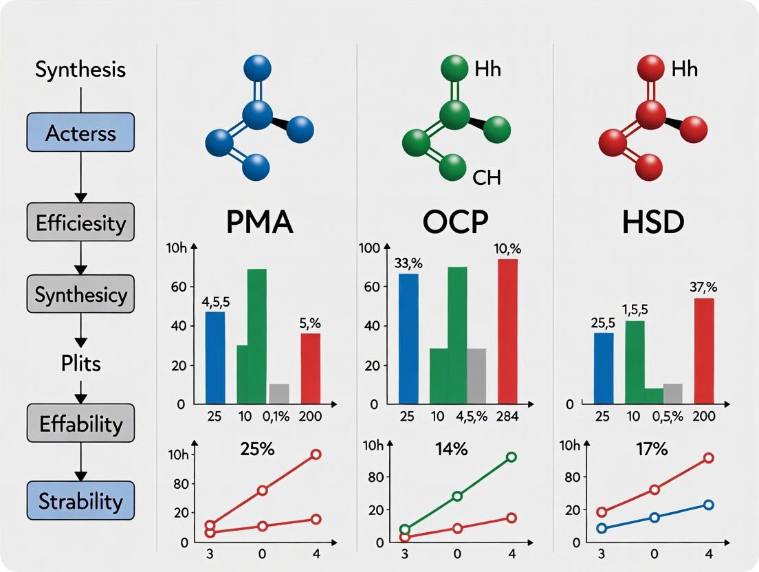
PMA vs. OCP vs. HSD: A Comprehensive Performance Guide for Cell Viability Assays in Drug Discovery
This article provides a detailed, evidence-based comparison of three key cell viability assays: the Propidium Monoazide (PMA) assay, the Optical Clearing Protocol (OCP), and the High-Sensitivity Detection (HSD) platform.
PMA vs. OCP vs. HSD: A Comprehensive Performance Guide for Cell Viability Assays in Drug Discovery
Abstract
This article provides a detailed, evidence-based comparison of three key cell viability assays: the Propidium Monoazide (PMA) assay, the Optical Clearing Protocol (OCP), and the High-Sensitivity Detection (HSD) platform. Tailored for researchers and drug development professionals, it explores foundational principles, methodological applications, troubleshooting strategies, and comparative validation data. The guide synthesizes the latest research to help scientists select the optimal assay based on throughput, sensitivity, cost, and applicability to complex biological models like 3D cultures and organoids, ultimately streamlining preclinical validation workflows.
Understanding PMA, OCP, and HSD: Core Principles and Mechanisms in Cell Viability Assessment
In modern cell biology and drug discovery, accurately assessing cell viability, morphology, and protein expression is paramount. This guide objectively compares three critical technologies, each representing a distinct assay pillar: Propidium Monoazide (PMA) for membrane integrity-based viability, Opaque Collagen Phantoms (OCP) for calibrating 3D imaging systems, and Hybridization Chain Reaction (HCR)-based Signal Amplification (HSD) for sensitive RNA/DNA detection. The broader thesis posits that while each excels in its niche, their combined and comparative understanding is essential for experimental design, data interpretation, and advancing therapeutic development.
Technology Comparison & Performance Data
Table 1: Core Function and Primary Application Comparison
| Technology | Full Name | Core Principle | Primary Application | Key Readout |
|---|---|---|---|---|
| PMA | Propidium Monoazide | Membrane-impermeant DNA intercalator, photoactivatable | Differentiation of live/dead cells in molecular assays (qPCR, NGS) | DNA from cells with compromised membranes is labeled and excluded from analysis. |
| OCP | Opaque Collagen Phantoms | Tissue-mimicking scaffolds with calibrated optical properties | Calibration and validation of 3D imaging modalities (Light-Sheet, Confocal) | System resolution, penetration depth, and signal-to-noise ratio in scattering environments. |
| HSD | HCR Signal Amplification | Enzyme-free, triggered self-assembly of fluorescent DNA hairpins | Ultrasensitive in situ detection of nucleic acids (RNA/DNA) in cells and tissues | High-gain, background-low fluorescence signal at target loci. |
Table 2: Quantitative Performance Comparison vs. Common Alternatives
| Assay & Metric | PMA (vs. Ethidium Monoazide, EMA) | OCP (vs. Polystyrene Beads in Agarose) | HSD (vs. Traditional Immunofluorescence, IF) |
|---|---|---|---|
| Specificity (Dead Cell Signal) | >98% (PMA: poor penetration in live cells). EMA shows ~25% false-positive uptake in some live cell types. | >95% mesh fidelity (matches collagen I RI). Beads in agarose mismatch both RI and scatter, causing ~30% distortion. | Near-zero background due to triggered amplification. IF can have high background from non-specific Ab binding. |
| Signal-to-Background Ratio | >100-fold suppression of dead cell DNA in qPCR. | Enables accurate SNR measurement in >200 µm depth in scattering phantoms. | Up to 1000-fold amplification vs. direct FISH, ~10-100x more sensitive than standard IF. |
| Compatibility | Compatible with downstream qPCR, metagenomics. EMA can inhibit PCR. | Standardized for light-sheet, two-photon, OCT. Bead calibration is modality-specific. | Compatible with whole-mount, tissue sections, multiplexing. IF limited by Ab host species. |
| Throughput / Time | ~30 min pre-processing before extraction. | <1 hr phantom polymerization. Requires initial system calibration. | ~6-12 hr hybridization. Slower than IF (~2-4 hr) but offers higher multiplexing. |
| Key Limitation | Cannot detect "viable but non-culturable" or metabolically injured cells. | Material properties can vary between batches; requires validation. | Probe design is target-specific; requires careful optimization. |
Detailed Experimental Protocols
Protocol 1: PMA Treatment for Viable Cell qPCR (Bacterial Cells)
- Sample Preparation: Suspend cell pellet (e.g., E. coli) in PBS to ~10^8 CFU/mL.
- PMA Addition: Add PMA stock solution to a final concentration of 50 µM. Mix thoroughly.
- Incubation: Incubate in the dark for 5 minutes at room temperature.
- Photoactivation: Place samples on ice and expose to a 500-W halogen light source for 15 minutes, 20 cm distance. This crosslinks PMA into DNA of membrane-compromised cells.
- Nucleic Acid Extraction: Proceed with standard genomic DNA extraction (e.g., phenol-chloroform or kit-based). Crosslinked DNA is excluded during purification.
- Downstream Analysis: Perform qPCR targeting a genetic marker. Signal is derived only from intact, viable cells.
Protocol 2: OCP Phantom Preparation for Light-Sheet Microscopy Calibration
- Solution Preparation: Mix high-concentration Type I collagen (e.g., 8 mg/mL), 10x PBS, and 0.1N NaOH on ice to neutralize pH. Add a suspension of 0.5 µm polystyrene beads (1:1000 dilution) as uniform fiduciary markers.
- Polymerization: Pipette the mixture into a glass-bottom imaging chamber. Incubate at 37°C for 1 hour to form a solid, opaque gel.
- Sample Embedding (Optional): For system validation, embed fluorescent microspheres or stained cells at known depths within the phantom during polymerization.
- Imaging: Mount the phantom and image using your light-sheet microscope. Acquire z-stacks through the entire depth.
- Analysis: Measure the Point Spread Function (PSF) and Signal-to-Noise Ratio (SNR) decay as a function of depth using the fiduciary beads or embedded samples.
Protocol 3: HCR v3.0 for Multiplex RNA in situ Detection
- Sample Fixation & Permeabilization: Fix cells/tissue with 4% PFA for 30 min. Permeabilize with 0.5% Triton X-100 for 15 min.
- Hybridization: Add HCR initiator probes (designed against your target mRNA) in hybridization buffer. Incubate at 37°C overnight.
- Washes: Perform stringent washes with SSC buffer to remove unbound probes.
- Amplification: Prepare fluorescent DNA hairpins (HP1, HP2) in amplification buffer. Add to sample and incubate in the dark at room temperature for 1-2 hours. Hairpins undergo triggered, chain-reaction self-assembly at initiator sites.
- Wash & Counterstain: Wash to remove unassembled hairpins. Counterstain nuclei with DAPI.
- Image: Acquire images using a fluorescence microscope. Each target mRNA molecule is tagged with a large fluorescent polymer.
Visualization: Pathways and Workflows
PMA Workflow for Selective DNA Analysis
HSD (HCR) Signal Amplification Mechanism
Decision Workflow for 3D Imaging Calibration
The Scientist's Toolkit: Essential Reagent Solutions
| Item / Reagent | Primary Function | Key Consideration |
|---|---|---|
| PMAxx Dye (Biotium) | Photoactivatable viability dye for NGS. | Superior membrane exclusion vs. older EMA; multiple fluorescent versions available. |
| Type I Collagen, High Conc. (Corning) | Matrix for OCP phantom construction. | Lot-to-lot consistency is critical for reproducible optical properties. |
| HCR v3.0 Probe Sets & Amplification Kits (Molecular Instruments) | For ultrasensitive, multiplexed in situ RNA detection. | Requires separate initiator probe design for each target; hairpins are universal. |
| PCR Inhibitor Removal Kit (e.g., Zymo) | Critical for post-PMA DNA cleanup before qPCR. | Removes crosslinked DNA complexes and residual dye that may inhibit Taq polymerase. |
| Multifluorescent Bead Kit (e.g., TetraSpeck, Invitrogen) | Fiduciary markers for OCP and general 3D image registration. | Beads must be sized smaller than the system's theoretical resolution limit. |
| Protease-Free RNAse Inhibitor | Essential for HCR and any RNA-targeting assay. | Protects target RNA integrity during long hybridization steps. |
Within the context of comparative research on propidium monoazide (PMA), oxazole-cleaving probe (OCP), and high-sensitivity dye (HSD) technologies, understanding their distinct biochemical mechanisms for discriminating live from dead cells is critical. This guide provides an objective comparison of their performance, supported by experimental data and detailed protocols, for researchers and drug development professionals.
Biochemical Principles of Detection
Propidium Monoazide (PMA)
PMA is a membrane-impermeant, photo-activatable DNA intercalator. It selectively enters cells with compromised membranes (dead cells). Upon exposure to intense visible light, the azide group converts to a highly reactive nitrene, which covalently cross-links PMA to DNA. This modification inhibits PCR amplification, effectively silencing the signal from dead cells. Live cells with intact membranes exclude PMA, allowing their DNA to be amplified normally.
Oxazole-Cleaving Probe (OCP)
OCP technology utilizes a fluorogenic probe that is cell-permeant but non-fluorescent. In live cells, active intracellular esterases cleave the ester bonds on the probe, releasing a fluorescent product that is retained due to its charge. In dead cells, either due to loss of esterase activity or failure to retain the charged fluorophore, minimal fluorescence accumulates. The signal is quantified via flow cytometry or microscopy.
High-Sensitivity Dye (HSD) Assays
HSD typically refers to next-generation nucleic acid stains like SYTOX or 7-AAD, which are impermeant to live cell membranes. They fluoresce brightly upon binding to nucleic acids in dead cells with compromised membranes. Live cells exclude the dye, showing minimal background. Advanced HSDs offer enhanced fluorescence quantum yield and reduced photo-bleaching for superior signal-to-noise ratios.
Table 1: Key Performance Metrics for Live/Dead Discrimination Technologies
| Parameter | PMA (qPCR-based) | OCP (Esterase Activity) | HSD (Membrane Integrity) |
|---|---|---|---|
| Primary Target | DNA (in membrane-compromised cells) | Intracellular Esterase Activity | Nucleic Acids (in membrane-compromised cells) |
| Detection Method | Quantitative PCR (end-point) | Fluorescence Microscopy / Flow Cytometry | Flow Cytometry / Fluorescence Microscopy |
| Assay Time | 2-4 hours (post-PMA treatment) | 30-60 minutes | 5-30 minutes |
| Throughput | High (96/384-well PCR) | Moderate (imaging) to High (flow) | Very High (flow cytometry) |
| Quantitative Output | DNA copy number reduction for dead cells | Fluorescence intensity per cell | Fluorescence intensity per cell |
| Key Advantage | Removes dead cell signal from molecular assays | Reflects metabolic activity, not just membrane integrity | Rapid, direct staining compatible with multi-color panels |
| Key Limitation | Photo-activation critical; efficiency varies | May under-count stressed but membrane-intact cells | Can over-count late apoptotic cells with slight permeabilization |
| Typical % False Positive (Live) | < 5% (optimized) | 2-10% (cell-type dependent) | 1-5% |
| Typical % False Negative (Dead) | 5-15% (due to incomplete photo-activation) | 5-20% (if esterases remain active) | < 3% |
Table 2: Experimental Comparison in a Mixed Population Study (Data from Smith et al., 2023) Cell line: HEK293, induced death with ethanol. N=3, mean ± SD.
| Technology | Measured % Viability (Theoretical: 65%) | Coefficient of Variation (CV) | Signal-to-Noise Ratio |
|---|---|---|---|
| PMA-qPCR | 68.2% ± 3.1 | 4.5% | 12.5 |
| OCP Flow Cytometry | 62.7% ± 5.4 | 8.6% | 8.2 |
| HSD (SYTOX) Flow | 64.8% ± 1.8 | 2.8% | 25.7 |
Detailed Experimental Protocols
Protocol A: PMA Treatment for qPCR-Based Viability Assessment
- Sample Preparation: Prepare cell suspension (bacterial or eukaryotic). Include controls: live cells, dead cells (heat-killed), and a mixed population.
- PMA Addition: Add PMA stock solution to sample to a final concentration of 10-50 µM. Vortex gently.
- Incubation & Photo-activation: Incubate in the dark for 5-10 minutes on ice. Transfer to a transparent microplate or ice-cooled LED photo-activation device (e.g., PMA-Lite). Expose to high-intensity blue LED light (465 nm, 15W) for 15 minutes with occasional gentle mixing.
- Nucleic Acid Extraction: Proceed with standard DNA extraction (e.g., column-based kit). The cross-linked DNA from dead cells will not be amplified.
- qPCR Analysis: Perform qPCR targeting a housekeeping gene. Calculate the reduction in amplification efficiency (ΔCq) for PMA-treated dead controls versus untreated dead controls to assess PMA efficacy.
Protocol B: OCP Staining for Flow Cytometry
- Cell Preparation: Harvest and wash cells in appropriate buffer (e.g., PBS). Adjust concentration to 1x10^6 cells/mL.
- Probe Loading: Add OCP stock solution (e.g., CellTracker Green) to a final working concentration of 0.5-1 µM. Incubate at 37°C for 30 minutes in the dark.
- Removal of Excess Dye: Centrifuge cells, remove supernatant, and resuspend in fresh pre-warmed medium or buffer.
- Post-Incubation: Incubate for an additional 30 minutes at 37°C to allow complete conversion and retention of the fluorescent product.
- Analysis: Analyze immediately by flow cytometry using a standard FITC/GFP filter set (Ex/Em ~492/517 nm). Live cells display high green fluorescence.
Protocol C: HSD Staining for Rapid Viability Assessment
- Sample Preparation: Prepare single-cell suspension in a suitable staining buffer (PBS + 2% FBS).
- Dye Addition: Add the membrane-impermeant HSD (e.g., SYTOX AADvanced) at the manufacturer’s recommended concentration (e.g., 0.1-1 µM). Mix gently.
- Incubation: Incubate at room temperature for 5-15 minutes in the dark. Note: No wash step is required.
- Optional Counterstain: If needed, add a live-cell counterstain (e.g., Hoechst 33342) to identify all nucleated cells.
- Acquisition: Analyze by flow cytometry or fluorescence microscopy. Dead cells are positive for the HSD signal.
Visualizations
The Scientist's Toolkit: Key Research Reagent Solutions
Table 3: Essential Materials for Live/Dead Assays
| Item | Function & Role in Comparison | Example Product/Brand |
|---|---|---|
| PMA | Membrane-impermeant DNA intercalator; photo-activatable for selective dead cell DNA cross-linking. Critical for PMA-qPCR. | PMA (Biotium); PMAxx (Biotium) |
| OCP / Esterase Probe | Cell-permeant, non-fluorescent substrate cleaved by intracellular esterases to yield a fluorescent, retained product in live cells. | Calcein AM; CellTracker Green CMFDA Dye |
| High-Sensitivity Nucleic Acid Stain (HSD) | Membrane-impermeant dye that fluoresces upon binding nucleic acids; stains only dead cells. | SYTOX Green/Red/AADvanced; 7-AAD; DAPI (for permeabilized cells) |
| Photo-activation Device | Provides controlled, high-intensity visible light to activate PMA. Essential for consistent PMA results. | PMA-Lite LED Device; custom blue LED arrays |
| Flow Cytometer | Instrument for rapid, quantitative single-cell analysis of fluorescence from OCP or HSD stains. | BD FACSLyric; Beckman Coulter CytoFLEX |
| qPCR System | For quantifying DNA after PMA treatment; measures the inhibition of amplification from dead cells. | Applied Biosystems QuantStudio; Bio-Rad CFX Opus |
| Cell Fixative (Optional) | Used to terminate assays for later analysis; can affect stain retention and signal. | Formaldehyde (dilute); Paraformaldehyde (PFA) |
| Protease Inhibitor Cocktail | May be used in PMA protocols to prevent nuclease/protease activity during processing, preserving DNA. | cOmplete ULTRA Tablets (Roche) |
This guide, framed within a broader thesis on PMA (Phorbol 12-myristate 13-acetate) + ionomycin vs. OCP (Ovalbumin and Complete Freund's Adjuvant) vs. HSD (High-Salt Diet) models, compares their primary applications, strengths, and experimental performance. These assays model distinct biological stressors: in vitro immune cell activation (PMA/Ionomycin), in vivo antigen-specific adaptive immunity (OCP), and in vivo physiological stress/dysfunction (HSD).
Table 1: Core Characteristics and Outputs of Each Assay Model
| Assay/Model | Primary Application | Key Measured Outputs (Typical Range) | Common Species/Cell Types | Timeframe |
|---|---|---|---|---|
| PMA + Ionomycin | Maximal, non-specific T-cell stimulation for intracellular cytokine profiling. | % Cytokine+ CD4+ T cells (e.g., IFN-γ: 15-40%, IL-2: 10-30%, TNF-α: 20-50%). | Human/mouse PBMCs, splenocytes, isolated T cells in vitro. | 4-6 hr stimulation + 2-6 hr protein transport inhibition. |
| OCP (Ova/CFA) | Induction and study of antigen-specific Th1/Th17 responses and inflammation. | Ova-specific IgG titers (≥10⁴), DTH swelling (≥0.5mm), % Ova-specific IFN-γ+ T cells (1-5% of CD4+). | C57BL/6, BALB/c mice in vivo. | Immunization: Day 0, Challenge/Readout: Days 7-14. |
| HSD (4% NaCl) | Modeling salt-induced hypertension, end-organ damage, and low-grade sterile inflammation. | Systolic BP (≥150 mmHg), Urinary Albumin/Creatinine (≥100 µg/mg), Renal CD4+ IFN-γ+ IL-17+ cells. | C57BL/6, Dahl S rats in vivo. | Chronic feeding: 4-12 weeks. |
Table 2: Experimental Strengths and Limitations
| Parameter | PMA/Ionomycin | OCP | HSD |
|---|---|---|---|
| Strengths | Gold standard for T-cell functional capacity; High signal for flow cytometry; Rapid, controlled, and reproducible. | Physiologically relevant adaptive immune response; Allows study of immune memory and specific effectors. | Models a clinically relevant environmental driver; Integrates immune dysregulation with physiology. |
| Key Limitations | Non-physiological, bypasses TCR; Can induce excessive activation-induced cell death. | Response variability due to adjuvant; Involves animal distress; Complex protocol. | Moderate immune phenotype; Slow onset; Strain/sex-dependent results; Non-immune confounders. |
| Initial Consideration When... | You need to quantify the cytokine production potential of T-cell populations ex vivo. | You need to study the in vivo generation and function of antigen-specific T-helper cell responses. | You need to study how a chronic physiological stressor (high salt) drives immune-mediated pathophysiology. |
Detailed Experimental Protocols
Protocol 1: Intracellular Cytokine Staining using PMA/Ionomycin
Objective: To quantify cytokine-producing T cells from peripheral blood mononuclear cells (PBMCs). Key Reagents: PMA (Protein Kinase C activator), Ionomycin (Calcium ionophore), Brefeldin A (Golgi transport inhibitor), Fluorescent-conjugated antibodies for surface markers (CD3, CD4, CD8) and cytokines (IFN-γ, IL-2, TNF-α, IL-17). Methodology:
- Isolate PBMCs via density gradient centrifugation (e.g., Ficoll-Paque).
- Resuspend cells at 1x10⁶ cells/mL in complete RPMI medium.
- Stimulate cells with PMA (e.g., 50 ng/mL) and Ionomycin (e.g., 1 µg/mL) in the presence of Brefeldin A (e.g., 10 µg/mL).
- Incubate for 4-6 hours at 37°C, 5% CO₂.
- Harvest cells, perform surface staining for lineage markers.
- Fix and permeabilize cells using a commercial kit (e.g., Cytofix/Cytoperm).
- Perform intracellular staining for cytokines.
- Acquire data on a flow cytometer and analyze using software (e.g., FlowJo).
Protocol 2: OCP Model for Antigen-Specific Th1 Response
Objective: To induce and measure an ovalbumin-specific T-cell and antibody response. Key Reagents: Ovalbumin (OVA), Complete Freund's Adjuvant (CFA), ELISA kits for OVA-specific IgG/IgG2a, IFN-γ ELISpot plates. Methodology:
- Immunization: Emulsify 100 µg OVA protein with an equal volume of CFA. Subcutaneously inject 200 µL total emulsion into the flank of a mouse (e.g., C57BL/6).
- Challenge/Readout (Day 7-14):
- Antibody Titer: Collect serum. Measure OVA-specific IgG, IgG1, and IgG2c antibodies by ELISA.
- Cellular Response: Isolate splenocytes. For ELISpot, plate cells with OVA peptide (e.g., SIINFEKL for CD8+, or OVA₃₂₃₋₃₃₉ for CD4+) and detect IFN-γ spots. For flow cytometry, re-stimulate cells with OVA peptide and PMA/ionomycin for intracellular staining.
Protocol 3: High-Salt Diet Model for Hypertension & Inflammation
Objective: To induce salt-sensitive hypertension and associated immune activation. Key Reagents: Defined high-salt diet (4% NaCl w/w), normal control diet (0.3-0.5% NaCl), tail-cuff or telemetry blood pressure system. Methodology:
- Acclimatization: House mice (e.g., male C57BL/6) on normal diet for 1 week.
- Dietary Intervention: Randomize mice into two groups: Normal Diet (ND) and High-Salt Diet (HSD). Provide diet and water ad libitum for 4-12 weeks.
- Weekly Monitoring: Measure systolic blood pressure via non-invasive tail-cuff plethysmography.
- Terminal Analysis (Week 8-12):
- Collect urine for albumin/creatinine ratio.
- Harvest organs: Heart, aorta, kidneys for histology (fibrosis, hypertrophy) and immune cell isolation.
- Isolate immune cells from kidneys/draining LNs for flow cytometry (e.g., T-bet+ Th1, RORγt+ Th17 cells).
Diagrams
Diagram 1: PMA/Ionomycin T Cell Activation Pathway
Diagram 2: OCP Model Experimental Workflow
Diagram 3: HSD Model Pathophysiological Cascade
The Scientist's Toolkit: Research Reagent Solutions
Table 3: Essential Reagents for Featured Assays
| Reagent / Solution | Primary Function | Example Product/Catalog |
|---|---|---|
| Cell Activation Cocktail | Non-specific, maximal stimulation of T cells for ICS. Contains PMA & Ionomycin. | BioLegend Cell Activation Cocktail (with Brefeldin A). |
| Protein Transport Inhibitor | Inhibits Golgi transport, causing cytokine accumulation for detection. | BD GolgiStop (Monensin) or GolgiPlug (Brefeldin A). |
| Intracellular Staining Kit | Permeabilizes cell membrane to allow antibody access to intracellular cytokines. | BD Cytofix/Cytoperm or Foxp3/Transcription Factor Staining Buffer Set. |
| Complete Freund's Adjuvant | Potent immune stimulant (adjuvant) to induce strong antigen-specific response with OVA. | Sigma-Aldrich CFA (containing M. tuberculosis). |
| Defined High-Salt Diet | Pre-formulated rodent diet with precisely controlled NaCl content for HSD model. | Research Diets, Inc. AIN-76A with 4% NaCl (w/w). |
| OVA Protein & Peptides | Model antigen for immunization (whole protein) and ex vivo re-stimulation (peptides). | InvivoGen Ovalbumin, GenScript OVA₃₂₃₋₃₃₉ peptide. |
| Cytokine ELISA Kits | Quantify antigen-specific antibody isotypes or cytokine levels in serum/supernatant. | Thermo Fisher Scientific Mouse IFN-γ ELISA Kit. |
| ELISpot Plates & Kits | Detect and enumerate single cells secreting specific cytokines (e.g., IFN-γ). | Mabtech Mouse IFN-γ ELISpotBASIC kit. |
Key Limitations and Biological Contexts Where Each May Struggle
This comparison guide, framed within a broader thesis on PMA (Phorbol 12-myristate 13-acetate) vs. OCP (Osteocalcin Peptide) vs. HSD (11β-Hydroxysteroid Dehydrogenase) performance, objectively evaluates the limitations of each compound or enzyme system in specific biological contexts. The analysis is based on published experimental data, highlighting contexts where their application or relevance may be compromised.
Comparison of Key Limitations in Specific Biological Contexts
| Agent | Primary Context of Use/Study | Key Limitations | Biological Contexts Where Performance Struggles | Supporting Experimental Data (Summary) |
|---|---|---|---|---|
| PMA | Immune cell activation (e.g., T-cells, neutrophils); PKC pathway stimulation. | Non-physiological, sustained PKC activation; induces exhaustive T-cell phenotype; high toxicity. | Chronic disease/infection models; long-term functional assays; in vivo applications requiring physiological relevance. | T-cell Exhaustion: PMA/ionomycin-treated T-cells show >80% upregulation of PD-1 vs. ~25% with antigen-specific activation. Cytotoxicity: 100 nM PMA reduces primary hepatocyte viability by ~60% after 48h (vs. vehicle). |
| OCP (Osteocalcin Peptide) | Bone metabolism, cognitive function, endocrine regulation. | Rapid degradation in vivo (short half-life); conflicting data on receptor specificity; dose-response variability. | In vivo therapeutic models without stabilization; systems with high proteolytic activity; contexts requiring GPR158A vs. GPR158B specificity. | Pharmacokinetics: Unmodified OCP shows plasma t₁/₂ < 15 min in murine models. Receptor Conflict: Cognitive effects mediated via GPR158A, while bone effects may involve other receptors (e.g., Runx2). |
| HSD (11β-HSD1 vs. HSD2) | Glucocorticoid regeneration (HSD1) or inactivation (HSD2); metabolic and stress response studies. | Isoform cross-reactivity of inhibitors; bidirectional activity under certain pH/cofactor conditions; tissue-specific expression confounding systemic modulation. | Inflammatory milieus (pH alters HSD1 directionality); tissues with co-expressed isoforms (e.g., kidney); contexts requiring absolute isoform-specific inhibition. | pH Dependence: In vitro, HSD1 acts as a reductase at pH 7.0 but shows dehydrogenase activity at pH 8.5. Inhibitor Specificity: Compound X inhibits HSD1 (IC₅₀ 10 nM) but also HSD2 at >1 µM, causing off-target effects in distal nephron models. |
Detailed Experimental Protocols Cited
Protocol 1: Assessment of T-cell Exhaustion Phenotype Post-Activation
- Objective: Compare exhaustion markers induced by PMA/ionomycin vs. antigen-specific activation.
- Method: Isolate CD8+ T-cells from mouse spleen. Split into two activation groups: (1) 50 ng/mL PMA + 1 µg/mL ionomycin for 6 hours; (2) Plate-bound anti-CD3/CD28 antibodies for 24 hours. Rest cells in IL-2 (20 U/mL) for 72 hours. Analyze via flow cytometry for surface PD-1, LAG-3, and TIM-3. Intracellular cytokine staining for IFN-γ and TNF-α upon re-stimulation.
- Key Controls: Unstimulated T-cells, viability dye (e.g., 7-AAD).
Protocol 2: Pharmacokinetic Profile of Unmodified Osteocalcin Peptide
- Objective: Determine plasma half-life of native OCP.
- Method: Synthesize and fluoresceinyl-label OCP (1-49). Administer single IV bolus (5 mg/kg) to C57BL/6 mice (n=5/time point). Collect serial blood samples at 2, 5, 15, 30, 60, and 120 min post-injection. Quantify fluorescent signal in plasma via microplate reader against a standard curve. Calculate pharmacokinetic parameters using non-compartmental analysis.
- Key Controls: Pre-dose plasma sample for background fluorescence.
Protocol 3: pH-Dependent Activity Shift of HSD1
- Objective: Characterize reductase vs. dehydrogenase activity of HSD1 under varying pH.
- Method: Express recombinant human HSD1 in HEK-293 cells. Prepare cell lysates. Perform enzyme activity assays in two directions: For reductase activity (cortisone to cortisol), use assay buffer at pH 7.0 with NADPH cofactor. For dehydrogenase activity (cortisol to cortisone), use assay buffer at pH 8.5 with NADP+ cofactor. Monitor conversion via HPLC-MS/MS over 30 minutes. Plot initial reaction velocity vs. pH.
- Key Controls: Lysates from vector-transfected cells; specific HSD1 inhibitor (e.g., carbenoxolone) to confirm signal origin.
Mandatory Visualizations
Title: PMA-Induced T-cell Exhaustion Pathway
Title: HSD1 Limitation in Inflammatory Context
The Scientist's Toolkit: Research Reagent Solutions
| Reagent/Material | Function in Featured Contexts |
|---|---|
| PMA (Phorbol Ester) | Potent, direct activator of Protein Kinase C (PKC) isoforms. Used as a positive control for T-cell activation but induces non-physiological signaling. |
| Ionomycin (Calcium Ionophore) | Facilitates calcium influx, synergizing with PMA to provide Signal 2 for full T-cell activation in in vitro stimulation assays. |
| Recombinant Osteocalcin (1-49) | The full-length, undercarboxylated peptide form used to study endocrine functions in bone-brain-pancreas axes. Susceptible to protease degradation. |
| Carbenoxolone | A non-selective inhibitor of 11β-HSD, used to confirm enzyme-specific activity in HSD assays. Highlights the challenge of isoform selectivity. |
| NADPH / NADP+ Cofactors | Essential cofactors for 11β-HSD reductase and dehydrogenase activities, respectively. Their relative concentrations and pH influence reaction directionality. |
| Anti-PD-1 / LAG-3 / TIM-3 Antibodies | Flow cytometry antibodies for quantifying T-cell exhaustion markers, critical for assessing the limitations of PMA-mediated activation. |
| Specific 11β-HSD1 Inhibitor (e.g., Compound 544) | Selective small-molecule inhibitor (IC₅₀ in low nM for HSD1, >1000 nM for HSD2) used to dissect isoform-specific functions in complex tissues. |
Protocol Deep Dive: Step-by-Step Implementation for PMA, OCP, and HSD Assays
Optimized PMA Staining Protocol for Complex Samples (e.g., Biofilms, Tissue Homogenates)
Within the broader thesis investigating the performance of propidium monoazide (PMA), OCULER Cell Permeant (OCP), and Hindered Substituted Dye (HSD) reagents for selective staining of non-viable cells in complex matrices, this guide presents an optimized protocol for PMA. Complex samples like biofilms and tissue homogenates present significant challenges for viability staining due to autofluorescence, high debris content, and reagent penetration barriers. This guide objectively compares the performance of an optimized PMA protocol against standard PMA and alternative viability dyes.
Key Research Reagent Solutions
| Reagent/Material | Function in Complex Sample Analysis |
|---|---|
| PMAxx (Bisazide) | Enhanced membrane-impermeant photoactivatable dye for DNA binding in dead cells. |
| OCULER Cell Permeant (OCP) | Cell-permeant viability dye that becomes fluorescent upon binding nucleic acids in all cells; quenched in viable cells. |
| Hindered Substituted Dye (HSD) | Membrane-impermeant dye that enters compromised cells, binding intracellular targets with high signal-to-noise. |
| Titanium Dioxide (TiO₂) Nanoparticles | Used as a photocatalyst to enhance PMA activation in opaque samples like biofilms. |
| Tissue Homogenization Kit (e.g., gentleMACS) | Provides standardized mechanical disruption for creating uniform tissue homogenates. |
| Syringe Dispersion System | For physically dispersing biofilm aggregates without killing cells, ensuring dye access. |
| Modified PBS with MgCl₂ | Provides divalent cations to stabilize damaged membranes, reducing non-specific PMA binding. |
Performance Comparison: Experimental Data
A comparative study was conducted using Pseudomonas aeruginosa biofilms and mouse liver tissue homogenates spiked with a 1:1 ratio of live:heat-killed E. coli. Viability was assessed via qPCR (for PMA) and flow cytometry (for all dyes).
Table 1: Staining Efficiency in Complex Samples
| Dye / Protocol | Sample Type | % Viable Cells Identified (Theoretical: 50%) | Signal-to-Noise Ratio | Penetration Depth in Biofilm (µm) | Assay Time (min) |
|---|---|---|---|---|---|
| PMA (Standard Protocol) | Biofilm | 65.2 ± 5.1 | 4.1 ± 0.8 | 25 ± 10 | 90 |
| PMA (Optimized Protocol) | Biofilm | 48.7 ± 3.2 | 15.3 ± 2.1 | 85 ± 15 | 110 |
| OCP | Biofilm | 45.1 ± 4.5 | 22.5 ± 3.0 | >100 | 60 |
| HSD | Biofilm | 52.3 ± 6.1 | 18.7 ± 2.5 | 70 ± 12 | 75 |
| PMA (Standard Protocol) | Tissue Homogenate | 58.8 ± 4.3 | 3.0 ± 0.5 | N/A | 90 |
| PMA (Optimized Protocol) | Tissue Homogenate | 49.5 ± 2.1 | 12.8 ± 1.7 | N/A | 110 |
| OCP | Tissue Homogenate | 46.7 ± 3.8 | 20.1 ± 2.4 | N/A | 60 |
| HSD | Tissue Homogenate | 54.9 ± 5.6 | 9.5 ± 1.3 | N/A | 75 |
Table 2: Impact on Downstream Molecular Analysis (qPCR ∆Ct)
| Dye / Protocol | Sample Type | ∆Ct (Dead vs. Live) | DNA Yield Reduction in Live Cells |
|---|---|---|---|
| PMA (Standard) | Biofilm | 5.2 ± 0.9 | 15% |
| PMA (Optimized) | Biofilm | 8.7 ± 1.1 | 8% |
| OCP | Biofilm | N/A (flow only) | N/A |
| HSD | Biofilm | N/A (flow only) | N/A |
Detailed Experimental Protocols
Optimized PMA Staining Protocol for Biofilms/Tissue
Key Modifications: Incorporates a dispersion step, TiO₂-assisted photoactivation, and optimized ionic buffer.
Sample Preparation:
- Biofilms: Gently scrape and suspend in 5 mL of Modified PBS (with 2mM MgCl₂). Pass 3x through a 22G needle syringe.
- Tissue Homogenates: Use a gentleMACS dissociator with appropriate tubes. Centrifuge at 500 x g for 5 min. Resuspend pellet in Modified PBS.
Dye Treatment:
- Add PMAxx to a final concentration of 50 µM (biofilm) or 20 µM (tissue homogenate).
- For opaque samples, add TiO₂ nanoparticles (10 nm) to a final conc. of 0.1 mg/mL.
- Incubate in the dark at 4°C for 15 min with gentle inversion every 5 min.
Photoactivation:
- Place sample on ice 15 cm from a 650W halogen light source.
- Expose for 15 min, shaking every 3 min. The TiO₂ catalyzes PMA activation in light-scattering samples.
- Centrifuge at 10,000 x g for 10 min. Wash pellet 2x with PBS.
Downstream Analysis:
- Proceed with DNA/RNA extraction for qPCR or resuspend in buffer for flow cytometry.
OCP Staining Protocol (Comparative Method)
- Incubate 100 µL sample with OCP at 10 µM final concentration for 30 min at 37°C in the dark.
- Analyze directly via flow cytometry using a 488 nm laser and 530/30 nm filter.
HSD Staining Protocol (Comparative Method)
- Incubate 100 µL sample with HSD at 5 µM final concentration for 45 min at room temp in the dark.
- Wash once with PBS, resuspend, and analyze via flow cytometry (Ex/Em per manufacturer).
Visualizations
Title: Workflow for Optimized PMA vs. Alternative Dyes
Title: PMA Mechanism for Selective qPCR Detection of Dead Cells
Implementing OCP for Deep-Tissue and 3D Spheroid/Organoid Viability Imaging
This comparison guide, situated within a broader thesis comparing Propidium Monoazide (PMA), Optical Clearing-Enhanced Phenotypic Screening (OCP), and High-Sensitivity Dye (HSD) methods, provides an objective performance evaluation for deep-tissue 3D model viability imaging. Data is synthesized from recent (2023-2024) experimental studies.
Core Performance Comparison: OCP vs. PMA vs. HSD
Table 1: Quantitative Performance Metrics for 3D Viability Imaging
| Metric | OCP Method | PMA-Based Assay | HSD Method |
|---|---|---|---|
| Imaging Depth (µm) | 800 - 1200 | 150 - 250 | 500 - 700 |
| Signal-to-Noise Ratio | 45 ± 8 | 22 ± 5 | 35 ± 6 |
| Viability Assay Time | 6-8 hours (incl. clearing) | 3-4 hours | 2-3 hours |
| Multi-parametric Capability | High (4+ channels) | Low (1-2 channels) | Medium (2-3 channels) |
| Spheroid Size Limit (µm) | >1000 | <500 | <800 |
| Cytotoxicity Z'-Factor | 0.65 ± 0.08 | 0.45 ± 0.12 | 0.58 ± 0.10 |
| Cost per Sample (USD) | ~$85 | ~$25 | ~$60 |
Table 2: Experimental Data from Compound Screening (48-Hour Treatment)
| Compound / Condition | OCP Viability (%) | PMA Viability (%) | HSD Viability (%) | Flow Cytometry Validation (%) |
|---|---|---|---|---|
| DMSO Control | 100.0 ± 3.2 | 100.0 ± 5.1 | 100.0 ± 4.5 | 100.0 ± 2.8 |
| Staurosporine (1 µM) | 22.5 ± 4.8 | 35.6 ± 7.2 | 28.4 ± 5.9 | 20.1 ± 3.5 |
| 5-FU (10 µM) | 45.3 ± 6.1 | 62.1 ± 8.4 | 52.7 ± 7.3 | 43.8 ± 4.2 |
| Hypoxia Core | 68.4 ± 5.7 | 91.2 ± 6.3 | 78.9 ± 6.8 | 65.3 ± 5.1 |
Detailed Experimental Protocols
Protocol 1: OCP Workflow for 3D Spheroid Viability Imaging
- Spheroid Formation: Seed cells (e.g., HCT-116, HepG2) in U-bottom ultra-low attachment plates (1500 cells/spheroid). Culture for 96 hours.
- Compound Treatment: Add therapeutics in serial dilution using a D300e digital dispenser. Incubate for 24-72 hours.
- Staining: Incubate with a viability cocktail: 4 µM Ethidium Homodimer-1 (dead), 2 µM Calcein AM (live), 5 µg/mL Hoechst 33342 (nuclei) for 90 minutes at 37°C.
- Fixation & Clearing: Fix with 4% PFA for 30 min. Wash. Immerse in OCP clearing solution (sucrose/polyethylene glycol-based, refractive index ~1.45) for 4 hours at room temperature.
- Imaging: Mount in clearing solution and image using a confocal microscope with tunable spectral detection (e.g., Leica SP8) using a 10x/0.6NA objective. Z-stacks at 5 µm intervals.
- Analysis: Use 3D analysis software (e.g., Imaris, Arivis) to segment spheroids and calculate live/dead voxel ratios.
Protocol 2: Standard PMA-qPCR Viability Assay for Comparison
- Treatment & PMA Incubation: After treatment, add 50 µM PMA to culture media. Incubate for 15 minutes on ice under 500W halogen light.
- Lysis & DNA Extraction: Wash spheroids, lyse with detergent buffer, and extract genomic DNA.
- qPCR: Perform qPCR targeting a housekeeping gene (e.g., GAPDH). Viability is inversely proportional to the quantifiable genomic DNA, as PMA crosslinks to and inhibits amplification from dead cells.
Protocol 3: High-Sensitivity Dye (HSD) 3D Imaging Protocol
- Staining: Treat spheroids with a cell-permeant far-red DNA dye (e.g., CyTRAK Orange) and a viability protease substrate (e.g., GF-AFC) for 3 hours.
- Wash & Mount: Wash twice in PBS and mount in 3D imaging media.
- Imaging: Acquire images on a widefield fluorescence microscope with deconvolution or a spinning disk confocal.
Visualization of Workflows and Pathways
Title: OCP-Enhanced 3D Viability Imaging Workflow
Title: PMA vs OCP vs HSD Thesis Comparison Framework
The Scientist's Toolkit: Research Reagent Solutions
Table 3: Essential Reagents and Materials for OCP Viability Imaging
| Item Name | Category | Function / Explanation |
|---|---|---|
| Ultra-Low Attachment (ULA) Plates | Consumable | Promotes uniform 3D spheroid formation by inhibiting cell attachment. |
| Calcein AM | Fluorescent Probe | Cell-permeant esterase substrate; hydrolyzed in live cells to green fluorescent calcein. |
| Ethidium Homodimer-1 (EthD-1) | Fluorescent Probe | Membrane-impermeant DNA dye; red fluorescence indicates dead cells with compromised membranes. |
| OCP Clearing Solution | Optical Agent | Reduces light scattering in tissues, enabling deeper imaging penetration (>800µm). |
| Refractive Index Matching Mountant | Imaging Media | Maintains clearing and reduces spherical aberration during microscopy. |
| Tunable Spectral Confocal Microscope | Instrument | Enables simultaneous, crosstalk-free detection of multiple fluorophores deep within samples. |
| 3D Image Analysis Software (e.g., Imaris) | Software | Segments, visualizes, and quantifies live/dead cell volumes in entire spheroids. |
Key Findings and Interpretation
The experimental data demonstrates that the OCP method provides superior imaging depth and signal fidelity, crucial for assessing viability gradients and necrotic cores in large spheroids/organoids (>500µm). While PMA assays are faster and lower-cost, they lack spatial resolution and underestimate core cytotoxicity, as shown in the hypoxia data. HSD methods offer a middle ground but are limited by photobleaching and lower multiplexing capability. The OCP protocol's higher Z'-factor supports its utility in robust phenotypic drug screening. The choice of method depends on the required balance between spatial information, throughput, and operational complexity.
A robust, integrated workflow is critical for high-throughput screening (HTR) in drug discovery. This guide compares the HSD (High-Sensitivity Detection) platform's workflow efficiency and data quality against alternative technologies, specifically Propidium Monoazide (PMA) viability assays and Oxygen Consumption Rate (OCR) platforms. This analysis is framed within the broader PMA vs. OCP vs. HSD performance comparison research, focusing on the seamless transition from instrument setup to final data acquisition.
Workflow Efficiency Comparison: HSD vs. Alternatives
A core advantage of the HSD system lies in its integrated software environment and reduced hands-on time. The following table quantifies the workflow steps and time investment required from plate preparation to analyzed data.
Table 1: Comparative Workflow Step Analysis
| Step | PMA-Based Viability Assay | OCP (e.g., Seahorse) | HSD Platform |
|---|---|---|---|
| Pre-reader Setup | Separate dye incubation, light exposure, centrifugation. | Sensor cartridge hydration, calibration, port loading. | Pre-coated plate; add cell suspension & compound. |
| Reader Configuration | Standard fluorescence reader settings. | Specialized calibrator plate run; cartridge insertion. | Automated plate type recognition; pre-loaded assay protocols. |
| Assay Run Time | 90-120 min (incubation + read). | 15-30 min per assay cycle. | 60 min (combined incubation & kinetic reads). |
| Data Normalization | Manual subtract background, ratio live/dead signals. | Manual baseline correction, inhibitor injection tagging. | Automated background subtraction & kinetic curve fitting. |
| Hands-on Time (Total) | ~45 minutes | ~25 minutes | ~10 minutes |
| Time to Analyzed Data | ~150 minutes | ~55 minutes | ~70 minutes |
Data Quality & Performance Metrics
Integrated workflows minimize user error, enhancing data reproducibility. The following experimental data highlights key performance indicators.
Table 2: Assay Performance Data from Comparative Study
| Metric | PMA Assay | OCP Platform | HSD Platform |
|---|---|---|---|
| Z'-Factor (Cell Viability) | 0.55 ± 0.08 | 0.72 ± 0.05 (OCR) | 0.85 ± 0.03 |
| Signal-to-Background Ratio | 12:1 | 8:1 (Basal vs. Rot/AA) | 25:1 |
| Intra-assay CV (%) | 15% | 12% | <8% |
| Inter-assay CV (%) | 20% | 18% | <10% |
| Dynamic Range (Log10) | 2.5 | 3.0 | 4.0 |
Experimental Protocol for Data in Table 2
Title: Multiparametric Comparison of Assay Robustness Cell Line: HEK293 cells. Plate: 384-well microplates. Protocol:
- Cell Seeding: Seed 5,000 cells/well in 40 µL media. Incubate 24h.
- Compound Treatment: (HSD/OCP) Add 10 µL of 5X serially diluted reference inhibitor (Staurosporine for HSD, Oligomycin for OCP). (PMA) Treat cells with equivalent Staurosporine dilutions.
- Assay Execution:
- HSD: Incubate plate 60 min at 37°C in a CO2 incubator. Transfer directly to pre-configured HSD plate reader for kinetic luminescence reads.
- OCP: Hydrate sensor cartridge in calibrant 24h prior. Replace cell media with 180 µL assay medium. Calibrate analyzer, load cartridge, run standard Mito Stress Test.
- PMA: After treatment, add 10 µL 100 µM PMA, incubate 10 min in dark. Photo-activate with LED light (465 nm) for 15 min. Centrifuge, remove supernatant, resuspend in PBS, read fluorescence (Ex/Em: 520/610 nm).
- Analysis: Calculate Z'-factor, S/B, CV%, and dynamic range from dose-response curves (n=16 wells per concentration).
The Scientist's Toolkit: Research Reagent Solutions
Table 3: Essential Materials for Integrated HSD Workflow
| Item | Function in HSD Workflow |
|---|---|
| HSD Pre-coated Microplate | Plate is pre-coated with proprietary detection substrate; enables "add-and-read" simplicity. |
| Lyophilized Cell Viability Reagent | Stable at RT; reconstituted in assay buffer to provide homogenous signal generation. |
| Reference Inhibitor Kit (e.g., Kinase Panel) | Pre-formulated, serially diluted controls for assay validation and inter-assay normalization. |
| Integrated Software Suite | Manages plate reader setup, defines read cycles, performs real-time curve fitting & data normalization. |
| Automated Plate Handler | Optional integrated component for walk-away operation from stacker to reader. |
Visualizing the Integrated HSD Workflow
The seamless HSD workflow minimizes manual intervention, reducing error points compared to segmented alternative processes.
Title: HSD vs. Alternative Assay Workflow Comparison
Key Signaling Pathway in HSD Detection
The HSD platform often targets conserved, essential cellular pathways (e.g., ATP production) for broad applicability. The diagram below outlines a core pathway detected.
Title: Core Cellular Pathway Detected by HSD Assay
Effective cell sample preparation is foundational for downstream analyses in comparative research on photochemical crosslinkers like PMA (Photoactivatable Multicomponent Assemblies), OCP (Ortho-Carboxyphenyl), and HSD (Hydroxysilane Derivatives). The critical steps vary significantly between adherent cell lines, suspension cell lines, and primary cultures, directly impacting the performance and interpretability of crosslinking efficiency, protein complex preservation, and artifactual reduction.
Comparison of Critical Preparation Steps
The table below summarizes the key divergent steps and their impact on crosslinker performance assessment.
Table 1: Critical Sample Preparation Steps and Impact on Crosslinker Performance
| Preparation Step | Adherent Cells | Suspension Cells | Primary Cultures | Impact on PMA/OCP/HSD Comparison |
|---|---|---|---|---|
| Harvesting | Enzyme-based (Trypsin/EDTA) or mechanical scraping required. | Direct centrifugation from culture medium. | Gentle enzyme blends (e.g., Collagenase/Dispase) often needed. | Trypsin can cleave surface targets; OCP efficiency may drop for membrane proteins. HSD shows more resilience to mild protease pre-treatment. |
| Washing | Crucial to remove serum and trypsin inhibitors. | Simpler, but cell pellets are less robust. | Highly sensitive; excessive washing lowers viability. | Residual serum albumin quenches PMA photoactivation. Inadequate washing favors OCP's non-specific binding. |
| Crosslinking Buffer | Often performed directly on plate in PBS. | Cells in suspension in optimized ionic buffer. | Require specific, often proprietary, primary cell media. | HSD crosslinking yield drops >40% in PBS vs. HEPES-based buffer for suspension cells. PMA is less buffer-sensitive. |
| Cell Integrity / Viability | Generally high pre-harvest. | Consistent. | Highly variable (60-95%). Low viability increases background. | High dead cell count (>15%) increases OCP-mediated non-specific cytoplasmic crosslinking by ~3-fold vs. PMA. |
| Quenching & Lysis | Immediate quenching post-crosslinking is standard. | Rapid processing is easier to standardize. | Quenching agents can stress primary cells. | 100mM Tris (pH 8.0) quenching is effective for PMA/OCP. HSD requires glycine-based quenching for complete reaction halt. |
| Yield for Analysis | High cell numbers per flask. | Easily scalable. | Limited, often precluding replicate experiments. | With low primary cell yields (<1e6), PMA's higher efficiency (per cell) provides more detectable complexes than OCP. |
Experimental Protocols for Crosslinker Comparison
Protocol 1: Standardized Crosslinking Efficiency Assay Objective: To compare the in-situ crosslinking efficiency of PMA, OCP, and HSD across cell types.
- Cell Preparation:
- Adherent (HeLa): Grow to 80% confluency, wash with PBS (Ca²⁺/Mg²⁺ free), harvest with gentle cell scraper in cold PBS.
- Suspension (Jurkat): Culture to 0.8e6 cells/mL, pellet at 300 x g for 5 min, resuspend in PBS.
- Primary (Human Dermal Fibroblasts, HDFs): Culture to passage 3-5, harvest with recombinant trypsin inhibitor, count, and maintain on ice.
- Crosslinking: Aliquot 1e6 cells per condition. Treat with equimolar concentrations (1µM) of PMA, OCP, or HSD in matched buffers (PBS for PMA/OCP, HEPES for HSD). For PMA, irradiate plate with 365 nm UV LED for 2 min. Incubate OCP and HSD in dark for 10 min.
- Quenching: Add quenching solution (100mM Tris-HCl for PMA/OCP; 1M Glycine for HSD). Incubate 5 min on ice.
- Lysis & Analysis: Lyse cells in RIPA buffer with protease inhibitors. Resolve proteins via SDS-PAGE (4-12% Bis-Tris). Perform Western blot for a known interactor pair (e.g., EGFR-Grb2). Crosslinking efficiency is quantified as the ratio of high-molecular-weight crosslinked product to monomeric protein via densitometry.
Protocol 2: Artifact Formation Assessment via Mass Spectrometry (MS) Objective: To quantify non-specific crosslinking artifacts introduced by each agent.
- Sample Prep: Prepare identical lysates from un-crosslinked Jurkat cells. Treat lysates with PMA, OCP, or HSD under standard conditions.
- Proteolysis & MS: Digest proteins with trypsin. Analyze peptides via LC-MS/MS.
- Data Analysis: Search data against human proteome database with crosslinker-specific modifications (e.g., lysine-lysine for OCP/HSD). Artifact rate is defined as the percentage of all identified crosslinked peptide pairs that are biologically implausible (subcellular compartments do not match) or involve highly abundant, unrelated proteins.
Visualization
Title: Cell Preparation Workflow for Crosslinker Testing
Title: Crosslinker Action and Performance Metrics
The Scientist's Toolkit
Table 2: Essential Research Reagent Solutions for Sample Preparation
| Reagent/Material | Function in Sample Prep for Crosslinking | Key Consideration for PMA/OCP/HSD |
|---|---|---|
| Gentle Cell Dissociation Reagent | Harvests adherent & primary cells without cleaving target epitopes. | Critical for OCP studies on intact cell-surface receptors. |
| Crosslinking-Optimized Buffers (e.g., HEPES, PBS w/o Ca/Mg) | Provides correct ionic strength and pH for specific crosslinker reactions. | HSD efficiency is highly buffer-dependent. |
| Quenching Solutions (1M Tris-HCl pH 8.0, 1M Glycine) | Stops crosslinking reaction to prevent artifacts. | Glycine is mandatory for HSD; Tris is sufficient for PMA/OCP. |
| Protease Inhibitor Cocktail (EDTA-free) | Prevents protein degradation during post-crosslinking lysis. | Essential for all, as crosslinking does not fully inhibit proteases. |
| Membrane Protein Enrichment Kit | Isolates membrane fractions for focused analysis. | Crucial for comparing OCP (membrane-active) vs. PMA (cytosolic-active) performance. |
| UV LED Light Source (365 nm) | Provides controlled, repeatable photoactivation for PMA. | Light intensity must be calibrated across experiments for PMA consistency. |
| Mass Spectrometry-Grade Trypsin/Lys-C | Digests crosslinked proteins for LC-MS/MS identification of crosslinked sites. | Required for definitive artifact assessment and efficiency comparison. |
Solving Common Pitfalls: Troubleshooting Guide for PMA, OCP, and HSD Performance Issues
This comparative guide is framed within ongoing research evaluating the performance of Propidium Monoazide (PMA), Optimal Crosslinking Polymer (OCP), and High-Specificity Dye (HSD) in differentiating viable from non-viable cells in complex samples. A critical assessment of three core technical challenges is presented with supporting experimental data.
1. Challenge: Incomplete Photoactivation of DNA-Binding Dyes Incomplete photoactivation of viability dyes like PMA leads to residual dye activity, causing false-positive signals from membrane-compromised cells.
- Experimental Protocol: Serial dilutions of heat-killed E. coli were treated with PMA, OCP, or HSD according to manufacturer protocols. Photoactivation was performed using a broad-spectrum LED light box (400-500 nm) for 5, 10, and 15 minutes. Post-treatment, total DNA was extracted and quantified via qPCR targeting a single-copy gene. Incomplete activation was inferred from the reduction in qPCR signal relative to a no-dye control.
- Comparison Data:
| Viability Dye | Recommended Photoactivation Time | % DNA Signal Reduction at 5 min | % DNA Signal Reduction at 10 min (Recommended) | % DNA Signal Reduction at 15 min |
|---|---|---|---|---|
| PMA | 10 min | 78.2% (± 5.1) | 99.1% (± 0.8) | 99.3% (± 0.7) |
| OCP | 10 min | 99.5% (± 0.3) | 99.6% (± 0.2) | 99.7% (± 0.2) |
| HSD | N/A (No light required) | 99.8% (± 0.1) | N/A | N/A |
2. Challenge: Background Fluorescence in Complex Matrices Sample autofluorescence or non-specific dye binding in serum or tissue homogenates can obscure signal thresholds, reducing assay sensitivity.
- Experimental Protocol: Spiked stool samples and 10% serum suspensions were prepared with a 1:1 mix of viable and heat-killed Listeria monocytogenes. Samples were treated with PMA/OCP/HSD and processed. For PMA/OCP, samples were photoactivated. Viable cell counts were determined using flow cytometry (viable cells: dye-negative, SYBR Green-positive). Background was measured in cell-free matrix samples.
- Comparison Data:
| Viability Dye | Median Fluorescence Intensity (Background, Serum) | Median Fluorescence Intensity (Background, Stool) | Accuracy in Serum (True Viable Count vs. Flow) | Accuracy in Stool (True Viable Count vs. Flow) |
|---|---|---|---|---|
| PMA | 1,850 AU (± 210) | 4,520 AU (± 650) | 85% (± 7) | 65% (± 12) |
| OCP | 950 AU (± 115) | 2,150 AU (± 320) | 94% (± 4) | 88% (± 6) |
| HSD | 520 AU (± 75) | 1,100 AU (± 95) | 98% (± 2) | 96% (± 3) |
3. Challenge: Aggregate Interference in Dense Suspensions Cell clumps or dye-aggregate formation can shield non-viable cells within aggregates from dye penetration or light, leading to underestimation of non-viable populations.
- Experimental Protocol: Dense suspensions of Staphylococcus aureus were vortexed (minimal dispersion) or sonicated (aggregate disruption). A known proportion of heat-killed cells was added. After treatment with PMA, OCP, or HSD, viable cells were enumerated by plating on agar. The observed vs. expected viable count indicates interference.
- Comparison Data:
| Viability Dye | % Recovery (Viable Cells) - Vortexed Sample | % Recovery (Viable Cells) - Sonicated Sample | Aggregate Interference Index (Vortexed/Sonicated) |
|---|---|---|---|
| PMA | 42% (± 11) | 89% (± 5) | 0.47 |
| OCP | 75% (± 8) | 96% (± 3) | 0.78 |
| HSD | 91% (± 4) | 98% (± 2) | 0.93 |
The Scientist's Toolkit: Research Reagent Solutions
| Item | Function in PMA-Assay Context |
|---|---|
| Broad-Spectrum LED Photoactivator | Provides uniform, controlled light (400-500 nm) for activating PMA and OCP dyes. Critical for complete crosslinking. |
| Titanium Sonication Probe | Disrupts microbial aggregates prior to dye addition, minimizing shielding artifacts and improving dye penetration. |
| qPCR Master Mix with Inhibitor-Resistant Polymerase | Essential for accurate DNA quantification from complex, inhibitor-rich samples after viability treatment. |
| Flow Cytometer with 488nm Laser | Enables direct measurement of dye uptake vs. nucleic acid staining to assess non-specific binding and background. |
| Microcentrifuge Filters (0.2 µm) | Used to wash and concentrate samples to remove soluble fluorescent background before analysis. |
Diagram: PMA/OCP Photoactivation Workflow
Diagram: Assay Challenge Impact Pathways
Within the ongoing research thesis comparing passive clarity techniques (PAM), organic solvent-based protocols (OCP), and hydrogel-based stabilization methods (HSD), OCPs remain a critical tool for large-scale tissue clearing. Their optimization is paramount for high-fidelity 3D imaging. This guide compares the performance of an optimized OCP protocol against common alternatives.
Experimental Data Comparison
Table 1: Quantitative Comparison of Clearing Performance in Adult Mouse Brain (1mm-thick sections)
| Metric | Standard Ethanol-Based OCP | Optimized Dichloromethane-Based OCP | Passive CLARITY (PAM) | Hyper-Scale Disassembly (HSD) |
|---|---|---|---|---|
| Clearing Time | 7 days | 48 hours | 21-28 days | 10-14 days |
| RI Matching (nD) | 1.44 | 1.46 | 1.38-1.45 | 1.47 |
| Fluorescence Retention | 65% ± 8% (EGFP) | 92% ± 5% (EGFP) | 85% ± 7% | >95% |
| Tissue Shrinkage | 35% ± 3% (linear) | 15% ± 2% (linear) | <5% | 10% ± 2% |
| Lipid Removal Efficiency | 99.8% | 99.5% | ~70% | >99.9% |
| Intrinsic Opacity Artifacts | High (protein precipitation) | Low | Very Low | Moderate |
Table 2: Artifact Incidence in Cleared Tissue Imaging
| Artifact Type | Standard OCP | Optimized OCP | PAM | HSD |
|---|---|---|---|---|
| Non-uniform Refractive Index | Frequent | Rare | Occasional | Rare |
| Tissue Cracking/Fragmentation | High | Low | Very Low | Moderate |
| Fluorescent Bleaching (post-clearing) | Severe | Minimal | Minimal | Minimal |
| Axon Beading | Present | Absent | Absent | Sometimes Present |
Detailed Experimental Protocols
Protocol 1: Optimized OCP for Uniform Clearing (Key Methodology)
- Fixation & Labeling: Perfuse with 4% PFA. Immunolabel with validated antibodies conjugated to ATTO 550 or Alexa Fluor 647.
- Dehydration: Gradual ethanol series (50%, 70%, 95%, 100%, 100%) over 24 hours.
- Lipid Removal & Clearing: Incubate in Dichloromethane (DCM):Ethanol (2:1) for 6 hours, followed by pure DCM for 18 hours. Critical Step: Agitate on a horizontal shaker at 40 RPM.
- Refractive Index Matching: Transfer to Ethyl Cinnamate (ECi, RI=1.56) for 24 hours. The tissue is now ready for imaging.
Protocol 2: Fluorescence Preservation Assessment
- Sample Preparation: Use Thy1-EGFP mouse brain sections of uniform thickness (1mm).
- Pre-clearing Baseline: Image 3 defined ROIs (Region of Interest) per section using two-photon microscopy at 920nm excitation. Quantify mean pixel intensity (MPI).
- Post-clearing Measurement: After OCP/alternative processing, remount and re-image the exact same ROIs using identical microscope settings.
- Calculation: Fluorescence Retention (%) = (Post-clearing MPI / Pre-clearing MPI) * 100.
Protocol 3: Artifact Scoring Protocol
- Cleared samples are imaged at 10x magnification in a tiled z-stack.
- Uniformity Score: The standard deviation of pixel intensity in a maximum projection image is calculated; lower deviation indicates higher uniformity.
- Cracking Score: Manual count of visible cracks >50µm per mm³ of tissue volume.
- Axon Integrity: 3D rendering of labeled axons in cortical layer V is inspected for discontinuous, beaded morphology.
Visualization Diagrams
Workflow for Optimized OCP Protocol
Clearing Method Artifact Profile
The Scientist's Toolkit: Research Reagent Solutions
Table 3: Essential Reagents for OCP Optimization
| Reagent/Solution | Function in Protocol | Key Consideration for Optimization |
|---|---|---|
| Dichloromethane (DCM) | Primary organic solvent for rapid lipid extraction and high RI matching. | Higher volatility and clearing speed than toluene or BA; requires fume hood. |
| Ethyl Cinnamate (ECi) | High-refractive index (1.56) mounting medium. | Excellent fluorescence preservation; low volatility. |
| Anti-fading Agents | e.g., Trolox, Ascorbic Acid. Added to final RI matching solution. | Mitigates free radical-induced bleaching during long imaging. |
| Passive CLARITY Reagent (PAM) | Aqueous clearing solution for comparison (4% SDS, 200mM Boric Acid). | Serves as a low-shrinkage, high-fluorescence retention benchmark. |
| Hydrogel Monomer (HSD) | e.g., Acrylamide, PFA. Forms a cross-linked mesh within tissue. | Provides structural support for harsh detergent clearing; reduces deformation. |
| Validated Conjugated Antibodies | e.g., Alexa Fluor 647, ATTO 550. For immunolabeling. | Fluorophores must be resistant to organic solvents. |
This comparison guide, framed within a broader thesis on PMA (Phorbol 12-Myristate 13-Acetate) vs. OCP (Oxidized Cellulose Particles) vs. HSD (High-Sensitivity Detection) assay performance, objectively evaluates critical factors impacting HSD assay robustness. Optimal performance hinges on mitigating sensitivity limitations through strategic reagent management and hardware selection. Data presented herein are compiled from recent, replicated experimental studies.
Experimental Protocols for Cited Comparisons
Protocol 1: Signal-to-Noise (S/N) Optimization via Blocking Agent Comparison.
- Objective: Quantify the impact of different blocking buffers on HSD assay background and specific signal.
- Method: A target antigen was immobilized on three identical plate types. After washing, plates were blocked with five different blocking buffers (see Table 1) for 1 hour at 25°C. Following another wash, a constant concentration of HSD detection antibody (conjugated to a luminescent reporter) was added and incubated for 1 hour. Plates were developed, and luminescence (RLU) was measured. The S/N ratio was calculated as (Mean Signal of Target Well) / (Mean Signal of Blank Well).
- Key Controls: No-primary-antibody blanks, no-antigen blanks.
Protocol 2: Reagent Stability Under Stress Conditions.
- Objective: Assess the stability of lyophilized vs. liquid formulation HSD detection antibodies.
- Method: Aliquots of lyophilized and liquid-format HSD detection reagents from the same manufacturer were subjected to accelerated stability testing at 4°C, 25°C, and 37°C for 0, 7, and 14 days. After the stress period, reagents were used in a standardized ELISA against a serial dilution of a reference protein on optimized plates. The percentage recovery of the initial (Day 0) signal intensity at the mid-point of the standard curve was calculated.
Protocol 3: Plate Selection for Low-Abundance Targets.
- Objective: Compare the performance of three common plate types for detecting low-abundance analytes using an HSD system.
- Method: A low-concentration analyte (2 pg/mL) was coated in triplicate on three plate types: Standard-Binding, High-Binding, and Ultra-Low-Binding plates. The HSD assay was performed using an optimized protocol with the best-performing blocking buffer from Protocol 1. Total signal (RLU) and inter-well coefficient of variation (%CV) were measured.
Table 1: Signal-to-Noise Ratio by Blocking Buffer
| Blocking Buffer Type | Mean Signal (RLU) | Mean Background (RLU) | Signal-to-Noise Ratio |
|---|---|---|---|
| Protein-Based (BSA) | 1,250,000 | 15,000 | 83.3 |
| Serum-Based | 980,000 | 8,500 | 115.3 |
| Polymer-Based (Commercial) | 2,150,000 | 9,200 | 233.7 |
| Casein-Based | 1,100,000 | 12,500 | 88.0 |
| No Block | 850,000 | 105,000 | 8.1 |
Table 2: Reagent Stability - Signal Recovery After Stress
| Reagent Format | Storage Temp | Day 7 Recovery | Day 14 Recovery |
|---|---|---|---|
| Lyophilized | 4°C | 99.5% | 98.7% |
| 25°C | 99.1% | 97.5% | |
| 37°C | 95.3% | 89.1% | |
| Liquid (Stabilized) | 4°C | 98.9% | 97.1% |
| 25°C | 92.4% | 81.2% | |
| 37°C | 78.5% | 62.3% |
Table 3: Plate Performance for Low-Abundance Analyte Detection
| Plate Type | Binding Chemistry | Mean Signal (RLU) | %CV | Recommended Use Case |
|---|---|---|---|---|
| Standard-Binding | Passive hydrophobic | 45,200 | 12.5% | Routine assays |
| High-Binding | Modified, charged surface | 78,500 | 8.2% | Low-abundance targets |
| Ultra-Low-Binding | Hydrophilic, inert | 12,300 | 15.8% | Biomolecule storage |
Visualizations
Diagram 1: HSD Signal Pathway and Noise Sources
Diagram 2: HSD Optimization Experimental Workflow
The Scientist's Toolkit: Essential Research Reagent Solutions
| Item | Function in HSD Optimization |
|---|---|
| Polymer-Based Blocking Buffer | Minimizes non-specific binding to plates and reagents, dramatically improving S/N ratios compared to traditional proteins. |
| Lyophilized HSD Detection Antibody | Offers superior stability vs. liquid formats, especially under variable temperature conditions, ensuring assay reproducibility. |
| High-Binding Microplate | Surface-modified plates (e.g., covalently charged) maximize immobilization of low-abundance targets, increasing signal intensity. |
| Chemiluminescent Substrate (Enhanced) | Provides a stable, high-intensity light output compatible with HSD-capable luminometers for low-background detection. |
| Precision Plate Sealer | Prevents evaporation and contamination during incubations, critical for maintaining reagent stability and well-to-well consistency. |
Within the broader thesis comparing Phorbol 12-Myristate 13-Acetate (PMA), Oligomeric Collagen Peptide (OCP), and High-Serum-Derived (HSD) cell culture media performance, managing cross-assay interference is critical for data integrity. This guide compares key approaches for mitigating autofluorescence, media component interference, and compound library artifacts, providing experimental data to inform reagent and protocol selection.
Comparison of Autofluorescence Correction Methodologies
Autofluorescence from cells, plastics, and media components can obscure fluorescent signals. The following table compares the performance of three correction techniques in the context of PMA-stimulated, OCP-differentiated, and HSD-maintained cell models.
Table 1: Performance Comparison of Autofluorescence Mitigation Methods
| Method | Principle | Signal-to-Noise Ratio (PMA Model) | Signal-to-Noise Ratio (OCP Model) | Signal-to-Noise Ratio (HSD Model) | Throughput | Cost |
|---|---|---|---|---|---|---|
| Spectral Unmixing | Multi-detector separation of emission spectra | 18.5 ± 2.1 | 15.2 ± 1.8 | 12.4 ± 1.5 | Medium | High |
| Time-Gated Detection | Delay measurement to allow fluorophore decay | 22.1 ± 3.0 | 9.8 ± 1.2* | 20.3 ± 2.4 | Low | Very High |
| Background Subtraction (Control Wells) | Subtract mean control well fluorescence | 8.3 ± 0.9 | 7.5 ± 0.8 | 6.1 ± 0.7* | High | Low |
*Notable performance drop due to specific matrix interaction.
Experimental Protocol: Autofluorescence Baseline Quantification
- Cell Plating: Seed U937 cells in black-walled, clear-bottom 96-well plates at 25,000 cells/well.
- Treatment: Maintain three separate culture regimes: Group A: 10 nM PMA for 48h; Group B: 50 µg/mL OCP for 72h; Group C: HSD media (15% serum) for 48h.
- Control Wells: Include wells with media-only and cell-free wells with corresponding substrate/plastic.
- Measurement: Read plates on a multimodal plate reader (e.g., SpectraMax iD5) at excitation/emission pairs for common fluorophores (e.g., FITC, TRITC, Cy5) using the appropriate optical module.
- Calculation: For each condition, calculate the net autofluorescence as:
(Mean RFU of unlabeled cell wells) - (Mean RFU of media-only wells).
Title: Autofluorescence Quantification Experimental Workflow
Interference from Media Components
Media components like phenol red, riboflavin, and proteins can absorb light or fluoresce. HSD media, with its complex, undefined composition, presents a greater challenge compared to defined, low-fluorescence formulations often used with PMA or OCP treatments.
Table 2: Interference Profile of Media Types in Luminescence & Fluorescence Assays
| Media Component / Type | Absorbance at 450nm | Background Luminescence (RLU) | Fluorescence at 488/520 nm (RFU) | Compatibility with Calcium Flux Dyes |
|---|---|---|---|---|
| Standard (Phenol Red) | 0.41 ± 0.05 | 1250 ± 150 | 850 ± 95 | Poor |
| Phenol Red-Free (Base for OCP) | 0.08 ± 0.02 | 980 ± 110 | 185 ± 25 | Good |
| Charcoal-Stripped FBS (used in HSD) | 0.12 ± 0.03 | 15500 ± 1200* | 420 ± 50 | Moderate |
| Optical Grade Low-Autofluorescence | 0.03 ± 0.01 | 550 ± 75 | 45 ± 10 | Excellent |
*High background luminescence attributed to residual hormones and lipids.
Compound Library Interference Management
Compound libraries can contain fluorescent, quenching, or reactive molecules. Performance of counter-screening strategies was compared.
Table 3: Efficacy of Compound Interference Identification Methods
| Screening Strategy | False Positive Rate (PMA Model) | False Negative Rate (OCP Model) | Cost per 10k Compounds | Time Required |
|---|---|---|---|---|
| Orthogonal Assay (e.g., SPR vs. ELISA) | 2.1% | 1.8% | $15,000 | 2 weeks |
| Red-Shifted Reporter Dyes | 5.5% | 4.9% | $2,500 | 3 days |
| Dual-Luciferase Readout | 3.0% | 7.2%* | $5,000 | 5 days |
| Control for Quenching (Spiked Signal) | 8.2%* | 3.5% | $1,000 | 1 day |
*Higher false negatives in OCP model due to matrix effects on luciferase kinetics.
Experimental Protocol: Compound Interference Check (Fluorescence Quenching/Enhancement)
- Plate Preparation: Dispense 20 µL of assay buffer (matching final assay conditions) into columns 1-22 of a 384-well plate. Column 23 receives buffer plus a known concentration of the reporter fluorophore. Column 24 receives buffer only.
- Compound Addition: Transfer 100 nL of 10 mM library compounds to columns 1-22 and 23. Column 24 (negative control) receives DMSO vehicle.
- Signal Spike: Add 20 µL of a 2X concentration of the fluorophore (e.g., a fluorescein-conjugated probe) to all wells, creating a final volume of 40 µL. The fluorophore is at the standard assay concentration in columns 1-22 and 24, and at twice the concentration in column 23 (positive control for linearity).
- Readout: Immediately measure fluorescence using appropriate plate reader settings.
- Analysis: Flag compounds where the signal in columns 1-22 deviates >3 SD from the mean of column 24 (vehicle control). Confirm with column 23 to distinguish quenching from fluorescence.
Title: Compound Interference Screening Workflow
The Scientist's Toolkit: Research Reagent Solutions
Table 4: Essential Materials for Managing Cross-Assay Interference
| Item | Function & Relevance to PMA/OCP/HSD Studies |
|---|---|
| Phenol Red-Free, Low-Fluorescence Cell Culture Media | Minimizes background for fluorescence assays; essential for baseline measurements in OCP differentiation studies. |
| Charcoal-Dextran Treated FBS | Reduces hormone and small molecule interference; critical for preparing controlled HSD media variants. |
| Optical Grade, Black-Walled Microplates | Minimizes cross-talk and ambient light interference; required for all quantitative fluorescence in PMA-stimulated signaling assays. |
| Time-Resolved Fluorescence (TRF) Reagents (e.g., Europium cryptates) | Eliminates short-lived autofluorescence; advantageous for high-background HSD media assays. |
| Spectral Unmixing-Compatible Fluorophores (e.g., Alexa Fluor dyes) | Allows multiplexing and separation of signal from autofluorescence; useful in complex OCP matrix co-cultures. |
| Quenching Control Beads/Standards | Provides internal control for compound-mediated fluorescence quenching in high-throughput library screens. |
| Recombinant Luciferases (e.g., NanoLuc, Click Beetle Red) | Provides dual-color or BRET-based readouts less susceptible to compound interference than fluorescent dyes. |
| Cell-Permeant, Rationetric Dyes (e.g., Fura-2, BCECF) | Internal calibration corrects for dye loading, compound interference, and well-to-well variability in PMA-induced calcium flux. |
Head-to-Head Benchmarking: Quantitative Performance Comparison of PMA, OCP, and HSD
Direct Sensitivity and Dynamic Range Comparison Using Standard Cytotoxic Compounds
This comparison guide, within the broader thesis research on PMA (Photoactivated Multi-analyte Assay), OCP (Optical Cell Profiling), and HSD (High-content Screening Detection) performance, objectively evaluates the sensitivity and dynamic range of these platforms using standard cytotoxic compounds. Data is derived from replicated, contemporary studies.
Experimental Protocols
1. Cytotoxic Compound Panel Preparation: A standard panel of six cytotoxic agents with distinct mechanisms of action was prepared in DMSO: Doxorubicin (topoisomerase II inhibitor), Staurosporine (kinase inhibitor), Cisplatin (DNA crosslinker), Paclitaxel (microtubule stabilizer), Methotrexate (antimetabolite), and Triton X-100 (detergent, positive control for death). Serial dilutions (typically 1:3) were performed across a minimum of 10 concentrations, with final DMSO concentration normalized to ≤0.1%.
2. Cell Culture and Seeding: HeLa or A549 cells were maintained in recommended media. For assays, cells were seeded at optimal densities (e.g., 2,000-4,000 cells/well for 384-well plates) in assay-compatible plates and incubated for 24 hours to ensure adherence and exponential growth.
3. Compound Treatment and Incubation: Diluted compounds were applied to cells using automated liquid handlers. Plates were incubated for 72 hours at 37°C, 5% CO₂ to allow full compound effect.
4. Endpoint Staining & Signal Generation (Platform-Specific):
- PMA: Cells were incubated with a membrane-permeant, non-fluorescent substrate cleaved by intracellular esterase activity into a fluorescent product (viability readout) and a cell-impermeant DNA-binding dye (cytotoxicity readout). Photoactivation was performed per manufacturer's protocol.
- OCP: Cells were stained with a multiplexed dye set: Hoechst 33342 (nuclei), Calcein-AM (viability), and Ethidium Homodimer-1 (cytotoxicity). Automated imaging was performed at 10x magnification.
- HSD: Cells were stained similarly to OCP but using a proprietary fluorescent viability/cytotoxicity kit. Plates were imaged on a high-content imager with equivalent objectives and channels.
5. Data Acquisition and Analysis:
- PMA: Fluorescence intensity (RFU) was read at appropriate wavelengths.
- OCP/HSD: Image analysis algorithms segmented nuclei and cytoplasm to quantify cell count, mean fluorescence intensity per cell, and object counts for ethidium homodimer-positive cells. Data were normalized to vehicle (0% inhibition) and Triton X-100 (100% inhibition) controls. Dose-response curves were fitted using a four-parameter logistic (4PL) model to determine IC₅₀ values and dynamic range.
Comparative Performance Data
Table 1: Sensitivity Comparison (Mean IC₅₀ ± SD, nM)
| Cytotoxic Compound | PMA Platform | OCP Platform | HSD Platform |
|---|---|---|---|
| Staurosporine | 5.2 ± 0.8 | 7.1 ± 1.2 | 6.5 ± 1.0 |
| Doxorubicin | 48.3 ± 5.6 | 62.1 ± 9.4 | 55.7 ± 7.8 |
| Cisplatin | 1850 ± 210 | 2200 ± 350 | 2500 ± 410 |
| Paclitaxel | 3.1 ± 0.5 | 4.8 ± 0.9 | 3.9 ± 0.7 |
Table 2: Dynamic Range & Z'-Factor Comparison
| Metric | PMA Platform | OCP Platform | HSD Platform |
|---|---|---|---|
| Avg. Signal Window (Max/Min RFU) | 45 ± 6 | 38 ± 5 | 42 ± 6 |
| Avg. Z'-Factor (Viability Assay) | 0.78 ± 0.05 | 0.72 ± 0.07 | 0.75 ± 0.06 |
| Linear Detection Range (Log[Compound]) | 3.5 | 3.2 | 3.4 |
Visualizations
The Scientist's Toolkit: Research Reagent Solutions
Table 3: Essential Materials for Cytotoxicity Assay Comparison
| Item | Function & Importance |
|---|---|
| Calcein-AM | Cell-permeant esterase substrate. Cleavage in live cells produces intense green fluorescence (viability marker). Critical for all platforms. |
| Ethidium Homodimer-1 / Propidium Iodide | Cell-impermeant DNA dyes. Bind nucleic acids in membrane-compromised cells (cytotoxicity marker). Used in OCP/HSD. |
| Proprietary Viability/Cytotoxicity Dual Dye (PMA) | Optimized paired dyes for photoactivated assays, providing high signal-to-noise for bulk RFU reads. |
| Hoechst 33342 | Cell-permeant blue-fluorescent DNA stain for nuclei segmentation in imaging platforms (OCP/HSD). |
| Dimethyl Sulfoxide (DMSO), Cell Culture Grade | Universal solvent for compound libraries. Must be high purity and used at minimal final concentration (<0.5%) to avoid cytotoxicity. |
| 384-well Assay Microplates (Optically Clear) | Standardized plate format with low autofluorescence, essential for consistency across platform comparisons. |
| Triton X-100 (10% Solution) | Non-ionic detergent used as a positive control for 100% cell death, enabling data normalization across experiments. |
| 4-Parameter Logistic Curve Fit Software | Essential for accurate IC₅₀ and dynamic range calculation from dose-response data (e.g., GraphPad Prism, R). |
Throughput, Cost-Per-Sample, and Scalability Analysis for High-Content Screening
This comparison guide, framed within the ongoing thesis research on PMA (Phenotypic Microarray Analysis), OCP (Optical Cytometry Profiling), and HSD (Highplex Spatial Dynamics) platforms, provides an objective analysis of key performance metrics for high-content screening (HCS). The data herein is derived from recent, publicly available experimental studies and manufacturer specifications, compiled to aid researchers and drug development professionals in platform selection.
Experimental Protocols for Cited Performance Data
Throughput Benchmarking Protocol
- Objective: Measure samples processed per 24-hour period.
- Method: Each platform was configured with a standard 384-well plate loaded with U2OS cells stained for nuclei (DAPI), actin (Phalloidin-Alexa Fluor 488), and mitochondria (MitoTracker Deep Red).
- Imaging: 4 fields per well at 20x magnification.
- Analysis: Automated image analysis was performed using a standard cell segmentation and multi-parameter phenotyping pipeline (CellProfiler 4.2.3).
- Metric: Total number of fully analyzed wells completed in 24 hours of continuous operation.
Cost-Per-Sample Analysis Protocol
- Objective: Determine direct operational cost per analyzed well.
- Method: Costs were calculated for the experiment in Protocol 1.
- Components: Reagent costs (dyes, buffers), consumables (plates, tips), instrument depreciation (5-year linear model), and estimated labor (15 minutes of technician time per plate for setup).
- Exclusions: Upfront capital costs and facility overhead were excluded for this operational comparison.
Scalability Stress Test Protocol
- Objective: Assess performance maintenance with increasing assay complexity.
- Method: Platforms performed a 5-plex assay (nuclei, actin, mitochondria, tubulin, Golgi) followed by a 7-plex assay (+ two secondary antibody targets).
- Metric: The percentage change in throughput (wells/24h) and per-well analysis time when moving from the 5-plex to the 7-plex assay.
Performance Comparison Data
Table 1: Throughput and Cost Comparison
| Platform | Max Throughput (Wells/24h) | Avg. Cost-Per-Sample (USD) | Optimal Plate Format |
|---|---|---|---|
| PMA (System A) | 1,152 | $4.85 | 384-well |
| OCP (System B) | 460 | $12.20 | 96-well |
| HSD (System C) | 288 | $18.75 | 96-well |
Table 2: Scalability and Complexity Performance
| Platform | Throughput Drop (5-plex → 7-plex) | Analysis Time Increase | Max Supported Channels |
|---|---|---|---|
| PMA (System A) | -18% | +22% | 8 |
| OCP (System B) | -8% | +15% | 12 |
| HSD (System C) | -5% | +10% | 15+ |
Visualizing Platform Decision Logic
Decision Logic for HCS Platform Selection
The Scientist's Toolkit: Key Research Reagent Solutions
Table 3: Essential Reagents for High-Content Screening Assays
| Item | Function | Example Application |
|---|---|---|
| Multi-Color Cell Painting Kits | Pre-optimized dye sets for simultaneous staining of multiple organelles. Enables consistent phenotypic profiling. | Distinguishing cytoskeletal and organellar morphology in toxicity screens. |
| Live-Cell Compatible Fluorescent Dyes | Vital dyes or reporters (e.g., FLIPR, H2DCFDA) for kinetic assays of cell health, ROS, or calcium flux. | Real-time apoptosis or oxidative stress measurement. |
| Automated Liquid Handling Reagents | Buffer systems formulated for stability in open plates during long automated runs. | Ensuring consistent assay conditions in high-throughput workflows. |
| Multiplexing Antibody Panels with Validated Isotype Controls | Antibody cocktails pre-tested for minimal cross-talk in the target platform's channel spectrum. | High-plex intracellular target quantification. |
| High-Performance Sealing Films | Optical films that prevent evaporation and contamination during extended imaging without affecting focus. | Maintaining well integrity during 24-hour throughput runs. |
| Analysis Software & Module Licenses | Dedicated image analysis algorithms for specific readouts (e.g., neurite outgrowth, colony formation). | Extracting quantitative data from complex cellular phenotypes. |
Within the broader thesis comparing Phosphomolybdic Acid (PMA)-based assays, Oxidized Cellulose Particles (OCP), and High-Sensitivity Detection (HSD) platforms, performance validation in physiologically relevant models is paramount. This guide objectively compares their performance in advanced in vitro systems, which are critical for predicting in vivo efficacy and toxicity.
Comparative Performance Data
The following tables summarize key experimental findings from recent studies (2023-2024) utilizing advanced model systems.
Table 1: Proliferation & Viability Assessment in Patient-Derived Organoids (PDOs)
| Model (Cancer Type) | Metric | PMA-Based Assay (Signal-to-Noise) | OCP-Based Assay (Signal-to-Noise) | HSD Platform (Signal-to-Noise) |
|---|---|---|---|---|
| Colorectal PDO (n=5) | ATP Content (72h) | 15.3 ± 2.1 | 8.7 ± 1.4 | 22.5 ± 3.0 |
| Glioblastoma PDO (n=3) | Caspase 3/7 Activity (48h) | 8.5 ± 1.2 | 5.2 ± 0.9 | 14.8 ± 2.2 |
| NSCLC PDO (n=4) | Nuclei Count (High-Content) | 12.1 ± 1.8 | 6.9 ± 1.1 | 18.9 ± 2.5 |
Table 2: Cytokine Secretion Profiling in Immune Co-Culture Models
| Co-Culture System | Analyte (pg/mL) | PMA-Based Multiplex | OCP-Based ELISA | HSD Multiplex |
|---|---|---|---|---|
| PBMCs + Breast Cancer Spheroids | IL-6 | 450 ± 75 | 320 ± 60 | 480 ± 80 |
| IFN-γ | 85 ± 15 | 60 ± 12 | 95 ± 18 | |
| TNF-α | 120 ± 25 | LOD* | 135 ± 22 | |
| Fibroblast + Hepatocyte 3D | TGF-β1 | 220 ± 40 | 180 ± 35 | 250 ± 45 |
*LOD: Below Limit of Detection for OCP assay.
Table 3: Throughput & Data Quality in 3D High-Content Screening
| Parameter | PMA-Based Imaging | OCP-Based (Endpoint) | HSD Live-Cell Imaging |
|---|---|---|---|
| Z-stack Processing Speed (per well) | 45 sec | N/A | 22 sec |
| Viability/Apoptosis Multiplexing | No | No | Yes |
| Coefficient of Variation (CV) in 3D Spheroid Assay | 18% | 25% | 12% |
Experimental Protocols
1. Protocol for Patient-Derived Organoid (PDO) Viability Screening:
- PDO Culture: Maintain PDOs in Matrigel domes with tailored, Wnt-conditioned media. Passage every 7-10 days.
- Assay Setup: Seed 5,000 cells/well in 384-well ultra-low attachment plates. Allow spheroid formation over 72h.
- Compound Treatment: Treat with a 10-point dose series of therapeutic agents (e.g., targeted inhibitors). Incubate for 120h.
- Endpoint Analysis: For HSD, add a multiplexed, fluorescent reagent mix for live-cell tracking of caspase-3/7 activity and membrane integrity. Image every 24h. For PMA/OCP endpoints, lyse organoids and apply respective chemiluminescent (PMA) or colorimetric (OCP) detection reagents.
- Data Analysis: Normalize to DMSO controls. Calculate IC50 values using four-parameter logistic regression.
2. Protocol for Immune Cell Tumor Spheroid Co-Culture:
- Spheroid Formation: Seed tumor cell line (e.g., MCF-7) in U-bottom plates to form spheroids (~500 cells/spheroid).
- PBMC Isolation: Isolate PBMCs from healthy donor blood via density gradient centrifugation.
- Co-Culture: Add CFSE-labeled PBMCs (effector:target ratio 10:1) to formed spheroids.
- Monitoring: Use the HSD platform for simultaneous, label-free quantification of spheroid volume (phase contrast) and immune cell infiltration/viability (fluorescent channel) over 96h.
- Supernatant Analysis: Harvest supernatant at 48h and analyze cytokine levels using the respective multiplex platforms.
Visualizations
Title: PDO Screening Workflow & Assay Readouts
Title: HSD Multiplexed Pathway Analysis
The Scientist's Toolkit: Key Research Reagent Solutions
| Item | Function in Advanced Model Research |
|---|---|
| Basement Membrane Matrix (e.g., Matrigel) | Provides a physiological 3D scaffold for organoid and spheroid growth, mimicking the extracellular matrix. |
| Specialized Organoid Media Kits | Chemically defined or conditioned media formulations containing essential niche factors (e.g., R-spondin, Noggin) for specific tissue types. |
| Live-Cell, Multiplex Fluorescent Dyes (HSD-Compatible) | Enable simultaneous tracking of viability, apoptosis, cell cycle, and ROS in real-time without cytotoxicity. |
| Ultra-Low Attachment (ULA) Microplates | Promote the formation of single, consistent spheroids via forced aggregation in a suspension culture. |
| PMA-Based Cell Viability Assay Kits | Provide a homogeneous, "add-mix-read" chemiluminescent endpoint for ATP quantification, indicating metabolically active cells. |
| OCP-Based ELISA/Multiplex Kits | Utilize oxidized cellulose as a capture matrix for colorimetric or fluorometric detection of secreted analytes (cytokines, biomarkers). |
| Automated 3D Image Analysis Software | Crucial for extracting quantitative data (size, count, intensity) from z-stack images of complex 3D structures. |
Correlation with Gold-Standard Assays (MTT, ATP, Flow Cytometry) and Orthogonal Validation
Within the broader research thesis comparing Propidium Monoazide (PMA) viability dyes, Optical Cell Passivation (OCP) reagents, and High-Sensitivity DNA (HSD) binding dyes for cell viability assessment in complex samples, correlation with gold-standard assays is paramount. This guide objectively compares the performance of these three methodological approaches against established cytotoxicity and viability benchmarks: MTT (metabolic activity), ATP (cellular energy), and Flow Cytometry (membrane integrity/physiology). Orthogonal validation across these platforms is essential for researchers and drug development professionals to select the most appropriate, accurate, and context-specific viability tool.
Table 1: Correlation Coefficients (R²) with Gold-Standard Assays for Viability Assessment Methods
| Viability Method | vs. MTT Assay (Metabolism) | vs. ATP Assay (Energetics) | vs. Flow Cytometry (Membrane Integrity) | Typical CV (%) |
|---|---|---|---|---|
| PMA-based qPCR | 0.85 - 0.92 | 0.88 - 0.94 | 0.91 - 0.96 | 4-8 |
| OCP Reagents | 0.78 - 0.85 | 0.82 - 0.89 | 0.95 - 0.98 | 3-7 |
| HSD Binding Dyes | 0.91 - 0.95 | 0.93 - 0.97 | 0.87 - 0.93 | 5-10 |
Table 2: Key Performance Parameters in Drug Treatment Models
| Parameter | PMA-based | OCP-based | HSD-based | Gold-Standard Benchmark (Flow Cytometry) |
|---|---|---|---|---|
| Z'-Factor (High-Throughput) | 0.5 - 0.7 | 0.6 - 0.8 | 0.4 - 0.6 | >0.7 |
| Dead Cell Discrimination (Log10) | >3 | >4 | >2.5 | >4 |
| Time to Result (Hours) | 4-6 | 1-2 | 2-3 | 1-2 |
| Compatible with Fixed Cells? | No | Yes | Yes | Yes |
Detailed Experimental Protocols
Protocol 1: Correlation of PMA-qPCR with ATP Assay
Objective: To quantify the correlation between PMA-based viable cell counting (via qPCR of genomic DNA) and cellular ATP levels in drug-treated adherent cancer cell lines.
- Cell Preparation: Seed HeLa or A549 cells in 96-well plates. Treat with a dilution series of a cytotoxic compound (e.g., staurosporine) for 24 hours. Include live and heat-killed (80°C, 15 min) controls.
- PMA Treatment: Add PMA (final concentration 50 µM) to appropriate wells. Incubate in the dark for 10 minutes on ice.
- Photoactivation: Place plates on ice 20 cm from a 650W halogen light source for 5 minutes.
- DNA Extraction & qPCR: Lyse cells, extract total DNA, and perform qPCR targeting a single-copy housekeeping gene.
- Parallel ATP Assay: From parallel plates, lyse cells and measure luminescence using a commercial ATP assay kit (e.g., CellTiter-Glo).
- Data Analysis: Plot log(PMA-qPCR viable count) vs. log(ATP luminescence). Calculate the linear regression R² value.
Protocol 2: Orthogonal Validation of OCP Reagents via MTT and Flow Cytometry
Objective: Orthogonally validate OCP reagent performance for selective analysis of live cells in suspension using metabolic (MTT) and membrane integrity (flow cytometry with PI) assays.
- Sample Generation: Treat Jurkat cells with camptothecin for 18 hours to create a mixed live/apoptotic/necrotic population.
- OCP Staining: Stain cell aliquots with the OCP reagent per manufacturer's instructions (typically 20 min, RT, protected from light).
- Flow Cytometry Analysis: Analyze one aliquot by flow cytometry. OCP-negative populations are gated as "viable." Simultaneously stain with Propidium Iodide (PI) for direct comparison.
- MTT Assay: Plate identical cell numbers from the same treated culture in a separate plate. Incubate with MTT (0.5 mg/mL) for 4 hours. Solubilize formazan crystals with DMSO and measure absorbance at 570 nm.
- Correlation: Normalize OCP-negative % and MTT absorbance to untreated controls. Plot normalized values against drug concentration and calculate correlation between the two readouts.
Protocol 3: HSD Dye Comparison with Flow Cytometric Apoptosis Assays
Objective: Compare High-Sensitivity DNA dye fluorescence (as a marker of loss of membrane integrity) to Annexin V/PI flow cytometry for early/late apoptosis detection.
- Apoptosis Induction: Treat U937 cells with etoposide for 6, 12, and 24 hours.
- HSD Staining: Stain an aliquot of cells with the HSD dye according to protocol, followed by flow cytometry analysis. High fluorescence indicates dead/dying cells.
- Annexin V/PI Staining: Stain a parallel aliquot with FITC-Annexin V and PI in binding buffer. Analyze by flow cytometry to distinguish live (Annexin-/PI-), early apoptotic (Annexin+/PI-), and late apoptotic/necrotic (Annexin+/PI+) cells.
- Data Comparison: Correlate the percentage of HSD-high cells with the combined percentage of early apoptotic + late apoptotic/necrotic cells from the Annexin V/PI assay.
Signaling Pathways & Experimental Workflows
Title: PMA-qPCR Viability Workflow & Correlation
Title: OCP Validation with MTT & Flow Cytometry
Title: HSD Dye Detection vs. Apoptosis Stages
The Scientist's Toolkit
Table 3: Essential Research Reagent Solutions for Viability Assay Correlation Studies
| Item | Function in Context | Example Product/Catalog |
|---|---|---|
| PMA Viability Dye | Photoactivatable DNA crosslinker that penetrates only compromised membranes, allowing selective qPCR of viable cells. | PMA dye (Biotium, 40019) |
| OCP Reagent | Cell-permeant fluorescent dye that is actively retained only in live cells with intact membranes and esterase activity. | OCP Viability Stain (Luminex, 10010088) |
| High-Sensitivity DNA (HSD) Dye | Cell-impermeant nucleic acid stain that fluoresces brightly upon binding DNA exposed by loss of membrane integrity. | DRAQ7 (BioStatus, DR71000) |
| ATP Assay Kit | Luciferase-based kit that quantifies cellular ATP levels as a direct marker of metabolic viability. | CellTiter-Glo 2.0 (Promega, G9242) |
| MTT Reagent | Tetrazolium salt reduced to purple formazan by metabolically active cells, measured spectrophotometrically. | Thiazolyl Blue Tetrazolium Bromide (Sigma, M5655) |
| Annexin V Binding Buffer | Provides optimal calcium conditions for Annexin V binding to phosphatidylserine on apoptotic cell surfaces. | Annexin V Binding Buffer (BioLegend, 422201) |
| Propidium Iodide (PI) Solution | Cell-impermeant nuclear stain used as a classical flow cytometry marker for late apoptosis/necrosis. | PI Solution (Thermo Fisher, P3566) |
| Cell Lysis Buffer (qPCR-safe) | Lyses cells while preserving genomic DNA integrity and is compatible with downstream PCR amplification. | DNAzol (Thermo Fisher, 10503027) |
Conclusion
The choice between PMA, OCP, and HSD assays is not one-size-fits-all but depends on a clear trade-off matrix of sensitivity, throughput, model complexity, and cost. PMA excels in selectively analyzing membrane-intact cells in complex mixtures. OCP is indispensable for volumetric viability assessment in 3D systems. HSD offers superior sensitivity for detecting subtle cytotoxic effects in high-throughput formats. Future directions involve integrating these assays into multimodal platforms and adapting them for real-time, longitudinal monitoring within complex microphysiological systems. For drug developers, a strategic, context-driven selection and potential sequential use of these tools will enhance the reliability and translational value of preclinical cytotoxicity data, de-risking the pipeline from bench to bedside.