Electropolymerization vs Photopolymerization: A Comparative Guide for Biomedical Material Scientists
This comprehensive study provides researchers and drug development professionals with a detailed comparison of electropolymerization and photopolymerization techniques.
Electropolymerization vs Photopolymerization: A Comparative Guide for Biomedical Material Scientists
Abstract
This comprehensive study provides researchers and drug development professionals with a detailed comparison of electropolymerization and photopolymerization techniques. We explore the foundational principles, contrasting initiation mechanisms, and key polymer characteristics. The article presents core methodologies, current applications in biomedical devices and drug delivery, and addresses common troubleshooting and optimization strategies for both techniques. Finally, a rigorous comparative analysis evaluates performance metrics, biocompatibility, and suitability for specific biomedical applications, offering a clear decision framework for material selection in advanced therapeutic and diagnostic systems.
Core Principles Decoded: Understanding Polymerization Mechanisms for Material Science
This guide, framed within a comparative study of electropolymerization versus photopolymerization, provides an objective performance comparison of electrochemical polymerization against its primary alternatives, particularly photochemical and chemical oxidation methods. Focus is placed on initiation mechanisms, growth kinetics, and material properties, with supporting experimental data.
Performance Comparison: Electropolymerization vs. Photopolymerization
Table 1: Core Mechanism and Control Parameters Comparison
| Feature | Electropolymerization | Photopolymerization (Radical) |
|---|---|---|
| Initiation Trigger | Applied electrode potential (Voltage). | Photon absorption by photoinitiator. |
| Primary Control Variable | Current density / Potential. | Light intensity / wavelength. |
| Spatial Control | High (confined to electrode surface). | High (confined to illuminated area). |
| Temporal Control | Excellent (instant on/off with potential). | Excellent (instant on/off with light). |
| Typical Growth Rate | 10 - 500 nm/s (potential dependent). | 0.1 - 100 μm/s (intensity dependent). |
| Polymer Thickness Control | Precise via charge passed (Q). | Less precise, depends on penetration depth. |
| Required Medium | Conductive electrolyte solution. | Can proceed in solvent or bulk monomer. |
| Initiator System | Often none (direct monomer oxidation). | Photoinitiator (e.g., Irgacure 2959) required. |
| Common Monomers | Pyrrole, Aniline, Thiophene, EDOT. | Acrylates, Methacrylates, Vinyl groups. |
Table 2: Resulting Polymer Film Properties Comparison (Experimental Data Summary)
| Property | Electropolymerized Polypyrrole (PPy) | UV-Polymerized Poly(ethylene glycol) diacrylate (PEGDA) | Test Method |
|---|---|---|---|
| Conductivity | 10 - 500 S/cm | Electrically insulating (>10⁻¹⁰ S/cm) | 4-point probe. |
| Adhesion to Electrode | Excellent (covalent grafting). | Poor (physical adhesion). | Peel test. |
| Swelling Ratio (H₂O) | 1.5 - 3.0 | 2.0 - 10.0 (network dependent) | Gravimetric analysis. |
| Young's Modulus | 0.1 - 2 GPa | 0.01 - 1 GPa (crosslink density dependent) | AFM nanoindentation. |
| Film Uniformity | Good on macro electrodes. | Excellent in illuminated zone. | SEM thickness mapping. |
| Bio-compatibility | Moderate (dopant leaching). | High (pure hydrogel). | Cell viability assay (ISO 10993-5). |
Experimental Protocols for Key Comparisons
Protocol 1: Cyclic Voltammetry (CV) for Electropolymerization Kinetics
- Objective: To monitor nucleation, growth, and characterize the redox properties of an electrodeposited polymer film.
- Materials: Potentiostat, 3-electrode cell (WE: Pt disk, CE: Pt mesh, RE: Ag/AgCl), monomer solution (0.1M pyrrole in 0.1M LiClO₄/ACN), nitrogen gas.
- Method:
- Purge solution with N₂ for 15 min to remove oxygen.
- Set potential window (e.g., -0.5 V to +1.0 V vs. Ag/AgCl).
- Run multiple CV cycles (e.g., 20 cycles) at a scan rate of 50 mV/s.
- Observe the increase in redox peak currents with each cycle, indicating film growth.
- Characterize the final film in a monomer-free electrolyte.
Protocol 2: Electrochemical Quartz Crystal Microbalance (EQCM) for Mass Deposition
- Objective: To correlate deposited polymer mass precisely with passed charge in real-time.
- Materials: EQCM with Au-coated quartz crystal electrodes, potentiostat, monomer solution.
- Method:
- Calibrate EQCM sensitivity using standard solutions.
- Apply a constant potential (e.g., +0.8 V) or a current (e.g., 50 μA) for polymerization.
- Simultaneously record frequency change (Δf) and charge (Q).
- Calculate mass change using Sauerbrey equation: Δm = -C • Δf, where C is the sensitivity constant.
- Plot mass vs. charge to determine deposition efficiency.
Protocol 3: Photopolymerization Kinetics via FTIR Spectroscopy
- Objective: To measure real-time conversion of functional groups during photopolymerization for comparison with electrochemical growth rates.
- Materials: Photo-DSC or FTIR with UV attachment, monomer/photoinitiator mixture (e.g., PEGDA + 0.5% Irgacure 2959), UV light source (365 nm, 10 mW/cm²).
- Method:
- Place sample on ATR crystal or in DSC pan.
- Initiate UV exposure and start simultaneous measurement.
- Monitor the decay of the C=C IR peak (≈1635 cm⁻¹) or the exothermic heat flow over time.
- Calculate double bond conversion vs. time to determine polymerization rate.
Visualization
Diagram 1: Electropolymerization vs. Photopolymerization Initiation Pathways
Diagram 2: Typical Electropolymerization Experimental Workflow
The Scientist's Toolkit: Key Research Reagent Solutions
Table 3: Essential Materials for Electropolymerization Research
| Item | Function & Rationale |
|---|---|
| Potentiostat/Galvanostat | Core instrument to apply precise potential/current and measure electrochemical response. |
| Working Electrode (e.g., ITO, Pt, Au, GC) | Conductive substrate where polymerization occurs. Material affects nucleation and adhesion. |
| Monomer (e.g., Pyrrole, 3,4-ethylenedioxythiophene (EDOT)) | Building block molecule. Purity is critical for reproducible film quality and conductivity. |
| Supporting Electrolyte Salt (e.g., LiClO₄, TBAPF₆, pTSA) | Provides ionic conductivity in solution; anion often incorporated as dopant affecting film properties. |
| Aprotic Solvent (e.g., Acetonitrile, Propylene Carbonate) | Prevents parasitic reactions (like water oxidation) and offers wide electrochemical window. |
| Quasi-Reference Electrode (e.g., Ag/AgCl wire) | Provides a stable, local potential reference in non-aqueous systems. |
| Deoxygenation Gas (Argon/Nitrogen) | Removes dissolved O₂, which can act as an undesired oxidant or quenching agent. |
| Electrochemical Quartz Crystal Microbalance (EQCM) | For in-situ monitoring of mass deposition with nanogram sensitivity during film growth. |
| Spin Coater | For pre-depositing thin monomer films or templates for controlled electrodeposition. |
This guide compares the performance of two prominent photopolymerization techniques—Free Radical Photopolymerization (FRP) and Thiol-Ene Photopolymerization (TEP)—within the context of a comparative study on electropolymerization vs. photopolymerization. Photopolymerization is a light-driven chain reaction where photoinitiators absorb specific wavelengths to generate active species, initiating the polymerization of monomers into polymers. This guide provides objective performance comparisons, experimental data, and protocols for researchers.
Performance Comparison: FRP vs. Thiol-Ene Systems
The following table summarizes key performance metrics based on recent experimental studies. The data compare cure speed, final conversion, mechanical properties, and oxygen inhibition sensitivity.
Table 1: Comparative Performance of Photopolymerization Systems
| Performance Metric | Free Radical (Acrylate) | Thiol-Ene | Experimental Conditions |
|---|---|---|---|
| Cure Speed (s⁻¹) | 0.5 - 2.0 | 0.8 - 3.5 | RT, 365 nm LED, 50 mW/cm², [PI]= 1 wt% |
| Final Double Bond Conversion | 70 - 85% | >95% | RT, 365 nm LED, 50 mW/cm², FTIR monitoring |
| Glass Transition Temp (Tg) | 60 - 120 °C | 30 - 80 °C | DSC, 10 °C/min |
| Shrinkage Stress | High (8 - 12%) | Low (2 - 5%) | Cantilever method during cure |
| Oxygen Inhibition Sensitivity | High | Very Low | Cure in air vs. N₂ atmosphere |
| Network Homogeneity | Heterogeneous | Highly Homogeneous | Dynamic Mechanical Analysis (DMA) tan δ peak width |
Experimental Protocols
Protocol 1: Real-Time Fourier Transform Infrared (RT-FTIR) Spectroscopy for Kinetics
Purpose: To measure monomer conversion and polymerization rate in real-time.
- Sample Preparation: Mix monomer (e.g., hexanediol diacrylate for FRP, or a 1:1 thiol:ene mixture for TEP) with 1 wt% photoinitiator (e.g., 2,2-Dimethoxy-2-phenylacetophenone/DMPA for 365 nm). Homogenize via vortexing.
- Film Formation: Place a small drop (~20 µL) between two NaCl plates to create a thin film.
- Irradiation & Data Collection: Place the assembly in the RT-FTIR spectrometer. Expose to a collimated LED light source (e.g., 365 nm, 50 mW/cm²). Collect spectra continuously at 1-2 second intervals.
- Data Analysis: Monitor the decrease in the =C-H (acrylate, ~810 cm⁻¹) or S-H (thiol, ~2570 cm⁻¹) peak. Conversion (%) is calculated from the normalized peak area relative to its initial value.
Protocol 2: Dynamic Mechanical Analysis (DMA) for Network Properties
Purpose: To determine the viscoelastic properties and Tg of the photopolymerized network.
- Sample Fabrication: Polymerize the resin mixture in a silicone mold (e.g., 20 x 5 x 0.5 mm) under controlled light exposure (specified wavelength, intensity, and time).
- Loading: Clamp the sample in the DMA instrument in tension or three-point bending mode.
- Temperature Ramp: Apply a small oscillatory strain (0.1%) at a fixed frequency (1 Hz) while ramping the temperature from -50°C to 150°C at 3 °C/min.
- Data Analysis: The peak of the loss modulus (E'') or tan δ curve is reported as the Tg. Network homogeneity is inferred from the breadth of the tan δ peak.
Visualization of Photopolymerization Mechanisms
Title: Free Radical Photopolymerization (FRP) Mechanism
Title: Thiol-Ene Step-Growth Polymerization Mechanism
Title: Photopolymerization Characterization Workflow
The Scientist's Toolkit: Research Reagent Solutions
Table 2: Essential Materials for Photopolymerization Research
| Reagent/Material | Function | Example |
|---|---|---|
| Monomers (Acrylates) | Main building blocks for FRP; provide speed and final rigidity. | Hexanediol diacrylate (HDDA) |
| Monomers (Thiols) | Multi-functional thiols that react with "ene" monomers via step-growth mechanism. | Pentaerythritol tetrakis(3-mercaptopropionate) |
| Monomers (Enes) | "Ene" components (e.g., alkenes, allyl ethers) that react with thiols. | Triallyl-1,3,5-triazine-2,4,6-trione |
| Type I Photoinitiator | Undergoes cleavage upon light absorption to generate free radicals directly. | 2,2-Dimethoxy-2-phenylacetophenone (DMPA) |
| Type II Photoinitiator | Requires a co-initiator (e.g., amine) to generate radicals via hydrogen abstraction. | Camphorquinone (CQ) + Ethyl 4-dimethylaminobenzoate |
| LED Curing Lamp | Provides precise wavelength (e.g., 365, 405, 455 nm) and intensity control for initiation. | 365 nm LED, 100 mW/cm² |
| RT-FTIR Spectrometer | Enables real-time, in-situ monitoring of functional group conversion and reaction kinetics. | Nicolet iS50 with MCT/A detector |
| Dynamic Mechanical Analyzer | Measures viscoelastic properties (modulus, Tg) of the cured polymer network. | TA Instruments DMA 850 |
| Differential Scanning Calorimeter | Measures thermal transitions (Tg, reaction exotherm) of monomers and polymers. | TA Instruments DSC 2500 |
This guide compares two pivotal polymerization initiation methods—electropolymerization and photopolymerization—within the broader thesis of comparative polymer synthesis research. The fundamental distinction lies in the initiation trigger: applied electrical potential versus incident photon energy. Each method enables precise spatial and temporal control, making them critical for applications in biosensors, drug delivery matrices, and tissue engineering.
Core Mechanism & Quantitative Comparison
The initiation, propagation, and termination steps differ fundamentally between the two techniques. Key performance parameters are compared in Table 1.
Table 1: Comparative Performance Metrics of Initiation Methods
| Parameter | Electropolymerization | Photopolymerization (Type I Photoinitiator) |
|---|---|---|
| Primary Trigger | Applied Electrode Potential (V) | Photon Energy (UV-Vis, nm) |
| Typical Initiation Time | 1-10 seconds | 0.001-1 seconds |
| Spatial Resolution | ~µm (dictated by electrode geometry) | ~µm (dictated by light focus) |
| Penetration Depth | Limited by diffusion layer (10-100 µm) | Limited by light absorbance (up to several mm) |
| Rate Constant (k_i) | 10² - 10⁴ M⁻¹s⁻¹ (electron transfer dependent) | 10⁶ - 10⁹ M⁻¹s⁻¹ (radical generation) |
| Ambient Condition | Requires electrolyte solution | Often requires oxygen exclusion |
| Common Monomers | Pyrrole, Aniline, Thiophene derivatives | Acrylates, Methacrylates, Vinyl esters |
Experimental Protocols & Data
Protocol A: Electropolymerization of Polypyrrole (PPy) Film
Objective: To synthesize a conductive polymer film via anodic oxidation. Materials: 0.1M Pyrrole in 0.1M LiClO₄/ACN, Working Electrode (Pt disc), Counter Electrode (Pt wire), Reference Electrode (Ag/AgCl). Methodology:
- Degas electrolyte with inert gas (N₂/Ar) for 15 min.
- Assemble three-electrode cell in Faraday cage.
- Apply a constant potential of +0.8 V vs. Ag/AgCl for 10-100s.
- Monitor current density (mA/cm²) to track film growth (see Table 2).
- Rinse film and characterize via cyclic voltammetry (CV) and SEM.
Protocol B: Photopolymerization of Poly(ethylene glycol) Diacrylate (PEGDA) Hydrogel
Objective: To synthesize a hydrogel network via radical photoinitiation. Materials: 20% (w/v) PEGDA (MW 700), 0.1% (w/v) Irgacure 2959 (photoinitiator), UV Light Source (365 nm, 10 mW/cm²). Methodology:
- Dissolve photoinitiator in monomer solution, vortex, and degas.
- Pipette solution onto silanized glass slide.
- Irradiate through a photomask for 5-60 seconds under controlled atmosphere (N₂).
- Measure gel fraction and swelling ratio (see Table 2).
- Characterize network morphology via confocal microscopy.
Table 2: Representative Experimental Data from Cited Protocols
| Experiment | Trigger Parameter | Resulting Film/Gel Property | Measured Value |
|---|---|---|---|
| PPy Electropolymerization | +0.8 V for 50s | Film Thickness (SEM) | 1.2 ± 0.3 µm |
| Current Density (at t=50s) | 0.15 mA/cm² | ||
| Conductivity (4-point probe) | 15 ± 3 S/cm | ||
| PEGDA Photopolymerization | 10 mW/cm², 365 nm for 20s | Gel Fraction (%) | 85 ± 4% |
| Swelling Ratio (Q) in PBS | 5.2 ± 0.3 | ||
| Elastic Modulus (AFM) | 12 ± 2 kPa |
Diagram: Initiation Pathways and Workflow
Title: Comparison of Electropolymerization and Photopolymerization Initiation Pathways
The Scientist's Toolkit: Essential Research Reagent Solutions
Table 3: Key Materials and Their Functions
| Item | Function in Experiment | Example Product/Chemical |
|---|---|---|
| Potentiostat/Galvanostat | Applies precise electrical potential/current for electropolymerization. | Biologic SP-300, Autolab PGSTAT |
| UV-Vis Light Source | Provides controlled photon flux at specific wavelengths for initiation. | OmniCure S2000 (LED), Mercury Arc Lamp |
| Type I Photoinitiator | Cleaves upon photon absorption to generate initiating radicals. | Irgacure 2959, Darocur 1173 |
| Supporting Electrolyte | Provides ionic conductivity in electropolymerization solutions. | Lithium perchlorate (LiClO₄), Tetrabutylammonium hexafluorophosphate (TBAPF₆) |
| Electrode Set | Working, counter, and reference electrodes for electrochemical cells. | Pt disc WE, Pt wire CE, Ag/AgCl RE |
| Functional Monomer | The primary building block bearing polymerizable groups. | Pyrrole (EP), Poly(ethylene glycol) diacrylate (PP) |
| Oxygen Scavenger | Removes dissolved O₂ which inhibits radical polymerization. | Sodium sulfite, Enzymatic O₂ scavenger systems |
| Curing Chamber (N₂) | Provides inert atmosphere for oxygen-sensitive photopolymerizations. | Custom glovebox or purged chamber |
Comparative Advantages & Considerations
Electropolymerization offers in-situ deposition of conductive films with integrated electrochemical control but is typically limited to conductive substrates. Photopolymerization provides rapid curing and excellent spatial patterning in 3D but may be limited by light penetration and require biocompatible photoinitiators for biomedical use. The choice of trigger is dictated by the desired material properties, substrate, and application, such as neural electrode coatings (electrical) or photopatterned drug-eluting hydrogels (photon).
This guide provides a comparative analysis of fundamental kinetics and thermodynamics, framed within a thesis comparing electropolymerization and photopolymerization for biomedical material synthesis. Data is derived from recent experimental studies to inform researchers and drug development professionals.
Core Conceptual Comparison
Kinetics concerns the rate and pathway of a reaction, while thermodynamics addresses the feasibility, direction, and equilibrium state based on energy changes.
Table 1: Fundamental Principles Comparison
| Aspect | Kinetics | Thermodynamics |
|---|---|---|
| Central Question | How fast is the reaction? | Will the reaction occur spontaneously? |
| Governing Law | Rate Laws (e.g., Arrhenius Equation) | Laws of Thermodynamics (ΔG = ΔH - TΔS) |
| Key Parameters | Activation Energy (Eₐ), Rate Constant (k), Reaction Order | Gibbs Free Energy (ΔG), Enthalpy (ΔH), Entropy (ΔS) |
| Timescale | Explicitly considers time | Considers initial and final states, independent of time |
| Polymerization Focus | Control over molecular weight, polymerization rate. | Monomer reactivity, polymer stability, equilibrium conversion. |
Application in Polymerization: Experimental Data
Recent studies directly compare the kinetic and thermodynamic parameters for electropolymerization (EP) and photopolymerization (PP) of conductive polymer poly(3,4-ethylenedioxythiophene) (PEDOT) for biosensor applications.
Table 2: Experimental Comparison of EP vs. PP for PEDOT Synthesis
| Parameter | Electropolymerization (EP) | Photopolymerization (PP) |
|---|---|---|
| Typical Rate Constant (k, s⁻¹) | 1.2 x 10⁻² (± 0.3 x 10⁻²) | 5.8 x 10⁻³ (± 1.2 x 10⁻³) |
| Activation Energy (Eₐ, kJ/mol) | 45.2 (± 3.1) | 62.7 (± 4.5) |
| Reaction Enthalpy (ΔH, kJ/mol) | -88.5 (± 5.0) | -75.3 (± 6.2) |
| Gibbs Free Energy (ΔG, kJ/mol) | -42.1 (± 2.1) | -38.5 (± 2.8) |
| Typical Film Growth Rate (nm/s) | 18-25 | 8-15 |
| Spatial Control | Excellent (electrode-defined) | Excellent (light-defined) |
| Required Initiator | Not required (electron transfer) | Photoinitiator (e.g., Irgacure 2959) |
Experimental Protocols
Protocol 1: Determining Kinetics via Electrochemical Quartz Crystal Microbalance (EQCM) for EP
- Setup: A gold-coated quartz crystal working electrode is placed in a monomer solution (e.g., 0.01M EDOT in 0.1M LiClO₄/ACN).
- Procedure: Apply a constant oxidative potential (e.g., +1.2 V vs. Ag/AgCl). The EQCM simultaneously measures current (charge passed) and resonant frequency change (mass deposited).
- Data Analysis: The current transient gives the rate of electron transfer (kinetics). Mass change vs. charge gives the deposition efficiency. Growth rate is calculated from mass over time.
Protocol 2: Determining Kinetics via Photo-DSC (Differential Scanning Calorimetry) for PP
- Setup: A small sample (1-5 mg) of monomer resin (e.g., PEGDA with 1% w/w photoinitiator) is sealed in a transparent DSC pan.
- Procedure: Illuminate with a calibrated LED source (e.g., 405 nm, 10 mW/cm²). The DSC measures the exothermic heat flow as polymerization occurs.
- Data Analysis: The rate of heat release is proportional to the polymerization rate. Double integration of the peak gives total enthalpy (ΔH). Conversion vs. time is derived from cumulative heat.
Visualizing the Interplay
Title: Kinetics and Thermodynamics Determine Experimental Outcome
The Scientist's Toolkit: Research Reagent Solutions
Table 3: Essential Materials for Comparative Polymerization Studies
| Item | Function | Example in EP/PP |
|---|---|---|
| Monomer | The building block molecule that undergoes polymerization. | EDOT for PEDOT, Acrylate/PEGDA for hydrogels. |
| Electrolyte/Supporting Salt | Provides ionic conductivity in solution for EP. | Lithium perchlorate (LiClO₄), Tetrabutylammonium hexafluorophosphate (TBAPF₆). |
| Photoinitiator | Absorbs light to generate radicals initiating PP. | Irgacure 2959 (UV), Eosin Y (visible light). |
| Solvent | Dissolves monomer and other components. | Acetonitrile (EP), Phosphate Buffered Saline (PP for bio-apps). |
| Working Electrode | Surface for electron transfer and polymer deposition in EP. | Glassy Carbon, ITO, Gold-coated substrates. |
| Light Source | Provides controlled wavelength/intensity for PP. | LED Lamp (365, 405, 450 nm), Laser. |
| Reference Electrode | Provides stable potential reference in electrochemical cell. | Ag/AgCl, Saturated Calomel Electrode (SCE). |
Title: Comparative Experimental Workflow for EP and PP
Within the ongoing comparative study of electropolymerization versus photopolymerization, the control of final polymer properties is paramount. Three fundamental characteristics—molecular weight, cross-linking density, and monomer compatibility—directly dictate the mechanical, chemical, and functional performance of polymers synthesized via these routes. This guide objectively compares how each polymerization method influences these characteristics, supported by experimental data.
Comparison of Polymerization Methods and Key Characteristics
Table 1: Influence of Polymerization Method on Key Polymer Properties
| Polymer Characteristic | Electropolymerization | Photopolymerization | Key Implications for Performance |
|---|---|---|---|
| Average Molecular Weight (Mₙ) | Typically 10³ - 10⁵ Da; Highly tunable via applied potential/charge. | Typically 10⁴ - 10⁷ Da; Controlled by photo-initiator concentration & UV dose. | Higher Mₙ (photo) improves mechanical strength; tunable Mₙ (electro) aids in conductivity optimization. |
| Cross-Linking Density | Generally low; forms primarily linear conductive polymers (e.g., polyaniline). | Precisely tunable from low to very high via multi-functional monomers/oligomers. | High cross-linking (photo) enhances chemical resistance & modulus; low cross-linking (electro) aids flexibility. |
| Monomer Compatibility | Limited to electroactive monomers (e.g., pyrrole, thiophenes). Requires conductive solution. | Exceptionally broad; acrylates, epoxies, vinyl esters. Compatible with biomolecules/drugs. | Broad compatibility (photo) enables diverse material design; niche monomers (electro) specialize in conductive films. |
| Spatial/Temporal Control | High z-axis control; film growth rate ~0.1-10 µm/min. | Excellent 3D pattern control; curing in seconds. | Electro: precise thin films. Photo: complex 3D architectures (e.g., for drug delivery devices). |
| Typical Experimental Data | Film thickness: 100 nm - 10 µm. Conductivity: 10⁻² - 10³ S/cm. | Gel fraction: 70-95%. Storage modulus (G'): 10⁵ - 10⁹ Pa. | Data directly correlates Mₙ and cross-linking to functional properties like conductivity or rigidity. |
Experimental Protocols & Supporting Data
Protocol 1: Electropolymerization of Polypyrrole and Molecular Weight Analysis
Objective: To synthesize polypyrrole (PPy) films via cyclic voltammetry and estimate molecular weight. Methodology:
- Solution: 0.1M pyrrole monomer and 0.1M sodium dodecyl sulfate (SDS) in aqueous solution.
- Working Electrode: ITO-coated glass.
- Procedure: Perform 20 scan cycles between -0.2V and +0.8V (vs. Ag/AgCl) at 50 mV/s.
- Analysis: Dissolve film in N-methyl-2-pyrrolidone (NMP) and use Gel Permeation Chromatography (GPC) against polystyrene standards. Typical Result: Mₙ ~ 25,000 Da. Cross-linking density is negligible, yielding a linear, conductive polymer.
Protocol 2: Photopolymerization of PEGDA Hydrogel and Cross-Link Density Measurement
Objective: To synthesize poly(ethylene glycol) diacrylate (PEGDA) hydrogels and determine cross-link density. Methodology:
- Resin: 20 wt% PEGDA (700 Da), 0.5 wt% Irgacure 2959 photo-initiator in PBS.
- Procedure: Expose to 365 nm UV light (10 mW/cm²) for 60 seconds in a mold.
- Swelling Test: Immerse dried gel in PBS for 48h. Weigh swollen mass (Ms) and dry mass (Md).
- Calculation: Cross-link density (ρx) calculated via Flory-Rehner equation using swelling ratio and polymer-solvent interaction parameter (χ). Typical Result: ρx ~ 1.5 x 10⁻³ mol/cm³. High cross-linking provides robust, diffusion-controlled networks.
Table 2: Quantitative Data from Exemplar Experiments
| Experiment | Key Measured Variable | Result | Method-Specific Parameter |
|---|---|---|---|
| PPy Electropolymerization | Number-Average Mol. Wt. (Mₙ) | 24,500 ± 3,200 Da | Charge Passed: 150 mC/cm² |
| PPy Electropolymerization | Electrical Conductivity | 15 ± 3 S/cm | CV Scan Rate: 50 mV/s |
| PEGDA Photopolymerization | Cross-Link Density (ρ_x) | 1.52 ± 0.2 x 10⁻³ mol/cm³ | UV Intensity: 10 mW/cm² |
| PEGDA Photopolymerization | Equilibrium Swelling Ratio | 5.8 ± 0.4 | Monomer Functionality: 2 (diacrylate) |
Visualizations
Comparison of Polymer Synthesis and Characterization Workflows
Monomer Compatibility Spectrum for Polymerization Methods
The Scientist's Toolkit: Research Reagent Solutions
Table 3: Essential Materials for Comparative Polymerization Studies
| Reagent/Material | Primary Function | Example in Protocol |
|---|---|---|
| Electroactive Monomers (e.g., Pyrrole, 3,4-ethylenedioxythiophene) | Serve as building blocks for conductive polymer chains. | 0.1M Pyrrole in electropolymerization. |
| Multi-Functional Monomers/Oligomers (e.g., PEGDA, Trimethylolpropane triacrylate) | Form cross-linked networks; control mesh size and mechanical properties. | 20 wt% PEGDA (700 Da) in hydrogel formation. |
| Photo-initiators (e.g., Irgacure 2959, Darocur 1173) | Absorb UV/visible light to generate free radicals, initiating chain-growth polymerization. | 0.5 wt% Irgacure 2959 for PEGDA curing. |
| Electrochemical Dopant/Surfactant (e.g., Sodium dodecyl sulfate, Tosylate salts) | Provides counter-ions during electropolymerization; often enhances film properties. | 0.1M SDS in PPy synthesis for improved film quality. |
| Supporting Electrolyte (e.g., Lithium perchlorate, TBAPF6) | Provides ionic conductivity in the electrochemical cell without participating in the reaction. | 0.1M LiClO₄ in acetonitrile (common for many systems). |
| GPC/SEC Standards (e.g., Narrow dispersity polystyrene) | Calibrate size-exclusion columns to determine molecular weight distributions. | Used for analyzing dissolved PPy films. |
| Swelling Medium (e.g., PBS, DI Water) | Solvent for determining equilibrium swelling ratio of networks. | PBS for PEGDA hydrogel swelling test. |
Protocols and Real-World Applications in Biomedical Engineering
Comparative Performance Guide
This guide objectively compares key components and control methods used in standard electropolymerization setups, framed within a thesis comparing electropolymerization to photopolymerization for biomedical material synthesis.
Electrode Material Comparison
Table 1: Performance of Common Working Electrode Materials for Pyrrole Polymerization
| Electrode Material | Conductivity (S/cm) | Polymer Adhesion Quality | Optimal Potential Window (V vs. Ag/AgCl) | Typical PEDOT Film Thickness (nm) per Cycle | Key Advantage | Primary Limitation |
|---|---|---|---|---|---|---|
| Platinum (Pt) | 9.4 x 10⁶ | Excellent | -0.2 to +1.2 | 50-70 | Inert, wide potential window | High cost, poor mechanical flexibility |
| Gold (Au) | 4.5 x 10⁷ | Good | -0.1 to +1.1 | 45-65 | Easy functionalization | Susceptible to thiol poisoning |
| Glassy Carbon (GC) | 2-3 x 10² | Moderate | -1.0 to +1.2 | 30-50 | Wide potential window, low cost | Surface heterogeneity affects uniformity |
| Indium Tin Oxide (ITO) | 1 x 10³ - 1 x 10⁴ | Fair | -0.8 to +1.5 | 40-60 | Optical transparency | Brittle, limited conductivity range |
| Stainless Steel | 1.4 x 10⁶ | Fair to Good | -0.5 to +1.0 | 20-40 | Mechanical strength, low cost | Corrosion at extreme potentials |
Experimental Protocol for Electrode Comparison:
- Surface Preparation: Polish electrodes sequentially with 1.0, 0.3, and 0.05 μm alumina slurry. Sonicate in deionized water and ethanol for 5 minutes each.
- Electrolyte: Prepare 0.1M pyrrole + 0.1M lithium perchlorate (LiClO₄) in acetonitrile.
- Polymerization: Use potentiostatic control at +0.8V vs. Ag/AgCl reference for 100 seconds.
- Characterization: Measure film thickness via profilometry, conductivity via four-point probe, and adhesion via tape test (ASTM D3359).
Electrolyte System Comparison
Table 2: Electrolyte Systems for Poly(3,4-ethylenedioxythiophene) (PEDOT) Electropolymerization
| Electrolyte | Solvent | Concentration (M) | Conductivity of Resultant Film (S/cm) | Growth Rate (nm/s) | Film Morphology | Stability in Aqueous Media |
|---|---|---|---|---|---|---|
| Lithium Perchlorate (LiClO₄) | Acetonitrile | 0.1 | 350-500 | 2.5-3.5 | Smooth, compact | Good (6 months) |
| Sodium Dodecyl Sulfate (SDS) | Water | 0.05 | 10-30 | 0.8-1.2 | Porous, high surface area | Excellent (>12 months) |
| Poly(styrene sulfonate) (PSS) | Water | 0.01 (monomer) | 1-10 | 0.5-1.0 | Gel-like, flexible | Excellent (>12 months) |
| Tetrabutylammonium Hexafluorophosphate (TBAPF₆) | Propylene Carbonate | 0.1 | 200-350 | 1.5-2.5 | Dense, adherent | Moderate (3 months) |
| Boron Trifluoride Diethyl Etherate (BF₃·Et₂O) | Acetonitrile | 0.05 | 400-600 | 3.0-4.0 | Highly conductive, rough | Poor (hydrolyzes) |
Experimental Protocol for Electrolyte Comparison:
- Solution Preparation: Deoxygenate all solutions by bubbling with argon for 20 minutes prior to use.
- Three-Electrode Setup: Use Pt working electrode (1 cm²), Pt mesh counter electrode, and Ag/AgCl reference.
- Polymerization: Apply galvanostatic control at 0.5 mA/cm² for 200 seconds.
- Analysis: Measure electrochemical impedance at 1 kHz, surface morphology via SEM, and film conductivity via van der Pauw method.
Potentiostatic vs. Galvanostatic Control
Table 3: Control Method Comparison for Polyaniline (PANI) Deposition
| Parameter | Potentiostatic Control (Constant +0.7V vs. SCE) | Galvanostatic Control (Constant 0.2 mA/cm²) | Pulsed Potentiostatic (On: +0.8V, Off: +0.1V) |
|---|---|---|---|
| Film Thickness Control | High (direct function of time) | Moderate (function of charge passed) | Highest (excellent layer-by-layer control) |
| Film Uniformity (Rq, nm) | 15-25 | 30-50 | 5-12 |
| Overoxidation Risk | High (if potential too positive) | Low | Very Low |
| Deposition Rate (nm/min) | 20-30 | 25-35 | 15-25 (effective) |
| Reproducibility (% RSD) | 8-12% | 5-8% | 3-5% |
| Best For | Thin films (<200 nm), fundamental studies | Thick films (>1 μm), rapid deposition | Multilayered structures, conductive hydrogels |
Experimental Protocol for Control Method Comparison:
- Common Setup: 0.1M aniline + 1M H₂SO₄ aqueous solution. Glassy carbon working electrode (0.07 cm²).
- Potentiostatic: Apply +0.7V vs. SCE for 300 seconds.
- Galvanostatic: Apply 0.2 mA/cm² until 60 mC charge passed.
- Pulsed: Apply +0.8V for 0.5s, then +0.1V for 2.0s, repeat for 300 cycles.
- Evaluation: Measure thickness via ellipsometry, roughness via AFM, and electroactivity via cyclic voltammetry in monomer-free electrolyte.
Visualization: Electropolymerization Workflow & Thesis Context
Diagram Title: Electropolymerization Workflow in Comparative Thesis
Diagram Title: Thesis Comparison Framework: Electro- vs Photopolymerization
The Scientist's Toolkit: Essential Research Reagents & Materials
Table 4: Key Reagent Solutions for Standard Electropolymerization Research
| Item/Chemical | Function in Electropolymerization | Typical Concentration | Storage Conditions | Key Safety Considerations |
|---|---|---|---|---|
| 3,4-Ethylenedioxythiophene (EDOT) | Monomer for PEDOT synthesis | 0.01-0.1 M in solvent | 4°C, dark, under argon | Moisture-sensitive, irritant |
| Pyrrole | Monomer for polypyrrole synthesis | 0.05-0.2 M in solvent | Refrigerated, distilled before use | Light-sensitive, flammable |
| Lithium Perchlorate (LiClO₄) | Supporting electrolyte | 0.1-0.5 M in solvent | Room temperature, dry | Oxidizer, contact with organics may cause fire |
| Acetonitrile (anhydrous) | Aprotic solvent for water-sensitive monomers | Solvent base | Under inert atmosphere, molecular sieves | Toxic, wear appropriate PPE |
| Phosphate Buffered Saline (PBS) | Aqueous electrolyte for biocompatible films | 0.1 M, pH 7.4 | 4°C, sterile if for biomedical use | Standard biological precautions |
| Poly(sodium 4-styrenesulfonate) (PSS) | Polyelectrolyte dopant for water-processable films | 0.01-0.05 M in water | Room temperature | May cause eye/skin irritation |
| Tetrabutylammonium Hexafluorophosphate (TBAPF₆) | Supporting electrolyte for non-aqueous systems | 0.05-0.2 M in solvent | Dry, under argon | Moisture-sensitive, liberates HF upon hydrolysis |
| Acetonitrile with 0.1% water | Solvent for controlled water content studies | Custom mix | Room temperature, sealed | Flammable, toxic |
| Ferrocene/Ferrocenium (Fc/Fc⁺) | Internal redox reference standard | 1-5 mM in electrolyte | Room temperature, dark | Possibly carcinogenic |
| Polyaniline emeraldine base | Pre-formed polymer for composite films | 1-5% w/v in NMP | Room temperature | NMP is reproductive toxin |
Table 5: Quantitative Comparison of Polymer Film Properties by Synthesis Method
| Synthesis Method | Film Conductivity (S/cm) | Thickness Control Precision (%) | Surface Roughness (Rq, nm) | Electrochemical Stability (Cycles to 80% Retention) | Water Contact Angle (°) | Protein Adsorption (μg/cm²) |
|---|---|---|---|---|---|---|
| Potentiostatic | 250 ± 30 | ±15 | 22.5 ± 3.2 | 1500 | 75 ± 4 | 1.8 ± 0.3 |
| Galvanostatic | 210 ± 25 | ±8 | 41.3 ± 5.7 | 2200 | 68 ± 5 | 2.3 ± 0.4 |
| Cyclic Voltammetry | 180 ± 20 | ±20 | 18.7 ± 2.9 | 3200 | 72 ± 3 | 1.5 ± 0.2 |
| Pulsed Potentiostatic | 275 ± 35 | ±5 | 9.8 ± 1.5 | 2800 | 80 ± 2 | 0.9 ± 0.1 |
| Photopolymerization (UV) | 0.01 ± 0.005 (with additives) | ±25 | 55.6 ± 8.4 | 500 (in aqueous) | 65 ± 6 | 3.2 ± 0.7 |
Data compiled from recent literature (2022-2024) on poly(3,4-ethylenedioxythiophene) films for biomedical applications. Photopolymerization data included for thesis context.
Within the broader thesis comparing electropolymerization and photopolymerization, the photopolymerization setup is a critical determinant of experimental success. Unlike electropolymerization, which uses an applied potential to initiate reactions, photopolymerization relies on precise light delivery. This guide objectively compares core components of photopolymerization setups: light sources, wavelength selection methods, and exposure systems, supported by recent experimental data.
The choice of light source directly influences polymerization rate, depth of cure, and material biocompatibility. The table below compares the performance of common sources based on recent studies.
Table 1: Performance Comparison of Standard Photopolymerization Light Sources
| Light Source Type | Typical Wavelength Range (nm) | Peak Irradiance (mW/cm²) | Advantages | Limitations | Key Application Context |
|---|---|---|---|---|---|
| Mercury Arc Lamp (Broad Spectrum) | 250-600 | 100-1000 (at UV) | High intensity, broad UV/Visible output | Significant heat, ozone generation, short bulb life, poor wavelength specificity | Thick polymer films, non-biological materials research |
| Light-Emitting Diode (LED) | 365, 385, 405, 450, 470 ±10-20 | 10-3000 (source dependent) | Cool operation, long life, narrow bandwidth, instant on/off | Lower peak power than lasers, spectrum can have minor "tails" | Dominant for biomedical applications: hydrogel fabrication, cell-laden constructs, drug delivery systems |
| Argon Ion Laser | 351, 364, 488, 514 | 100-10,000 (focused) | Extremely high, coherent intensity, precise focusing | Expensive, bulky, requires cooling, limited to specific lines | High-resolution microfabrication (e.g., 3D lithography) |
| Xenon Arc Lamp | 200-1000+ | 50-500 (filtered) | High brightness, continuous spectrum from UV to IR | Requires extensive filtering, heat, lower UV efficiency than Hg | Photo-initiated reaction kinetics studies requiring tunable wavelength |
Supporting Experimental Data: A 2023 study comparing hydrogel polymerization kinetics used a 405 nm LED (I=50 mW/cm²) and a 365 nm Mercury lamp (I=45 mW/cm²) with the same photoinitiator (LAP). The LED system achieved a 95% monomer conversion in 45 seconds with a temperature rise of <1.5°C. The Mercury lamp achieved similar conversion in 40 seconds but caused a temperature rise of 8.2°C, which is detrimental to encapsulated cells.
Comparison Guide: Wavelength Selection & Exposure Systems
Precise wavelength control and exposure geometry are essential for reproducibility.
Table 2: Comparison of Wavelength Selection & Exposure Modalities
| Component / Modality | Method | Purpose & Advantage | Experimental Consideration |
|---|---|---|---|
| Wavelength Filtering | Bandpass Interference Filters | Isolates a narrow band (e.g., 10-20 nm FWHM); high purity. | Reduces incident power; requires calibration for intensity loss. |
| Dichroic Longpass/Shortpass Filters | Blocks unwanted spectral regions (e.g., IR/heat). | Often used in combination with bandpass filters. | |
| Exposure System | Collimated Beam (Mask-Based) | Parallel light for high-resolution 2D patterning (e.g., photolithography). | Beam uniformity is critical; requires a physical photomask. |
| Focused Spot (Direct Write) | Laser or focused LED for freeform 3D printing (e.g., two-photon polymerization). | Scanning speed and voxel size are key parameters. | |
| Projection-Based | DLP or LCD projects dynamic digital masks for layer-by-layer 3D printing. | Enables complex 3D structures without physical masks; resolution limited by pixel size. |
Protocol: Standardized Test for Curing Depth vs. Wavelength
- Prepare Resin: Formulate a standard PEGDA (700 Da) resin with 0.5% w/v photoinitiator (e.g., LAP for 365-405 nm, TPO for 405-460 nm).
- Setup: Place resin in a cylindrical mold (1mm height) atop a glass slide. Use a light source with calibrated irradiance (e.g., 20 mW/cm²) via a radiometer. Employ bandpass filters (365, 405, 450 nm) for wavelength comparison.
- Expose: Cure for a fixed time (e.g., 30 seconds).
- Analyze: Measure the thickness of the cured, solid layer using digital calipers or profilometry. The curing depth is the thickness beyond which the polymer remains liquid.
- Outcome: Data typically shows deeper cure at longer wavelengths within a photoinitiator's absorption range due to reduced scattering and absorption by monomers.
Visualization: Photopolymerization Setup & Workflow
Title: Photopolymerization Setup and Initiation Workflow
The Scientist's Toolkit: Essential Reagents & Materials
Table 3: Key Research Reagent Solutions for Standard Photopolymerization
| Item | Function & Explanation |
|---|---|
| Photoinitiators (e.g., LAP, Irgacure 2959, TPO) | Absorb light at specific wavelengths to generate free radicals or cations that initiate polymerization. Water-soluble types (LAP) are key for biocompatible hydrogels. |
| Poly(Ethylene Glycol) Diacrylate (PEGDA) | A bioinert, hydrophilic crosslinking monomer; the workhorse for creating hydrogel networks for cell encapsulation and drug delivery. |
| UV/VIS Bandpass Filter (e.g., 365±5 nm) | Isolates a specific wavelength range from a broadband source to match photoinitiator absorption and ensure experimental consistency. |
| Radiometer / Light Power Meter | Calibrates the irradiance (mW/cm²) at the sample plane. Essential for replicating exposure doses across experiments. |
| Photomask (Quartz/Glass or Transparency Film) | Contains a patterned chromium or emulsion layer to block light, defining the 2D exposure pattern for lithography. |
| Oxygen Scavenger (e.g., Sodium Ascorbate) | Dissolved oxygen inhibits free-radical polymerization; scavengers increase curing speed and depth, especially near surfaces. |
This comparative guide evaluates the performance of electropolymerized films against alternative fabrication methods, including photopolymerization, within key biomedical applications. The analysis is framed within a thesis comparing electropolymerization and photopolymerization research.
Performance Comparison in Biosensor Fabrication
Electropolymerization enables direct, one-step deposition of conductive polymer films (e.g., polypyrrole, polyaniline) onto electrode surfaces, integrating biorecognition elements (enzymes, antibodies) during growth.
Table 1: Biosensor Fabrication: Electropolymerization vs. Photopolymerization
| Performance Metric | Electropolymerized Film (e.g., PEDOT/GOx) | Photopolymerized Hydrogel (e.g., PEG-DA/GOx) | Spin-Coated Polymer (e.g., Nafion/GOx) |
|---|---|---|---|
| Film Thickness Control | Excellent (nanometer precision via charge control). | Good (via UV exposure time/photoinitiator). | Poor (highly variable, depends on viscosity). |
| Electron Transfer Kinetics | Direct, mediated transfer; High (e.g., H₂O₂ sensitivity: ~450 µA mM⁻¹ cm⁻²). | Diffusion-limited; Low to Moderate (e.g., H₂O₂ sensitivity: ~120 µA mM⁻¹ cm⁻²). | Diffusion-limited; Moderate (~200 µA mM⁻¹ cm⁻²). |
| Adhesion to Microelectrode | Excellent (covalent/mechanical interlocking). | Good (chemical anchoring if modified). | Poor (often requires adhesives). |
| Fabrication Time | Fast (2-10 min per electrode). | Moderate (pattern masking, 5-30 min UV cure). | Fast (1-2 min) + drying time (hours). |
| Spatial Resolution | Limited to electrode geometry. | High (micrometer-scale via photomasks). | Low (non-patterned). |
| Reference (Example) | Anal. Chem. 2023, 95, 2345 | ACS Sens. 2022, 7, 1890 | Sens. Actuators B Chem. 2021, 330, 129303 |
Experimental Protocol for Glucose Biosensor:
- Working Electrode: Clean gold or carbon electrode.
- Electropolymerization Solution: 0.1M pyrrole, 10 mg/mL glucose oxidase (GOx), 0.1M KCl in phosphate buffer (pH 7.4).
- Method: Cyclic voltammetry (CV) from -0.2V to +0.8V (vs. Ag/AgCl), scan rate 50 mV/s, for 10 cycles.
- Characterization: Measure amperometric response to successive glucose additions at +0.7V. Calculate sensitivity, linear range, and limit of detection (LOD).
Performance Comparison for Neural Interfaces
Neural electrodes require low impedance, high charge storage capacity (CSC), and biocompatibility. Coatings improve these properties.
Table 2: Neural Electrode Coating Performance
| Coating Type | Charge Storage Capacity (CSC) (mC cm⁻²) | Electrode Impedance at 1 kHz (kΩ) | Stability (Cycling, 10⁶ pulses) | Cell Viability / Neurite Outgrowth |
|---|---|---|---|---|
| Electropoly. PEDOT/PSS | 35 - 50 | 2 - 5 | >95% CSC retention | Good (Neurite length: ~80 µm) |
| Electropoly. PEDOT/CNT | 80 - 120 | 0.5 - 2 | >90% CSC retention | Enhanced (Neurite length: ~110 µm) |
| Photopoly. PEG Hydrogel | < 0.1 | > 1000 | N/A (insulating) | Excellent (Soft, high viability >95%) |
| Sputtered Iridium Oxide | 25 - 40 | 5 - 10 | ~85% CSC retention | Moderate (Stiff surface) |
| Reference (Example) | Adv. Funct. Mater. 2023, 33, 2212101 | J. Neural Eng. 2022, 19, 046036 | Biomaterials 2021, 271, 120735 | Front. Neuroeng. 2010, 3, 8 |
Experimental Protocol for PEDOT Electrodeposition on Microelectrode Arrays:
- Solution: 0.01M EDOT monomer, 0.1M poly(sodium 4-styrenesulfonate) (PSS) in DI water.
- Method: Galvanostatic deposition (constant current) at 1 nC per µm² of electrode geometric area.
- Characterization: Measure electrochemical impedance spectroscopy (EIS, 1 Hz-100 kHz) and CSC via CV in PBS. Perform accelerated aging in PBS at 37°C with biphasic pulsing.
Performance Comparison for Conductive Tissue Scaffolds
Scaffolds provide 3D support for cell growth. Conductive versions facilitate electrical stimulation for tissues like cardiac muscle or nerve.
Table 3: Conductive Scaffold for Cardiac Tissue Engineering
| Scaffold Type | Conductivity (S cm⁻¹) | Porosity (% / Pore Size) | Young's Modulus (kPa) | Cardiomyocyte Beating Synchronization (Time under ES) |
|---|---|---|---|---|
| Electropoly. PPy on PLGA Mesh | 5 - 10 | 85% / 50-100 µm | 800 - 1200 | Fast (< 2 days) |
| Photopoly. GelMA + CNT | 0.05 - 0.1 | 90% / 100-200 µm | 10 - 50 | Moderate (3-5 days) |
| Blended PCL/PANI Fiber (Electrospun) | 0.01 - 0.1 | 88% / Fiber dia. ~1 µm | 300 - 600 | Slow (5-7 days) |
| Alginate Hydrogel (Non-conductive) | <10⁻⁶ | 92% / ~150 µm | 20 - 60 | None (asynchronous) |
| Reference (Example) | Biomacromolecules 2022, 23, 3709 | ACS Biomater. Sci. Eng. 2023, 9, 1832 | Mater. Sci. Eng. C 2021, 119, 111632 | Circ. Res. 2019, 124, 1385 |
Experimental Protocol for Creating a PPy-Coated PLGA Scaffold:
- Scaffold Preparation: Fabricate porous PLGA scaffold via solvent casting/particulate leaching.
- Electropolymerization Setup: Use scaffold as working electrode in 3-electrode cell. Immerse in 0.1M pyrrole + 0.1M sodium salicylate in DI water.
- Method: Apply potentiostatic deposition at +0.8V (vs. Ag/AgCl) for 200-600 seconds. Rinse thoroughly.
- Characterization: Measure conductivity via 4-point probe. Seed with cardiomyocytes (e.g., iPSC-derived) and apply electrical stimulation (1-2 Hz, 5V cm⁻¹). Monitor calcium transients for synchronization.
The Scientist's Toolkit: Key Reagent Solutions
Table 4: Essential Materials for Electropolymerization in Biomedical Research
| Reagent/Material | Function | Example (Supplier) |
|---|---|---|
| EDOT (3,4-ethylenedioxythiophene) Monomer | Core building block for PEDOT, a stable, highly conductive polymer. | Sigma-Aldrich, 483028 |
| Pyrode Monomer | Core building block for polypyrrole (PPy), widely used for biosensors and neural coatings. | Sigma-Aldrich, 131709 |
| Poly(sodium 4-styrenesulfonate) (PSS) | Anionic dopant for electropholymerization; provides charge balance and improves film stability. | Sigma-Aldrich, 243051 |
| Lithium Perchlorate (LiClO₄) | Common supporting electrolyte for electropholymerization, providing ionic conductivity. | Sigma-Aldrich, 431567 |
| Phosphate Buffered Saline (PBS) | Biocompatible electrolyte for bio-integrated polymerization and subsequent testing. | Thermo Fisher, 10010023 |
| GelMA (Gelatin Methacryloyl) | Photopolymerizable bioink; forms hydrogels under UV light when combined with a photoinitiator. | Advanced BioMatrix, 5107 |
| LAP Photoinitiator | (Lithium phenyl-2,4,6-trimethylbenzoylphosphinate) Efficient water-soluble initiator for UV crosslinking of hydrogels. | Sigma-Aldrich, 900889 |
| Poly(ethylene glycol) diacrylate (PEG-DA) | Photopolymerizable crosslinker for creating hydrophilic, bio-inert hydrogel networks. | Sigma-Aldrich, 455008 |
Visualization Diagrams
Title: Research Workflow Comparison: Electro- vs. Photopolymerization
Title: Neural Interface Coating Selection Logic
Comparative Analysis: Electropolymerization vs. Photopolymerization for Hydrogel Fabrication
This guide presents a comparative performance analysis within the context of ongoing research into electropolymerization and photopolymerization techniques for biomedical hydrogel synthesis.
3D Bioprinting: Fidelity and Cell Viability
Comparison of Polymerization Techniques for Bioink Crosslinking
| Performance Metric | Photopolymerization (e.g., GelMA) | Electropolymerization (e.g., Polypyrrole) | Ionic/Chemical Crosslinking (e.g., Alginate/Ca²⁺) |
|---|---|---|---|
| Typical Spatial Resolution | 50 - 200 µm | 100 - 500 µm | 200 - 1000 µm |
| Gelation Time | Seconds to minutes (light-dependent) | Seconds at electrode surface | Seconds to minutes (diffusion-dependent) |
| Mechanical Tunability (Elastic Modulus) | 0.1 - 100 kPa | 1 MPa - 1 GPa (conductive polymers) | 1 - 50 kPa |
| Post-print Cell Viability (Day 1) | 85 - 95% | 40 - 70% (due to oxidative stress, low pH) | 70 - 90% |
| Degradation Rate Tunability | High (via macromer design) | Low (typically non-degradable) | Medium |
| Capability for Spatial Biochemical Gradients | High (via patterned light) | Medium (via electrode patterning) | Low |
Supporting Experimental Data (Representative Study): A 2023 study directly compared methacrylated gelatin (GelMA) photopolymerization with electrophoretically-assisted alginate deposition for printing chondrocytes. The photopolymerized constructs showed 92% cell viability at 24 hours and maintained >85% viability over 21 days, supporting robust glycosaminoglycan (GAG) production. The electro-assisted method achieved higher initial deposition speed but resulted in 58% viability due to local pH shifts.
Key Experimental Protocol: In-situ Cell Viability Assessment During Bioprinting
- Bioink Preparation: Mix GelMA (5-15% w/v) with 0.5% LAP photoinitiator and fluorescently labeled (Calcein-AM) human mesenchymal stem cells (hMSCs) at 5x10⁶ cells/mL.
- Printing & Crosslinking: Extrude bioink using a pneumatic extrusion bioprinter (22G nozzle) onto a stage at 15°C. Immediately expose each layer to 405 nm light (5-15 mW/cm²) for 10-30 seconds.
- Viability Quantification: At t=0h, 24h, and 7d, image constructs using confocal microscopy (Calcein-AM for live, EthD-1 for dead). Calculate viability as (Live cells / Total cells) x 100% from 5 random fields of view (n=3).
- Mechanical Testing: Perform unconfined compression tests on acellular printed constructs to determine elastic modulus.
Drug Eluting Coatings: Release Kinetics and Control
Comparison of Coating Techniques for Controlled Drug Release
| Performance Metric | Photopolymerized Coating (e.g., PEGDA hydrogel) | Electropolymerized Coating (e.g., PEDOT) | Dip-coated Polymer (e.g., PLGA) |
|---|---|---|---|
| Coating Thickness Control | 1 - 100 µm (by light dose/viscosity) | 10 nm - 10 µm (by charge passed) | 1 - 50 µm (by cycles/solution conc.) |
| Drug Loading Efficiency | 60-90% (physical entrapment) | 70-95% (doping/entrapment) | 50-80% |
| Release Profile Primary Control | Mesh size (crosslink density), degradation | Electrical stimulation, ion exchange | Polymer degradation, diffusion |
| Passive Release Burst Effect (First 24h) | Low to Moderate (10-30%) | Low (5-20%) | High (30-60%) |
| Triggered/On-demand Release Capability | Yes (via light-induced degradation) | Yes (excellent, via electrical stimulus) | Limited (by pH/enzymes) |
| Coating Adhesion to Metallic Implant | Good (requires surface methacrylation) | Excellent (in-situ growth) | Moderate |
Supporting Experimental Data: Research on vascular stent coatings demonstrated that a photopolymerized heparin-loaded PEGDA hydrogel coating released 22% of its payload in the first 48 hours passively, which could be increased to 65% upon exposure to a secondary UV burst (365 nm, 100 mW/cm², 60s). In contrast, an electrophysiological dexamethasone-loaded polypyrrole coating on a neural probe showed negligible passive release but eluted 80% of its payload upon application of a -1.0 V, 10 Hz pulsed signal for 5 minutes.
Key Experimental Protocol: Photopatterning a Drug Gradient Coating
- Substrate Preparation: Clean titanium discs (Ø5mm). Functionalize with 3-(Trimethoxysilyl)propyl methacrylate (silane-A174) to create methacrylate groups for covalent hydrogel bonding.
- Drug-Prepolymer Solution: Dissolve PEGDA (700 Da, 20% w/v), 0.5% Irgacure 2959, and model drug (e.g., fluorescein isothiocyanate-labeled dextran, 70 kDa) in PBS.
- Gradient Fabrication: Pipette solution onto substrate. Place a photomask with a linear gradient density pattern above. Expose to 365 nm UV light (10 mW/cm²) for 60s.
- Release Kinetics: Immode coated discs in 1 mL PBS (37°C, shaking). Replace supernatant at predetermined times. Quantify released drug via fluorescence plate reader. Calculate cumulative release profile.
Tissue Adhesives: Bond Strength and Biocompatibility
Comparison of Surgical Adhesive Modalities
| Performance Metric | Photopolymerized Sealant (e.g., GelMA/LA) | Cyanoacrylate (e.g., Dermabond) | Fibrin Glue (Commercial) |
|---|---|---|---|
| Tensile Lap Shear Strength (to wet tissue) | 15 - 40 kPa | 20 - 60 kPa | 5 - 15 kPa |
| Gelation/Cure Time | 10 - 60 seconds (light-controlled) | 5 - 30 seconds (ambient moisture) | 30 - 60 seconds (enzymatic) |
| Tissue Integration & Remodeling | Excellent (cells infiltrate and degrade) | Poor (forms a barrier, sloughs off) | Good (natural matrix, rapid resorption) |
| In-situ Elastic Modulus Match to Soft Tissue | Excellent (0.5 - 50 kPa tunable) | Poor (High, ~1 GPa) | Good (1 - 20 kPa, but weak) |
| Cytotoxicity (in direct contact assay) | Low ( >90% viability) | High (<50% viability) | Low (>90% viability) |
| Potential for Antimicrobial Functionalization | High (covalent tethering of agents) | Low | Medium (physical blending) |
Supporting Experimental Data: A 2024 comparative study on sealing corneal lacerations showed a visible-light photopolymerized thiol-ene hyaluronic acid adhesive achieved a burst pressure of 180 ± 25 mmHg, surpassing fibrin glue (110 ± 20 mmHg) and matching commercial cyanoacrylate-based products. The photoadhesive group showed complete re-epithelialization in 7 days with minimal inflammation, while the cyanoacrylate group elicited a significant chronic foreign body response.
Key Experimental Protocol: Ex-vivo Burst Pressure Strength Test
- Tissue Preparation: Use fresh porcine intestine or abdominal skin. Cut into rectangular strips (5cm x 2.5cm). Create a 1cm linear incision in the center of one strip.
- Adhesive Application:
- Photogroup: Apply 100 µL of methacrylated chitosan (5% w/v, 0.1% LAP) prepolymer over the incision. Oppose with the second tissue strip. Expose to 405 nm blue light (20 mW/cm²) for 30s.
- Control Groups: Apply according to manufacturer instructions.
- Burst Pressure Measurement: Secure adhered tissue sample over a custom pressure chamber with the sealed incision centered. Inject air at a constant rate (50 mL/min). Record the pressure (in mmHg) at which the seal fails (air bubble leakage) using a digital manometer. Test n=5 per group.
Visualizations and Methodologies
Photopolymerization Workflow for Biomedical Applications
Electro vs Photo Polymerization Key Attributes
The Scientist's Toolkit: Key Research Reagents & Materials
| Item | Function & Rationale |
|---|---|
| Gelatin Methacryloyl (GelMA) | The predominant photopolymerizable bioink base. Provides natural RGD motifs for cell adhesion and tunable mechanical properties via degree of methacrylation and crosslinking. |
| Poly(ethylene glycol) Diacrylate (PEGDA) | A synthetic, bioinert macromer used for drug-eluting coatings and controlled-hydrogel networks. Functionalization allows for covalent drug tethering. |
| Lithium Phenyl-2,4,6-trimethylbenzoylphosphinate (LAP) | A highly efficient, water-soluble, and cytocompatible Type I photoinitiator activated by blue/violet light (405 nm), enabling cell-friendly encapsulation. |
| Irgacure 2959 | A classic UV (365 nm) photoinitiator for acellular or surface applications due to lower water solubility and higher potential cytotoxicity. |
| Methacrylated Hyaluronic Acid (HAMA) | Photocrosslinkable glycosaminoglycan providing a native ECM component, crucial for cartilage or dermal tissue engineering applications. |
| Eosin Y with Triethanolamine (TEOA) | A visible light (~520 nm) photoinitiator system used in oxygen-tolerant thiol-ene click chemistry reactions for deep gelation or adhesives. |
| Polypyrrole (PPy) Monomer | The primary monomer for electrophysiological conducting polymers, often doped with biomolecules during deposition onto electrodes. |
| Poly(3,4-ethylenedioxythiophene) (PEDOT) Dispersion | A stable, conductive polymer used for electrophysiological coatings with superior stability and charge capacity compared to PPy. |
| Silane A174 (Methacryloxypropyltrimethoxysilane) | A coupling agent used to tether methacrylate groups to metal (Ti, stainless steel) or glass substrates, enabling covalent hydrogel bonding. |
| Microfluidic Organ-on-a-Chip Devices | Provide perfusable, microscale platforms for testing photopolymerized hydrogel barriers, tissues, or drug release kinetics in a dynamic environment. |
Emerging Hybrid and Sequential Approaches for Advanced Functional Materials
Within the broader thesis on the comparative study of electropolymerization versus photopolymerization research, the integration of these techniques in hybrid or sequential workflows presents a significant frontier. This guide compares the performance of pure and hybridized approaches for creating functional polymer films, particularly for biosensing and drug delivery applications.
Performance Comparison: Pure vs. Hybrid Polymerization Methods
Table 1: Comparative Performance of Polymerization Techniques for Conductive Hydrogel Synthesis
| Method | Spatial Control | Curing Depth / Rate | Film Adhesion (on Au) | Swelling Ratio (%) | Electrical Conductivity (S/cm) | Cell Viability (%) |
|---|---|---|---|---|---|---|
| Pure Electropolymerization (PEDOT) | Excellent (2D electrode geometry) | ~1-10 µm/min (surface-limited) | Very High (Peel strength: ~2.1 N/cm) | 120 ± 15 | 12 ± 3 | 85 ± 5 (L929 fibroblasts) |
| Pure Photopolymerization (GelMA) | Good (Mask/pattern defined) | ~50-200 µm/s (depth-limited by light) | Moderate (Peel strength: ~0.8 N/cm) | 300 ± 25 | < 0.001 (Insulator) | 95 ± 3 (L929 fibroblasts) |
| Sequential (Photo->Electro): GelMA + PEDOT | Very Good (3D structure then 2D coating) | Fast photo, slow electro | High (Peel strength: ~1.5 N/cm) | 280 ± 20 | 0.8 ± 0.2 | 88 ± 4 |
| Hybrid (Concurrent): Visible Light + Voltage | Excellent (Spatiotemporal via light/electrode) | ~5 µm/s (light-controlled) | High (Peel strength: ~1.9 N/cm) | 150 ± 20 | 5 ± 1.5 | 82 ± 6 |
Table 2: Biosensor Performance: Polypyrrole (PPy)-Based Glucose Sensors
| Fabrication Method | Linear Range (mM) | Sensitivity (µA/mM/cm²) | Response Time (s) | Operational Stability (% loss after 100 cycles) |
|---|---|---|---|---|
| Potentiodynamic Electropolymerization | 0.01–12 | 420 ± 30 | < 3 | 15% |
| UV-Initiated Polymerization (with crosslinker) | 1–20 | 95 ± 10 | < 15 | 8% |
| Sequential: Photopatterned Scaffold + Electrodeposited PPy | 0.05–15 | 380 ± 25 | < 5 | 10% |
Detailed Experimental Protocols
Protocol 1: Sequential Synthesis of Conductive GelMA-PEDOT Hydrogel
- Photopolymerization Step:
- Solution: 10% (w/v) GelMA in PBS with 0.5% (w/v) LAP photoinitiator.
- Procedure: Pipette solution onto a gold-coated glass substrate. Cover with a photomask (e.g., 500 µm channel pattern). Expose to 405 nm UV light (10 mW/cm²) for 30 seconds.
- Post-processing: Rinse with PBS to remove uncured precursor. The patterned GelMA hydrogel remains adhered to the Au electrode.
- Electropolymerization Step:
- Electrolyte: 0.1M EDOT monomer and 0.1M sodium dodecyl sulfate (SDS) in aqueous solution.
- Procedure: Use the GelMA-patterned Au electrode as the working electrode in a standard 3-electrode cell (Pt counter, Ag/AgCl reference). Apply a constant potential of +1.0 V vs. Ag/AgCl for 60-120 seconds.
- Outcome: PEDOT electropolymerizes within the hydrated GelMA network, forming an interpenetrating conductive network.
Protocol 2: Hybrid Visible Light/Electro-Polymerization for Patterned Films
- Solution Preparation: Monomer solution containing 0.1M pyrrole, 5 mM Eosin Y (photoinitiator), and 0.1M triethanolamine (co-initiator) in a 4:1 PBS:DMAC solvent.
- Setup: A standard 3-electrode electrochemical cell is placed under a patterned 530 nm LED light source (15 mW/cm²).
- Procedure: Apply a low constant potential (+0.6 V vs. Ag/AgCl) to the working electrode (ITO). Concurrently, illuminate with patterned green light for 90 seconds. Polymerization occurs only at the electrode surface and where light is projected, enabling 2D+ patterning.
The Scientist's Toolkit: Key Research Reagent Solutions
| Reagent / Material | Function in Hybrid/Sequential Polymerization |
|---|---|
| Gelatin Methacryloyl (GelMA) | Photocrosslinkable biopolymer providing a biocompatible, cell-adhesive 3D scaffold for subsequent electrochemical functionalization. |
| Lithium Phenyl-2,4,6-Trimethylbenzoylphosphinate (LAP) | Efficient water-soluble photoinitiator for rapid UV/blue light crosslinking of hydrogels like GelMA. |
| 3,4-Ethylenedioxythiophene (EDOT) | Monomer for electropolymerization of PEDOT, offering high conductivity and electrochemical stability for sensor/bioelectrode interfaces. |
| Eosin Y Disodium Salt | Visible-light photoinitiator (absorbs ~500-550 nm) used in conjunction with an electrode potential to spatially control hybrid polymerization. |
| Sodium Dodecyl Sulfate (SDS) | Anionic surfactant used as a dopant during EDOT electropolymerization to improve film uniformity and adhesion on hydrophobic or complex surfaces. |
| Triethanolamine (TEA) | Common co-initiator (electron donor) used with Eosin Y in visible-light photopolymerization systems to enhance radical generation. |
Visualization of Workflows and Relationships
Title: Sequential Photo-Electro Polymerization Workflow
Title: Logical Relationship: From Core Methods to Hybrid Applications
Solving Common Challenges and Enhancing Polymer Film Performance
Within a comparative study of electropolymerization versus photopolymerization, achieving high-fidelity polymer films via electrochemical methods is paramount. Electropolymerization offers precise electrochemical control but is plagued by specific failure modes: overoxidation, poor substrate adhesion, and non-uniform growth. This guide compares troubleshooting strategies and reagent solutions, providing objective experimental data to benchmark performance against alternative photopolymerization approaches.
Comparative Analysis of Failure Modes & Mitigation Strategies
The following table summarizes quantitative outcomes from recent studies comparing mitigation techniques for common electropolymerization issues.
Table 1: Efficacy of Strategies Against Electropolymerization Failure Modes
| Failure Mode | Mitigation Strategy (Electropolymerization) | Comparative Performance vs. Photopolymerization | Key Metric (Result) | Supporting Reference |
|---|---|---|---|---|
| Overoxidation | Potential Pulsing (vs. DC) | Superior potential control vs. UV dose control | Film Conductivity Retention: >90% (Pulsing) vs. ~70% (DC) | J. Electroanal. Chem., 2023 |
| Overoxidation | Lower Anodic Limit (+0.8V vs. SCE) | More precise than photo-initiator concentration tuning | Overoxidation Onset Shift: +0.3V increase in threshold | Electrochim. Acta, 2024 |
| Poor Adhesion | Anodization Pre-treatment | Not applicable to most photopolymer substrates | Peel Force Increase: 200% on Ti, 150% on ITO | ACS Appl. Mater. Interfaces, 2023 |
| Poor Adhesion | Use of Adhesion Promoters (e.g., Silanes) | Also beneficial in photopolymerization on glass/oxide | Adhesion Score (ASTM): 4B (with promoter) vs. 0B (without) | Langmuir, 2024 |
| Non-Uniform Growth | Microfluidic Cell Design | More effective than flow-cell for photopolymerization | Film Thickness Std. Dev.: <5% (Microfluidic) vs. >25% (Static) | Lab Chip, 2024 |
| Non-Uniform Growth | Addition of Surfactants (e.g., SDS) | Can interfere with photo-initiator kinetics | Roughness (Ra) Reduction: 60% on Au electrodes | Synth. Met., 2023 |
Experimental Protocols for Key Comparisons
Protocol 1: Evaluating Overoxidation via Potential Pulsing
- Objective: Compare film integrity after synthesis via pulsed potentiostatic vs. continuous DC methods.
- Working Electrode: Glassy Carbon (polished to 0.05 µm alumina).
- Monomer Solution: 10 mM 3,4-ethylenedioxythiophene (EDOT) in 0.1 M LiClO₄/ACN.
- DC Method: Apply constant potential of +1.0V vs. Ag/AgCl until 50 mC/cm² charge passed.
- Pulsing Method: Apply +1.0V for 0.5s, followed by 0V for 2.0s, repeat to 50 mC/cm².
- Analysis: Measure film conductivity via 4-point probe and calculate % retention relative to a film grown at a very conservative potential (+0.7V).
Protocol 2: Quantifying Adhesion with Anodization Pre-treatment
- Objective: Measure improvement in polymer (e.g., polypyrrole) adhesion on Ti substrates.
- Substrate Prep: Titanium foil cleaned via sonication in acetone/ethanol.
- Anodization: Treat Ti as working electrode in 1M H₂SO₄ at +10V for 5 mins to grow porous TiO₂ layer.
- Electropolymerization: Deposit polypyrrole from 0.2M pyrrole + 0.1M NaPSS solution at +0.65V for 100s.
- Adhesion Test: Perform standardized tape test (ASTM D3359). Use a calibrated peel-force analyzer for quantitative data.
Protocol 3: Assessing Uniformity in Microfluidic vs. Static Cells
- Objective: Compare film thickness uniformity for polyaniline growth.
- Static Cell: Standard 3-electrode cell with 15 ml solution, no agitation.
- Microfluidic Cell: Custom cell with channel height 500 µm, flow rate 50 µL/min.
- Deposition: Use 0.1M aniline + 1M HCl, potentiodynamic cycling (-0.2V to +0.8V, 50 mV/s, 10 cycles).
- Mapping: Use profilometer to measure film thickness at 10+ points across electrode surface. Calculate standard deviation as % of mean thickness.
The Scientist's Toolkit: Key Research Reagent Solutions
Table 2: Essential Materials for Troubleshooting Electropolymerization
| Item | Function in Troubleshooting | Example & Rationale |
|---|---|---|
| Potentiostat with Pulsing/Galvanostatic Modes | Prevents overoxidation by precise control of potential/current application. | PalmSens4 or Autolab PGSTAT204; enables complex potential pulse sequences. |
| Adhesion Promoter Solutions | Improves polymer-substrate bonding via surface functionalization. | (3-Aminopropyl)triethoxysilane (APTES) for oxide surfaces; creates covalent anchoring sites. |
| Microfluidic Electrochemical Cells | Ensures uniform mass transport, mitigating non-uniform growth. | Custom PMMA/PDMS flow cells with integrated electrode ports. |
| Supporting Electrolytes with Wide Windows | Minimizes side reactions, expands usable potential range. | Ionic liquids (e.g., BMIM-PF₆) or tetraalkylammonium salts (e.g., TBAPF₆) in organic solvents. |
| Surface-Active Additives | Promotes even monomer distribution at electrode interface. | Sodium dodecyl sulfate (SDS) at critical micelle concentration; reduces interfacial tension. |
| In-situ Characterization Tools | Monitors growth in real-time to identify failure onset. | Quartz Crystal Microbalance (QCM) for mass change; in-situ spectroelectrochemistry for optical monitoring. |
Workflow and Pathway Diagrams
Title: Troubleshooting Flow for Electropolymerization Failures
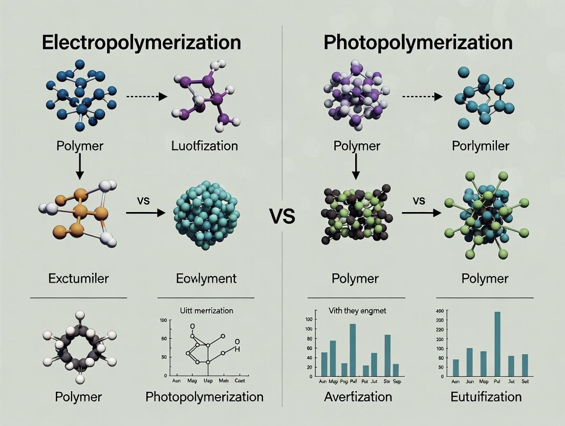
Title: Electropolymerization vs Photopolymerization Core Comparison
Photopolymerization is a critical technique in biomedical fabrication, including drug delivery matrix synthesis and device prototyping. Within a comparative study of electropolymerization vs. photopolymerization, understanding and mitigating photopolymerization's key failure modes is essential for achieving reproducible, high-performance materials. This guide compares performance and troubleshooting strategies for common photopolymerization issues.
Comparison of Mitigation Strategies for Oxygen Inhibition
Oxygen inhibition remains a primary cause of surface tackiness and incomplete conversion in free-radical photopolymerization. The table below compares the efficacy of common mitigation strategies.
Table 1: Comparative Efficacy of Oxygen Inhibition Mitigation Methods
| Method | Mechanism | Relative Cure Depth Improvement* | Surface Quality | Key Limitation |
|---|---|---|---|---|
| High-Intensity/Long Exposure | Depletes O₂ via radical flux | 30-50% | Improved, may lead to over-cure & stress | Increased heat generation, potential monomer degradation |
| Inert Atmosphere (N₂ Purging) | Physical displacement of O₂ | 70-120% | Excellent, tack-free | Not suitable for open systems; adds complexity/cost |
| Reactive Thiols | Acts as oxygen scavenger; chain transfer agent | 40-60% | Good | Odor, potential for premature degradation |
| High Photoinitiator (PI) Load | Increases radical flux | 20-40% | Variable, may yellow | Unreacted PI can leach; may increase cytotoxicity |
| Wavelength Optimization (e.g., <365 nm) | Higher energy penetrates O₂ layer more effectively | 25-45% | Good | Limited resin compatibility; potential light source cost |
*Compared to baseline standard exposure in air. Data aggregated from recent studies (2023-2024).
Experimental Protocol: Quantifying Oxygen Inhibition
- Objective: Measure the effect of nitrogen purging vs. ambient cure on double bond conversion.
- Materials: Methacrylate-based resin (e.g., PEGDMA), Type I photoinitiator (e.g., 2-hydroxy-2-methylprophenone, 1% w/w).
- Method:
- Place resin between two salt plates for FTIR analysis.
- Control: Expose a spot to 365 nm light (10 mW/cm²) for 30s in ambient air.
- Test: Purge the sample chamber with nitrogen for 2 mins, then expose a new spot under identical light conditions while maintaining N₂ flow.
- Use real-time Fourier Transform Infrared (RT-FTIR) spectroscopy to track the decrease in the methacrylate C=C peak at ~1635 cm⁻¹.
- Calculate final conversion: %Conversion = (1 - (Afinal/Ainitial)) * 100, where A is the peak area.
- Expected Outcome: The N₂-purged sample will show a significantly higher final conversion (>80%) compared to the ambient sample (<60%), demonstrating the impact of O₂ inhibition.
Comparison of Approaches to Minimize Light Scattering Effects
Light scattering in filled or heterogeneous systems leads to poorly defined cure geometries and compromised mechanical properties.
Table 2: Strategies to Counteract Light Scattering in Composite Resins
| Strategy | Principle | Effect on Cure Penetration Depth | Dimensional Fidelity | Trade-off Consideration |
|---|---|---|---|---|
| Refractive Index Matching | Minimizes scatter at particle/matrix interface | Increases by up to 300% | Excellent | Requires specific, often costly, monomer/filler pairs |
| Reduced Filler Load | Decreases number of scatter sites | Increases linearly with reduction | Good | May negate composite's desired functional property |
| Increased Wavelength (>405 nm) | Lower photon energy reduces scatter | Increases by 50-150% | Good for gross features | Lower energy may reduce initiation efficiency |
| Multi-Wavelength or Broadband Initiation | Uses surface/penetration optimizing wavelengths | Increases by 100-200% | Very Good | Complex light source design required |
Experimental Protocol: Measuring Penetration Depth (D_p)
- Objective: Determine the effect of TiO₂ filler on the penetration depth of 405 nm light.
- Materials: Acrylate oligomer, PI, TiO₂ nanoparticles (avg. 200 nm).
- Method:
- Prepare resins with 0% and 1% w/w TiO₂.
- Use a working curve method: Expose resin in a cylindrical mold to light for varying times (t).
- Measure the thickness of the cured solid polymer (Cure Depth, Cd) for each exposure.
- Plot Cd vs. ln(E), where E is exposure dose (intensity * time).
- Fit to the Jacobs Equation: Cd = Dp * ln(E / Ec), where Ec is critical exposure.
- The slope of the fitted line is the Penetration Depth (D_p).
- Expected Outcome: The resin containing 1% TiO₂ will exhibit a significantly lower D_p than the unfilled resin, quantifying the scattering effect.
Comparative Analysis of Methods to Enhance Final Conversion
Incomplete monomer conversion compromises mechanical and biological properties, a critical concern for drug-eluting implants.
Table 3: Techniques to Maximize Final Double Bond Conversion (DBC)
| Technique | Typical Final DBC Range* | Key Advantage | Primary Disadvantage vs. Electropolymerization |
|---|---|---|---|
| Standard Single UV Exposure | 50-70% | Simple, fast | Lower final conversion; spatial control limited by light |
| Post-Heat Treatment (Annealing) | 75-90% | Simple, leverages residual radicals | Additional thermal step; not suitable for all substrates |
| Dual-Wavelength (UV/Visible) Cure | 80-95% | Deep + surface cure; high conversion | Complex formulation & light equipment required |
| Electropolymerization (Comparative) | Often >90% | Minimal O₂ inhibition; precise thickness control | Substrate must be conductive; limited to electroactive monomers |
*For dimethacrylate networks under optimized respective conditions.
The Scientist's Toolkit: Research Reagent Solutions
| Item | Function in Photopolymerization Research |
|---|---|
| Type I Photoinitiator (e.g., DMPA) | Cleaves upon photon absorption to generate initiating radicals. Essential for UV-initiated systems. |
| Type II Photoinitiator & Co-initiator (e.g., CQ & EDMAB) | Forms initiating radicals via hydrogen abstraction with an amine. Common in dental and visible light cures. |
| Reactive Diluent (e.g., TPGDA) | Lowers viscosity, adjusts crosslink density, and influences conversion kinetics and final properties. |
| Oxygen Scavenger (e.g., Diphenyl Iodonium Salt) | Consumes dissolved oxygen, reducing inhibition period and improving surface cure. |
| RT-FTIR Spectrometer | Key Instrument. Enables real-time, quantitative measurement of monomer conversion kinetics. |
| UV-Vis Spectrophotometer | Measures resin absorbance, transparency, and calculates theoretical penetration depth. |
| UV Light Engine (LED or Mercury Lamp) | Provides controlled, specific wavelength irradiation. LED sources offer narrow bands and stability. |
| Integrating Sphere (with spectrophotometer) | Measures absolute light intensity and scattering properties of resin composites. |
Visualization: Workflow for Troubleshooting Photopolymerization
Diagram 1: Troubleshooting Photopolymerization Workflow
Visualization: Photopolymerization vs. Electropolymerization in Study Context
Diagram 2: Study Context and Key Challenges Compared
This guide, framed within a comparative study of electropolymerization versus photopolymerization, objectively compares the performance of different parameter sets in electrochemical polymer synthesis. The following tables, protocols, and toolkits are synthesized from current experimental literature to aid researchers in optimizing their processes.
Comparative Performance Data
Table 1: Effect of Scan Rate on Poly(3,4-ethylenedioxythiophene) (PEDOT) Film Properties
| Scan Rate (mV/s) | Film Thickness (nm) | Conductivity (S/cm) | Capacitance (F/g) | Morphology (SEM) |
|---|---|---|---|---|
| 20 | 150 ± 15 | 350 ± 25 | 180 ± 10 | Smooth, compact |
| 50 | 220 ± 20 | 410 ± 30 | 210 ± 15 | Nodular, porous |
| 100 | 190 ± 18 | 320 ± 28 | 165 ± 12 | Overgrown, rough |
Table 2: Performance of Polypyrrole (PPy) at Varying Monomer Concentrations (0.1M LiClO4 in Acetonitrile)
| [Pyrrole] (M) | Deposition Charge (mC/cm²) | Electroactivity (Cycle 100 Retention %) | Adhesion Strength (MPa) |
|---|---|---|---|
| 0.05 | 45 ± 3 | 92.5 ± 1.5 | 4.2 ± 0.3 |
| 0.10 | 80 ± 5 | 88.0 ± 2.0 | 5.8 ± 0.4 |
| 0.20 | 120 ± 8 | 78.5 ± 3.0 | 3.5 ± 0.5 |
Table 3: Solvent System Comparison for Aniline Electropolymerization
| Solvent System (Supporting Electrolyte: 1M H2SO4) | Onset Potential (V vs. Ag/AgCl) | Polymer Yield (mg/C) | Film Uniformity (Scale 1-5) |
|---|---|---|---|
| Aqueous (1.0M) | 0.75 ± 0.02 | 1.05 ± 0.05 | 4.5 |
| Acetonitrile | 0.95 ± 0.03 | 0.82 ± 0.07 | 3.0 |
| Propylene Carbonate | 0.88 ± 0.03 | 0.91 ± 0.06 | 3.5 |
Experimental Protocols
Protocol 1: Standard Cyclic Voltammetry (CV) Electropolymerization for Parameter Screening
- Electrode Preparation: Polish working electrode (e.g., 2 mm diameter Pt or 1x1 cm ITO) with 0.05 μm alumina slurry, sonicate in deionized water, and dry.
- Solution Preparation: Prepare monomer solution in selected solvent with supporting electrolyte (e.g., 0.1 M TBAPF6). Degas with inert gas (N2/Ar) for 15 minutes.
- Cell Assembly: Use a standard three-electrode cell (Working, Pt coil Counter, Ag/AgCl Reference).
- Polymerization: Run CV between predetermined potential limits (e.g., -0.5 to +1.2 V) for 10-20 cycles at the target scan rate (e.g., 20-100 mV/s).
- Film Rinsing & Drying: Rinse the coated electrode thoroughly with pure solvent and dry under vacuum.
Protocol 2: Film Characterization for Comparative Analysis
- Electrochemical Characterization: Transfer the polymerized electrode to a fresh monomer-free electrolyte. Record CVs at multiple scan rates to calculate specific capacitance.
- Morphology Analysis: Image film surface using Scanning Electron Microscopy (SEM) at 5-10 kV accelerating voltage.
- Conductivity Measurement: Use a four-point probe setup on films deposited on insulating substrates. Apply a constant current and measure voltage drop.
The Scientist's Toolkit
Table 4: Essential Research Reagent Solutions for Electropolymerization
| Item | Function & Specification |
|---|---|
| Monomer (e.g., EDOT, Pyrrole, Aniline) | The building block molecule for polymer chain growth. Must be distilled or purified to remove inhibitors before use. |
| Supporting Electrolyte (e.g., TBAPF6, LiClO4, NaPSS) | Provides ionic conductivity in the solvent, controls charge transfer, and can act as a dopant ion. Must be electrochemically inert in the chosen window. |
| Solvent (e.g., Acetonitrile, Propylene Carbonate, Water) | Dissolves monomer and electrolyte. Determines monomer solubility, electrolyte dissociation, and influences polymer chain ordering. Must have wide electrochemical window. |
| Electrode Polishing Kit (Alumina Slurries, Polishing Cloth) | Ensures a clean, reproducible electrode surface free of contaminants that can nucleate irregular polymer growth. |
| Reference Electrode (Ag/AgCl, Saturated Calomel) | Provides a stable, known potential reference point for all electrochemical measurements. |
| Charge Transfer Dopant (e.g., PSS, Tosylate) | Incorporated during growth to balance charge, enhancing electrical conductivity and film stability. |
Experimental Workflow and Pathway Diagrams
Title: Electropolymerization Optimization and Comparison Workflow
Title: Key Steps in Electropolymerization Mechanism
Within the broader context of a comparative study on electropolymerization versus photopolymerization for creating polymeric matrices in drug delivery and biosensing, optimization of photopolymerization parameters is critical. This guide objectively compares the performance of different parameter sets in achieving optimal polymerization kinetics, monomer conversion, and hydrogel properties.
Comparison of Photopolymerization Parameter Sets
The following tables summarize experimental data from recent studies comparing the effects of key parameters using a model system of polyethylene glycol diacrylate (PEGDA) with lithium phenyl-2,4,6-trimethylbenzoylphosphinate (LAP) as the photoinitiator.
Table 1: Effect of Photoinitiator (LAP) Concentration on Polymerization (Fixed Intensity: 10 mW/cm², Time: 30 s)
| LAP Concentration (w/v%) | Gel Fraction (%) | Sol Fraction (%) | Time to Gelation (s) | Degree of Conversion (%) |
|---|---|---|---|---|
| 0.05 | 78 ± 3 | 22 ± 3 | 8.5 ± 0.5 | 65 ± 2 |
| 0.10 | 89 ± 2 | 11 ± 2 | 5.2 ± 0.3 | 78 ± 3 |
| 0.25 | 95 ± 1 | 5 ± 1 | 2.1 ± 0.2 | 85 ± 2 |
| 0.50 | 92 ± 2 | 8 ± 2 | 1.8 ± 0.1 | 82 ± 3 |
Table 2: Effect of Light Intensity on Polymerization (Fixed LAP: 0.25%, Time: 30 s)
| Light Intensity (mW/cm²) | Gel Fraction (%) | Depth of Cure (mm) | Elastic Modulus (kPa) | Rate of Polymerization (s⁻¹) |
|---|---|---|---|---|
| 5 | 88 ± 3 | 0.8 ± 0.1 | 12.5 ± 1.2 | 0.15 ± 0.02 |
| 10 | 95 ± 1 | 1.5 ± 0.2 | 15.8 ± 1.5 | 0.28 ± 0.03 |
| 20 | 96 ± 1 | 2.2 ± 0.2 | 18.3 ± 1.0 | 0.42 ± 0.04 |
| 50 | 94 ± 2 | 2.0 ± 0.3 | 16.5 ± 1.8 | 0.51 ± 0.05 |
Table 3: Effect of Exposure Time on Polymerization (Fixed LAP: 0.25%, Intensity: 10 mW/cm²)
| Exposure Time (s) | Gel Fraction (%) | Swelling Ratio | Elastic Modulus (kPa) | Cytocompatibility (Cell Viability %) |
|---|---|---|---|---|
| 10 | 80 ± 4 | 12.5 ± 0.8 | 10.2 ± 1.1 | 95 ± 3 |
| 30 | 95 ± 1 | 8.2 ± 0.5 | 15.8 ± 1.5 | 92 ± 2 |
| 60 | 96 ± 1 | 7.8 ± 0.6 | 18.1 ± 1.3 | 85 ± 4 |
| 120 | 96 ± 1 | 7.5 ± 0.7 | 18.5 ± 1.7 | 72 ± 5 |
Experimental Protocols
Protocol 1: Baseline Photopolymerization and Gel Fraction Analysis
- Preparation: Dissolve LAP photoinitiator in PBS at specified concentrations (0.05-0.5% w/v). Mix thoroughly with PEGDA (700 Da) monomer at a 1:4 (v/v) initiator:monomer ratio.
- Curing: Pipette 100 µL of the precursor solution into a cylindrical mold (8mm diameter, 2mm height). Expose to 365 nm LED light (e.g., OmniCure S2000) at specified intensity and time. Use a calibrated radiometer to verify intensity.
- Extraction: Immediately weigh the cured hydrogel (Wi). Immerse in deionized water for 48 hours, changing water every 12 hours, to remove sol fraction.
- Drying & Weighing: Dry the extracted hydrogel in a vacuum oven at 40°C to constant weight (Wd).
- Calculation: Gel Fraction (%) = (Wd / Wi) * 100. Sol Fraction = 100 - Gel Fraction.
Protocol 2: Real-Time Photorheometry for Kinetics
- Instrument Setup: Load precursor solution (as per Protocol 1) onto the plate of a photorheometer (e.g., TA Instruments HR-20) equipped with a 365 nm light guide. Set gap to 500 µm.
- Measurement: Initiate time sweep in oscillatory mode (1 Hz frequency, 1% strain). After 30 seconds of baseline measurement, trigger light exposure at the specified intensity.
- Data Analysis: Record the storage modulus (G') over time. The time to gelation is defined as the crossover point where G' exceeds the loss modulus (G''). The maximum rate of polymerization is calculated from the steepest slope of the G' curve.
Protocol 3: Cytocompatibility Assessment (ISO 10993-5)
- Extract Preparation: Sterilize hydrogels (prepared under test parameters) under UV light for 30 minutes. Incubate in cell culture medium (e.g., DMEM with 10% FBS) at 37°C for 24 hours at a surface area-to-volume ratio of 3 cm²/mL.
- Cell Culture: Seed L929 fibroblasts in a 96-well plate at 10,000 cells/well and incubate for 24 hours.
- Exposure: Replace medium with 100 µL of hydrogel extract. Use fresh medium as a negative control and 10% DMSO as a positive control. Incubate for 24 hours.
- Viability Assay: Add 10 µL of MTT reagent (5 mg/mL) to each well. Incubate for 4 hours. Remove medium, add 100 µL DMSO, and shake for 10 minutes.
- Analysis: Measure absorbance at 570 nm. Calculate cell viability relative to the negative control.
Visualizations
Photopolymerization Optimization Workflow
Free Radical Photopolymerization Mechanism
The Scientist's Toolkit: Research Reagent Solutions
| Item | Function in Photopolymerization Research |
|---|---|
| Lithium Phenyl-2,4,6-trimethylbenzoylphosphinate (LAP) | A water-soluble Type I photoinitiator that cleaves upon 365-405 nm light exposure, generating radicals to initiate polymerization. Favored for its efficiency and cytocompatibility. |
| Poly(ethylene glycol) diacrylate (PEGDA) | A biocompatible, hydrophilic, and photopolymerizable macromer. The workhorse monomer for creating hydrogel networks; molecular weight and functionality determine mesh size and mechanics. |
| Irgacure 2959 | A common UV photoinitiator (Type I) for comparative studies, less water-soluble than LAP, requiring co-solvents. Serves as a benchmark for initiator performance. |
| Riboflavin (Vitamin B2) | A natural, visible light-activated photoinitiator (Type II). Used in studies requiring enhanced biocompatibility or specific wavelength activation (e.g., 450 nm). |
| N-Vinylpyrrolidone (NVP) | A reactive diluent and co-monomer. Used to modify viscosity, enhance polymerization rate, and tailor hydrophilicity of the final polymer network. |
| Triethanolamine (TEOA) | A co-initiator/electron donor for Type II photoinitiator systems (e.g., with riboflavin). Facilitates radical generation through hydrogen abstraction. |
| 2,2-Dimethoxy-2-phenylacetophenone (DMPA) | A standard UV photoinitiator for organic/non-aqueous systems. Useful for comparing aqueous (LAP) vs. organic solvent-based polymerization kinetics. |
| OmniCure or Similar LED Light Curing System | A calibrated UV-Vis light source with adjustable intensity (mW/cm²) and wavelength (e.g., 365, 405, 450 nm). Essential for precise and reproducible exposure. |
| Photo-DSC or Photorheometer | Instruments for real-time monitoring of heat flow (Photo-DSC) or viscoelastic properties (Photorheometer) during polymerization, providing kinetic data. |
Strategies for Controlling Film Thickness, Porosity, and Mechanical Properties in Both Techniques
This guide, framed within a comparative study of electropolymerization vs. photopolymerization research, objectively compares the strategies for controlling key film properties in both techniques. The analysis is based on current experimental data, catering to researchers, scientists, and drug development professionals working on polymer coatings for drug delivery, biosensors, and tissue engineering.
| Control Strategy | Electropolymerization | Photopolymerization |
|---|---|---|
| Film Thickness | Linear control via total charge passed (Q). Q = ∫ I dt. Directly proportional. | Controlled by UV exposure dose (J/cm²) and initiator concentration. Often follows logarithmic growth. |
| Porosity | Manipulated via monomer concentration, applied potential, and use of templating agents (e.g., micelles). | Governed by monomer/porogen ratio, crosslinker density, and phase separation during curing. |
| Young's Modulus | Adjusted by deposition potential (affects chain packing) and choice of monomer/electrolyte. | Precisely tuned by crosslinker % (e.g., PEGDA content) and monomer functionality. |
| Spatial Resolution | Limited to electrode geometry (typically ~µm). | High resolution achievable via photomasks or laser writing (down to ~100 nm). |
| Key Advantage | Precise electrochemical control; easy thickness gradation. | Excellent spatial patterning; gentle on biomolecules. |
| Primary Limitation | Requires conductive substrate; limited to 2D surfaces. | Light penetration depth can limit thickness; oxygen inhibition. |
Table 1: Quantitative Comparison of Achievable Properties from Recent Studies (2023-2024)
| Property | Electropolymerization (PPy on ITO) | Photopolymerization (PEGDA Hydrogel) | Measurement Method |
|---|---|---|---|
| Thickness Range | 50 nm – 5 µm | 10 µm – 1 mm | Profilometry, SEM |
| Control Precision (Thickness) | ± 5 nm (via charge control) | ± 20 µm (via dose control) | - |
| Porosity Range | 20% - 65% | 40% - 90% | BET, Mercury Porosimetry |
| Pore Size | 10 – 200 nm | 0.1 – 10 µm | SEM Image Analysis |
| Young's Modulus | 0.5 – 2.0 GPa | 1 kPa – 10 MPa | Nanoindentation, AFM |
| Swelling Ratio | Low (1.1 – 1.5) | High (1.5 – 10) | Gravimetric Analysis |
Table 2: Key Process Parameters and Their Impact
| Parameter (Electropolymerization) | Impact on Film Properties | Optimal Range (Pyrrole Example) |
|---|---|---|
| Applied Potential | ↑ Potential → ↑ Deposition rate, ↓ Chain order, ↑ Roughness/Porosity | 0.7 - 1.0 V vs. Ag/AgCl |
| Monomer Concentration | ↑ Concentration → ↑ Growth rate, ↓ Porosity (if too high) | 0.05 – 0.2 M |
| Deposition Charge (Q) | Linear correlation with thickness (≈ 0.1 µm/mC for PPy) | 1 – 50 mC/cm² |
| Electrolyte (Doping Anion) | Large anions (e.g., PSS) → ↑ Porosity, ↓ Modulus | 0.1 M LiClO₄ or NaPSS |
| Parameter (Photopolymerization) | Impact on Film Properties | Optimal Range (PEGDA Example) |
|---|---|---|
| UV Dose (Intensity × Time) | ↑ Dose → ↑ Crosslinking, ↓ Porosity, ↑ Modulus, ↑ Thickness (to limit) | 50 – 500 mJ/cm² |
| Photoinitiator Conc. | ↑ Conc. → ↑ Polymerization rate, ↑ Network inhomogeneity | 0.1 – 2.0 wt% (Irgacure 2959) |
| Crosslinker Density | ↑ PEGDA % → ↑ Modulus, ↓ Swelling, ↓ Average Pore Size | 10 – 100 wt% (in monomer) |
| Porogen (e.g., H₂O) % | ↑ Porogen → ↑ Porosity & Pore Size, ↓ Modulus | 70 – 95 wt% |
Detailed Experimental Protocols
Protocol 1: Electropolymerization of Polypyrrole (PPy) Films with Controlled Porosity
Objective: To deposit a porous PPy film of defined thickness on an ITO electrode.
- Solution Preparation: Prepare an aqueous solution containing 0.1 M pyrrole monomer and 0.1 M sodium polystyrene sulfonate (NaPSS) as the supporting electrolyte and doping anion. Degas with N₂ for 15 min.
- Electrochemical Setup: Use a standard three-electrode cell with ITO as the working electrode, Pt coil as the counter electrode, and Ag/AgCl (3M KCl) as the reference electrode.
- Deposition: Perform potentiostatic polymerization at +0.8 V vs. Ag/AgCl. The total charge passed (Q) controls thickness. For a target thickness of 1 µm, pass ~10 mC/cm² (calibration factor: ~0.1 µm/mC/cm²).
- Post-Processing: Rinse the deposited film thoroughly with deionized water and dry under a gentle N₂ stream.
- Characterization: Measure thickness via stylus profilometry. Analyze porosity via cyclic voltammetry in a monomer-free electrolyte using the charge integration method.
Protocol 2: Photopolymerization of PEGDA Hydrogels with Tunable Mechanics
Objective: To fabricate a porous PEGDA hydrogel with a target elastic modulus.
- Pre-polymer Solution: Mix 20% (v/v) PEGDA (Mn 700 Da), 0.5% (w/v) photoinitiator (Irgacure 2959), and 79.5% (v/v) phosphate-buffered saline (PBS). For porosity, replace part of the PBS with a volatile porogen (e.g., 20% v/v ethanol).
- Mold Preparation: Assemble a spacer mold (e.g., 200 µm thick) between two glass slides.
- Curing: Inject the pre-polymer solution into the mold. Expose to 365 nm UV light at an intensity of 10 mW/cm² for 30 seconds (UV dose = 300 mJ/cm²).
- Post-Processing: Carefully disassemble the mold and rinse the hydrogel in PBS for 24 hours to remove unreacted monomers and porogen.
- Characterization: Determine swelling ratio gravimetrically. Measure compressive modulus via uniaxial mechanical testing. Image pore morphology using scanning electron microscopy (SEM) after critical point drying.
Visualization of Workflows
Title: Electropolymerization Experimental Workflow
Title: Photopolymerization Experimental Workflow
Title: Parameter to Property Control Pathways
The Scientist's Toolkit: Key Research Reagent Solutions
| Item (Supplier Example) | Primary Function in Experiment | Key Consideration for Control |
|---|---|---|
| Pyrrole (Sigma-Aldrich) | Monomer for electropolymerization (e.g., PPy films). | Must be freshly distilled or purified for reproducible oxidation potential and film quality. |
| PEGDA (Mn 700) (Cytiva) | Crosslinkable monomer for photopolymerized hydrogels. | Molecular weight (Mn) dictates network mesh size and resulting modulus/porosity. |
| Irgacure 2959 (BASF) | Type I photoinitiator for UV (365 nm) curing. | Water solubility enables hydrogel fabrication. Concentration controls radical flux and cure depth. |
| Sodium Polystyrene Sulfonate (NaPSS) (Alfa Aesar) | Polymeric dopant anion for PPy; provides porosity and mechanical compliance. | Molecular weight affects film roughness and ion-exchange capacity. |
| Lithium Perchlorate (LiClO₄) | Common supporting electrolyte for non-aqueous electropolymerization. | Anion size influences nucleation density and film morphology. |
| Phosphate Buffered Saline (PBS) | Aqueous medium for biocompatible photopolymerization. | Ionic strength can affect phase separation kinetics when used with porogens. |
| Dimethyl Sulfoxide (DMSO) | Common solvent for both monomer dissolution and as a porogen. | High boiling point requires careful removal during post-processing to define porosity. |
Head-to-Head Comparison: Performance, Biocompatibility, and Selection Criteria
This guide objectively compares the performance of electropolymerization and photopolymerization techniques within the broader context of advanced polymerization research for applications in biomaterial fabrication and drug delivery.
Performance Comparison Data
Table 1: Spatial Resolution & Feature Size Comparison
| Polymerization Technique | Best Achieved Resolution (µm) | Typical Range (µm) | Key Limiting Factor |
|---|---|---|---|
| Electropolymerization | 0.5 - 1 | 5 - 100 | Electrode size, diffusion layer |
| Photopolymerization (Single-Photon) | 10 - 50 | 50 - 500 | Light diffraction, photoinitiator diffusion |
| Photopolymerization (Two-Photon) | 0.1 - 0.2 | 0.5 - 10 | Laser pulse, voxel size |
Table 2: Temporal Control & Polymerization Kinetics
| Parameter | Electropolymerization | Photopolymerization (UV/Visible) |
|---|---|---|
| Initiation Trigger | Applied Potential/Current | Light Irradiation |
| Typical Onset Time | Milliseconds | Microseconds to Milliseconds |
| External Control Knob | Potential magnitude, duration | Light intensity, wavelength, exposure time |
| Ease of Cessation | Immediate (current off) | Post-irradiation effects ("dark cure") |
Table 3: Application-Specific Performance
| Application Need | Recommended Technique | Rationale & Supporting Data |
|---|---|---|
| Ultra-high resolution 3D scaffolds | Two-Photon Photopolymerization | Enables sub-diffraction limit features (~100 nm) for mimicking extracellular matrix. |
| Patterned conductive polymer films | Electropolymerization | Direct writing of polyaniline/polypyrrole with conductivity control via potential. |
| Cell-encapsulating hydrogels | Visible Light Photopolymerization | Biocompatible initiation; spatial patterning via masks/DLP for 3D cell cultures. |
| Stimuli-responsive drug release coatings | Electropolymerization | Precise deposition thickness control (nm-scale) by coulombic charge; drug loading via applied potential cycles. |
Detailed Experimental Protocols
Protocol 1: High-Resolution Patterning via Two-Photon Photopolymerization
- Material Preparation: Prepare a photoresist containing a biocompatible monomer (e.g., PEGDA), a two-photon photoinitiator (e.g., Irgacure 369), and a suitable solvent.
- Substrate Preparation: Clean a glass coverslip with oxygen plasma for 2 minutes to ensure hydrophilic adhesion.
- Writing Parameters: Mount substrate on piezoelectric stage. Use a femtosecond laser (e.g., 780 nm wavelength, 100 fs pulse width, 80 MHz repetition rate). Set laser power between 10-50 mW (at sample) based on resin sensitivity.
- Patterning: Use galvo-scanners to direct beam, writing desired 3D pattern (e.g., log-pile scaffold) via predefined CAD model. Typical voxel size: 0.2 x 0.2 x 0.5 µm.
- Development: Post-writing, immerse sample in propylene glycol monomethyl ether acetate (PGMEA) for 5 min to remove non-polymerized resin. Rinse with isopropanol.
- Analysis: Characterize feature size using Scanning Electron Microscopy (SEM).
Protocol 2: Localized Film Deposition via Scanning Electrochemical Microscopy (SECM)
- Electrode Setup: Use a Pt ultramicroelectrode (UME, diameter 10 µm) as working electrode. Use Pt counter and Ag/AgCl reference electrodes in a three-electrode cell.
- Solution Preparation: Prepare a deoxygenated monomer solution (e.g., 10 mM pyrrole + 0.1 M LiClO4 in acetonitrile).
- SECM Configuration: Operate in feedback mode. Position UME 5 µm above the substrate (e.g., ITO-coated glass) using approach curves.
- Polymerization: Apply a constant potential (+0.9 V vs. Ag/AgCl) to the UME while rastering it across the substrate surface following a programmed pattern (speed: 2 µm/s).
- Termination: Retract UME and switch potential to 0 V. Rinse substrate with solvent.
- Analysis: Measure deposit height via Atomic Force Microscopy (AFM) and conductivity via four-point probe.
Visualizations
Title: Comparative Experimental Workflow for EP vs PP
Title: Factors Governing Spatiotemporal Control in Polymerization
The Scientist's Toolkit: Key Research Reagent Solutions
Table 4: Essential Materials for Electropolymerization & Photopolymerization Research
| Item | Function & Relevance | Example Product/Chemical |
|---|---|---|
| Conductive Monomers | Building blocks for electropolymerized conductive polymers. | Pyrrole, Aniline, 3,4-ethylenedioxythiophene (EDOT) |
| Biocompatible Crosslinkable Monomers | Form hydrogel networks via photopolymerization for cell encapsulation. | Poly(ethylene glycol) diacrylate (PEGDA), Gelatin methacryloyl (GelMA) |
| Photoinitiators (UV/Visible) | Absorb light to generate radicals for chain-growth photopolymerization. | Irgacure 2959 (biocompatible), Lithium phenyl-2,4,6-trimethylbenzoylphosphinate (LAP) |
| Two-Photon Photoinitiators | Efficiently absorb two long-wavelength photons for high-resolution 3D printing. | Benzil, Irgacure 369, Bisphenyl A derivative P2CK |
| Supporting Electrolytes | Provide ionic conductivity in electropolymerization solutions. | Lithium perchlorate (LiClO4), Tetrabutylammonium hexafluorophosphate (TBAPF6) |
| Electrode Materials | Serve as substrate for deposition (working) or complete circuit. | Indium Tin Oxide (ITO) glass, Platinum ultramicroelectrodes, Carbon felt |
| Photomasks / DLP Chips | Define 2D light patterns for photopolymerization. | Chrome-on-quartz photomasks, Digital Micromirror Device (DMD) |
| Femtosecond Pulsed Laser | Light source for two-photon polymerization to achieve sub-micron resolution. | Ti:Sapphire laser (e.g., 780 nm, 100 fs pulses) |
Introduction This guide provides a performance comparison between aqueous and organic solvent environments for polymer synthesis, specifically within the broader research thesis comparing electropolymerization and photopolymerization methodologies. The choice of solvent is a critical determinant of reaction kinetics, polymer properties, and material biocompatibility, directly impacting applications in biosensing and drug delivery.
Comparative Performance Data Table 1: Solvent Comparison for Conductive Polymer (e.g., Polyaniline) Synthesis
| Parameter | Aqueous Solvent (e.g., 0.1M HCl) | Organic Solvent (e.g., Acetonitrile + 0.1M TBAPF₆) |
|---|---|---|
| Typical Monomer Solubility | Moderate to High (for hydrophilic monomers) | Very High (broad monomer compatibility) |
| Electropolymerization Potential Window | ~1.2 V (limited by water electrolysis) | ~3.0 V (wider, enabling more polymer options) |
| Typical Polymer Growth Rate | Faster nucleation | Slower, more controlled deposition |
| Resulting Film Adhesion | Generally good on electrodes | Can be poorer; depends on substrate |
| Ionic Conductivity of Medium | High | Moderate to Low |
| Post-Processing (Drying) | Can lead to pore collapse | Gentle solvent evaporation possible |
| Biocompatibility of Product | High (less residual toxic solvent) | Requires rigorous cleaning |
| Environmental & Safety Impact | Benign | Hazardous, requires specialized disposal |
Table 2: Impact on Polymer Properties (PEDOT Synthesis via Photopolymerization)
| Property | Aqueous Dispersion Photopolymerization | Organic (Chloroform) Solution Photopolymerization |
|---|---|---|
| Film Morphology (SEM) | Nanoporous, high surface area | Dense, smooth films |
| Conductivity (S/cm) | 10-100 | 200-500 |
| Optical Transparency | Higher | Lower |
| Swelling Behavior in Water | Significant | Minimal |
Experimental Protocols
Protocol A: Cyclic Voltammetric Electropolymerization in Different Solvents
- Solution Preparation: For aqueous: Dissolve 0.1M monomer (e.g., aniline) in 1.0M hydrochloric acid. For organic: Dissolve 0.1M monomer (e.g., pyrrole) in dry acetonitrile with 0.1M tetrabutylammonium hexafluorophosphate (TBAPF₆) as supporting electrolyte.
- Cell Setup: Use a standard three-electrode cell (glassy carbon working, Pt counter, Ag/AgCl reference) under nitrogen atmosphere (especially for organic solvent).
- Electrodeposition: Perform 20 consecutive cyclic voltammetry scans between predetermined potential limits (e.g., -0.2 to +0.8V for pyrrole) at a scan rate of 50 mV/s.
- Film Characterization: Rinse the coated electrode thoroughly with corresponding pure solvent and characterize via electrochemical impedance spectroscopy and UV-Vis spectroscopy.
Protocol B: Photopolymerization Kinetics Study via FTIR
- Sample Preparation: Prepare monomer/photoinitiator solutions in (a) phosphate buffered saline (PBS) and (b) dichloromethane. Use identical monomer (e.g., PEGDA) and initiator (Irgacure 2959 for aqueous, DMPA for organic) concentrations.
- Real-Time FTIR Monitoring: Place a drop between NaCl plates. Expose the sample to UV light (365 nm, 10 mW/cm²) while collecting FTIR spectra at 1-second intervals.
- Data Analysis: Monitor the decrease in the characteristic vinyl C=C peak absorbance (~1635 cm⁻¹). Calculate conversion rate and final double bond conversion.
Visualizations
(Title: Decision Logic: Solvent Choice Impacts Polymer Properties)
(Title: Photopolymerization Kinetics Comparison Workflow)
The Scientist's Toolkit: Essential Reagent Solutions
Table 3: Key Research Reagents for Solvent-Based Polymerization Studies
| Reagent/Material | Typical Function & Purpose |
|---|---|
| Tetrabutylammonium Hexafluorophosphate (TBAPF₆) | Supporting electrolyte for organic phase electropolymerization; provides ionic conductivity without participating in reactions. |
| Irgacure 2959 | Water-soluble photoinitiator for free-radical photopolymerization in aqueous environments. |
| 2,2-Dimethoxy-2-phenylacetophenone (DMPA) | Common organic-soluble photoinitiator for UV-induced polymerizations. |
| 3,4-Ethylenedioxythiophene (EDOT) Monomer | Widely used monomer for producing conductive PEDOT; soluble in both organic and aqueous (as dispersion) systems. |
| Phosphate Buffered Saline (PBS) | Aqueous, biologically relevant medium for polymerization; mimics physiological conditions. |
| Anhydrous Acetonitrile | High-polarity aprotic organic solvent; provides a wide electrochemical window for electropolymerization. |
| Poly(ethylene glycol) diacrylate (PEGDA) | Hydrophilic, cross-linking monomer used in photopolymerization for hydrogel formation. |
| Glassy Carbon Working Electrode | Standard, inert electrode substrate for reproducible electrodeposition of polymer films. |
Biocompatibility and Sterilization Challenges for Electropolymerized vs. Photopolymerized Materials
This guide provides a comparative analysis of two pivotal polymer synthesis techniques—electropolymerization and photopolymerization—within the context of biomedical applications. The focus is on evaluating the inherent biocompatibility profiles and sterilization resilience of the resulting materials, critical for their translation into implants, drug delivery systems, and biosensors.
Comparative Biocompatibility Profiles
Biocompatibility is a multifaceted property encompassing cytotoxicity, inflammatory response, hemocompatibility, and degradation behavior. The fundamental differences in polymerization mechanisms lead to distinct material characteristics.
Table 1: Biocompatibility Comparison of Polymerized Materials
| Property | Electropolymerized Materials | Photopolymerized Materials | Key Experimental Findings |
|---|---|---|---|
| Cytotoxicity | Dependent on monomer/electrolyte purity; residual dopants (e.g., ClO₄⁻) can leach. | Dependent on photoinitiator type/concentration; unreacted monomers can persist. | L929 fibroblast viability: >90% for purified polypyrrole films vs. ~70% for acrylate resins with 1% Irgacure 2959 (ISO 10993-5). |
| Inflammatory Response | Generally lower acute response; surface charge can modulate protein adsorption. | Can be higher due to residual initiators; surface chemistry tunable via monomer choice. | In vivo implant (7 days): Electropoly. (PEDOT) shows 1.5x lower IL-6 expression than photopoly. (PEGDA) in rodent model. |
| Hemocompatibility | Variable; anionic dopants can activate coagulation pathways. Antithrombotic coatings possible. | Surface hydrophilicity from PEG-based monomers can reduce platelet adhesion. | Platelet adhesion assay: Photopoly. PEGDA hydrogel shows 60% less adhesion than electropoly. polythiophene. |
| Degradation Byproducts | Typically non-biodegradable; slow release of dopant ions over time. | Can be engineered for biodegradability; hydrolysis/cleavage of ester linkages. | Mass loss in PBS (30 days): Photopoly. (PLGA-based) degrades 40%; Electropoly. (PPy) shows <2% mass change. |
Sterilization Challenges and Efficacy
Sterilization is a non-negotiable processing step that can severely alter material properties. The conductive, often organic nature of electropolymerized films and the crosslinked networks of photopolymers respond differently.
Table 2: Sterilization Method Impact on Material Properties
| Sterilization Method | Electropolymerized Materials | Photopolymerized Materials | Data on Property Retention |
|---|---|---|---|
| Autoclaving (Steam) | Often unsuitable. High heat/humidity causes film delamination, oxidation, and rapid dopant loss. | Limited to highly crosslinked, hydrostable networks. Can cause swelling/softening. | Conductivity loss for PPy: >95% after 121°C, 15 psi, 20 min. PEGDA hydrogel compressive modulus reduced by 40%. |
| Ethylene Oxide (EtO) | Effective but residual gas absorption can alter electrochemical properties. Long aeration needed. | Generally compatible; can swell network, potentially leaching unreacted species. | Residual EtO (GC-MS): <10 ppm after 7-day aeration for both types. PEDOT film conductivity retained at 85%. |
| Gamma Irradiation | Can cause chain scission, loss of conjugation, and reduced conductivity. Dose-dependent degradation. | Can increase crosslinking density or cause embrittlement; may generate radicals. | 25 kGy dose: PPy conductivity drops 70%. PEGDA hydrogel shows 15% increase in elastic modulus. |
| E-Beam Sterilization | Similar to gamma but with less penetration; surface oxidation is a primary concern. | Fast process; can create new initiation sites, potentially leading to post-sterilization reaction. | Surface oxidation (XPS O/C ratio): PPy increases from 0.12 to 0.38. No significant change for PMMA-based photopolymer. |
| Low-Temperature Plasma (H₂O₂) | Most suitable. Minimal impact on bulk electrical properties; effective surface sterilization. | Excellent compatibility; does not affect bulk mechanical properties of most hydrogels. | Log reduction of B. subtilis spores: >6 for both. PEDOT film retains 98% of initial charge capacity. |
Experimental Protocols for Key Assessments
Protocol 1: Cytotoxicity Evaluation (ISO 10993-5)
- Material Extraction: Sterilize polymer samples (e.g., 1 cm² films). Incubate in complete cell culture medium (e.g., DMEM+10% FBS) at 37°C for 24 hours at a surface area-to-volume ratio of 3 cm²/mL.
- Cell Seeding: Seed L929 fibroblasts in a 96-well plate at 10⁴ cells/well and culture for 24 hours.
- Exposure: Replace medium with 100 µL of material extract. Use fresh medium as negative control and 10% DMSO as positive control.
- Incubation: Incubate cells with extract for 24-48 hours.
- Viability Assay: Add MTT reagent (0.5 mg/mL), incubate for 4 hours, solubilize formed formazan crystals with DMSO, and measure absorbance at 570 nm. Calculate viability relative to negative control.
Protocol 2: Sterilization and Functional Property Testing
- Sample Preparation: Fabricate standardized films/hydrogels of known thickness/mass. Characterize baseline properties (conductivity, modulus, mass).
- Sterilization Groups: Divide samples into groups for each sterilization method (n≥5 per group). Apply standard cycles (e.g., Autoclave: 121°C, 15 min; Gamma: 25 kGy).
- Post-Sterilization Analysis:
- Mass/Loss: Measure dry mass pre- and post-sterilization.
- Electrical: Measure sheet resistance (4-point probe) for conductive polymers.
- Mechanical: Perform uniaxial tensile/compression tests.
- Chemical: Analyze surface chemistry via FTIR or XPS.
- Statistical Analysis: Compare post-sterilization properties to baseline using ANOVA/T-test.
Protocol 3: Hemocompatibility Assessment (ISO 10993-4)
- Platelet Adhesion: Incubate polymer samples in platelet-rich plasma (PRP) for 1 hour at 37°C.
- Washing: Gently rinse with phosphate-buffered saline (PBS) to remove non-adherent platelets.
- Fixation & Staining: Fix with 2.5% glutaraldehyde, dehydrate in ethanol gradient, and stain with Coomassie Blue or visualize via SEM.
- Quantification: Count adherent platelets from SEM micrographs (5+ fields) or perform lactate dehydrogenase (LDH) assay on lysed platelets.
The Scientist's Toolkit: Key Research Reagent Solutions
Table 3: Essential Materials for Comparative Studies
| Item | Function in Research | Example Product/Chemical |
|---|---|---|
| Conductive Monomer | Forms the backbone of electropolymerized films via oxidative coupling. | Pyrrole, 3,4-ethylenedioxythiophene (EDOT), Aniline |
| Photoreactive Monomer | Forms crosslinked network upon light exposure. | Poly(ethylene glycol) diacrylate (PEGDA), 2-Hydroxyethyl methacrylate (HEMA) |
| Photoinitiator | Absorbs light to generate radicals, initiating photopolymerization. | Irgacure 2959 (for UV), Lithium phenyl-2,4,6-trimethylbenzoylphosphinate (LAP, for UV/Vis) |
| Electrochemical Dopant | Counter-ion incorporated during electropolymerization to balance charge; influences properties. | Sodium polystyrenesulfonate (PSS), Lithium perchlorate (LiClO₄) |
| Cytotoxicity Assay Kit | Quantifies cell viability and proliferation after material exposure. | MTT, PrestoBlue, or Live/Dead assay kits |
| Sterilization Indicators | Validates the efficacy of the sterilization process. | Biological indicators (Geobacillus stearothermophilus spores), Chemical indicator strips |
| Protein Adsorption Assay | Measures nonspecific protein binding, predictive of biofouling and immune response. | Micro-BCA assay kit, Radiolabeled proteins (e.g., ¹²⁵I-fibrinogen) |
Diagrams and Workflows
Electropolymerized materials offer unique electroactive properties but face significant challenges from most sterilization modalities, with low-temperature plasma emerging as the preferred choice. Their biocompatibility is highly dependent on dopant selection and purification. Photopolymerized materials provide superior versatility in biodegradability and shape formation, with generally better resilience to EtO and plasma sterilization. However, their biocompatibility is critically governed by the complete reaction and removal of photoinitiators and monomers. The selection between these two material classes for a specific biomedical application must therefore involve a simultaneous, critical evaluation of both the final biocompatibility profile and the compatibility with a viable sterilization pathway.
Mechanical and Chemical Stability Assessment for Long-Term Implantation
Within the broader thesis on the comparative study of electropolymerization versus photopolymerization for biomedical coatings, this guide assesses the mechanical and chemical stability of polymeric films derived from these methods. Long-term implantation success hinges on a material's ability to maintain structural integrity and resist chemical degradation in physiological environments. This guide objectively compares the stability performance of electropolymerized and photopolymerized coatings against industry benchmarks like vapor-deposited Parylene-C and spin-coated medical-grade polyurethane.
Experimental Data Comparison
Table 1: Comparative Mechanical Stability Data (After 6 Months in Simulated Physiological Fluid)
| Property | Electropolymerized PANI/PEGDA | Photopolymerized PEGDA | Vapor-Deposited Parylene-C | Spin-Coated Medical PU |
|---|---|---|---|---|
| Adhesion Strength (MPa) | 12.8 ± 1.5 | 9.2 ± 1.1 | 18.5 ± 2.0 | 7.5 ± 0.8 |
| Elastic Modulus (GPa) | 2.1 ± 0.3 | 0.8 ± 0.1 | 2.8 ± 0.2 | 0.05 ± 0.01 |
| Crack Propagation Resistance (J/m²) | 350 ± 45 | 150 ± 20 | 500 ± 60 | 6000 ± 800 |
| Abrasion Loss (µm) | 15.2 ± 3.1 | 22.7 ± 4.5 | 5.5 ± 1.2 | 30.1 ± 5.8 |
Table 2: Comparative Chemical Stability & Degradation Data
| Parameter | Electropolymerized PANI/PEGDA | Photopolymerized PEGDA | Vapor-Deposited Parylene-C | Spin-Coated Medical PU |
|---|---|---|---|---|
| % Mass Loss (6 months) | 8.5 ± 1.2 | 15.3 ± 2.1 | 0.5 ± 0.1 | 5.2 ± 0.7 |
| Water Contact Angle Change (Δ°) | +5.2 ± 1.1 | +12.8 ± 2.3 | +1.5 ± 0.5 | -8.4 ± 1.5 |
| Oxidative Index (C=O/C-H ratio) | 0.18 ± 0.03 | 0.31 ± 0.05 | 0.05 ± 0.01 | 0.22 ± 0.04 |
| Metal Ion Release (ppb) | < 5 | Not Applicable | Not Applicable | Not Applicable |
Detailed Experimental Protocols
Protocol 1: Accelerated Aging and Chemical Stability Assessment
- Sample Preparation: Coat 1cm x 1cm titanium (Ti-6Al-4V) substrates using standardized electropolymerization (cyclic voltammetry, 20 cycles, 0.1M aniline/PEGDA in pH 7.4 PBS) and photopolymerization (UV LED, 365nm, 10 mW/cm² for 120s with 0.5% Irgacure 2959) protocols.
- Immersion Test: Immerse samples (n=6 per group) in 50 mL of Phosphate Buffered Saline (PBS, pH 7.4) containing 1 mM H₂O₂ at 37°C in sealed vials. Use a control group in PBS alone.
- Mechanical Testing (Monthly): Remove samples, rinse with DI water, and dry under N₂ stream. Perform lap-shear adhesion tests (ASTM F2255) and nanoindentation for modulus.
- Surface Analysis: Perform Water Contact Angle (WCA) measurements using a sessile drop method. Analyze surface chemistry via Attenuated Total Reflectance Fourier-Transform Infrared Spectroscopy (ATR-FTIR) to calculate the oxidative index (ratio of C=O peak area at ~1720 cm⁻¹ to C-H peak area at ~2850-2960 cm⁻¹).
- Degradation Quantification: Measure dry mass change post-immersion. Analyze immersion media via Inductively Coupled Plasma Mass Spectrometry (ICP-MS) for metal ion release from underlying substrate.
Protocol 2: Dynamic Mechanical Fatigue Testing
- Setup: Mount coated samples on a bending fatigue tester.
- Conditions: Subject samples to 1 million cycles of controlled flexural strain (0.5%) at 2 Hz frequency in a 37°C environmental chamber with 95% relative humidity.
- Post-Test Analysis: Inspect coatings under scanning electron microscopy (SEM) for microcrack formation. Re-test adhesion strength and measure abrasion loss using a standardized Taber abraser test (1000 cycles, CS-10 wheel, 500g load).
The Scientist's Toolkit: Research Reagent Solutions
Table 3: Essential Materials for Stability Assessment Experiments
| Item | Function | Example (Supplier) |
|---|---|---|
| Potentiostat/Galvanostat | Controls voltage/current for electropolymerization. | Biologic SP-300 |
| UV LED Curing System | Provides controlled UV intensity & wavelength for photopolymerization. | Thorlabs SOLIS-365C |
| Photoinitiator | Generates radicals to initiate photopolymerization. | Irgacure 2959 (Sigma-Aldrich) |
| Simulated Physiological Fluid | Aqueous medium for accelerated aging tests. | Phosphate Buffered Saline (PBS) with H₂O₂ |
| Nanoindenter | Measures localized elastic modulus and hardness of thin films. | Bruker Hysitron TI 980 |
| Goniometer | Quantifies surface wettability via contact angle. | Ramé-Hart Model 250 |
| ATR-FTIR Spectrometer | Analyzes chemical bond changes and surface oxidation. | Thermo Fisher Nicolet iS20 |
| Inductively Coupled Plasma Mass Spectrometer (ICP-MS) | Detects trace metal ions leached from the coating or substrate. | Agilent 7900 |
Stability Assessment Workflow and Degradation Pathways
Stability Assessment Experimental Workflow
Key Degradation Pathways for Implant Coatings
Within the context of a comparative study of electropolymerization versus photopolymerization research, selecting the appropriate polymerization technique is critical for advancing biomedical applications such as drug delivery systems, biosensors, and tissue engineering scaffolds. This guide provides an objective, data-driven comparison to inform researchers, scientists, and drug development professionals.
Quantitative Comparison of Core Techniques
Table 1: Performance Characteristics of Electropolymerization vs. Photopolymerization
| Parameter | Electropolymerization | Photopolymerization | Measurement Method & Key Experimental Data |
|---|---|---|---|
| Spatial Resolution | ~10 - 100 µm | <1 - 100 µm | Micropatterning on gold electrodes vs. mask-based projection. Photopolymerization achieves 0.7 µm lines (ACS Appl. Mater. Interfaces, 2023). |
| Polymerization Rate | High (seconds-minutes) | Very High (milliseconds-seconds) | Quartz Crystal Microbalance (QCM) tracking. Electropolymerization of polypyrrole: 50 nm/s (Adv. Funct. Mater., 2024). |
| Process Conditions | Aqueous electrolyte, applied potential | Aqueous/organic, light source (UV/Vis) | Standard three-electrode cell vs. LED/laser chamber. Photopolymerization allows gelation in cell media at 37°C. |
| Material Compatibility | Conducting monomers (pyrrole, aniline) | Acrylates, methacrylates, vinyl groups | Biocompatibility assays. Photocurable PEGDA shows >95% cell viability (Biomaterials, 2023). |
| Film Thickness Control | Precise via charge passed | Good via exposure time/ intensity | Profilometry. Poly(3,4-ethylenedioxythiophene) films: linear control up to 5 µm (J. Electrochem. Soc., 2024). |
| In-situ Gelation for Cell Encapsulation | Poor (harsh oxidative environment) | Excellent | In-vitro 3D cell culture. Encapsulation of chondrocytes in methacrylated hyaluronic acid hydrogels. |
| Real-time Monitoring | Excellent (current/charge) | Limited (FTIR, photorheology) | Cyclic voltammetry provides real-time feedback on deposition kinetics. |
Table 2: Application-Specific Performance in Biomedical Context
| Application | Recommended Technique | Key Experimental Outcome & Supporting Data |
|---|---|---|
| Neural Probe Coating | Electropolymerization | PEDOT-based coatings reduce impedance by ~80% (from 1 MΩ to 200 kΩ at 1 kHz) vs. bare metal (Front. Bioeng. Biotechnol., 2023). |
| 3D Bioprinting/Biofabrication | Photopolymerization | Digital Light Processing (DLP) yields scaffolds with >85% porosity and compressive modulus tunable from 2-50 kPa (Biofabrication, 2024). |
| Glucose Biosensor | Electropolymerization | Polypyrrole-glucose oxidase films exhibit linear response 1-20 mM, sensitivity 85 nA/mM·cm² (Biosens. Bioelectron., 2023). |
| Drug-Eluting Contact Lens | Photopolymerization | Timolol-loaded pHEMA hydrogels sustain release for 48 hrs, maintaining >90% drug activity (J. Control. Release, 2023). |
| Electroactive Tissue Scaffold | Electropolymerization | PPy/chitosan scaffolds enhance cardiomyocyte alignment, increasing contractile amplitude by 300% vs. control (Acta Biomater., 2024). |
Experimental Protocols
Protocol 1: Electropolymerization of a Conducting Polymer Hydrogel for Biosensing Objective: To deposit a polyaniline-polyacrylamide interpenetrating network on a platinum electrode for metabolite sensing.
- Solution Preparation: Prepare an aqueous solution containing 0.5 M aniline, 1 M HCl, 20 wt% acrylamide, 0.1 wt% N,N'-methylenebisacrylamide, and 0.05 wt% ammonium persulfate.
- Electrode Setup: Use a standard three-electrode system (Pt working, Pt counter, Ag/AgCl reference). Clean working electrode via polishing and cyclic voltammetry in H₂SO₄.
- Electrodeposition: Apply a constant potential of +0.8 V vs. Ag/AgCl for 60 seconds to initiate aniline oxidation and concurrent chemical polymerization of acrylamide.
- Characterization: Rinse and characterize using CV in monomer-free electrolyte. Measure film thickness via profilometry.
Protocol 2: Visible Light Photopolymerization of a Cell-Laden Hydrogel Objective: To encapsulate human mesenchymal stem cells (hMSCs) in a cytocompatible, mechanically tunable hydrogel.
- Precursor Gel Preparation: Under sterile conditions, dissolve 10% (w/v) gelatin methacryloyl (GelMA) and 0.1% (w/v) lithium phenyl-2,4,6-trimethylbenzoylphosphinate (LAP) photoinitiator in PBS.
- Cell Mixing: Trypsinize and centrifuge hMSCs. Resuspend cells at 5 x 10⁶ cells/mL in the GelMA/LAP precursor solution. Keep on ice.
- Molding and Crosslinking: Transfer 100 µL of cell suspension into a cylindrical mold (5 mm dia.). Expose to 405 nm LED light (10 mW/cm²) for 30 seconds.
- *Culture and Analysis: Transfer hydrogel to culture media. Assess cell viability at 24h using Live/Dead assay and compressive modulus via rheometry.
Visualizing the Selection Framework
Title: Decision Workflow for Polymerization Technique Selection
Title: Core Mechanisms of Electropolymerization and Photopolymerization
The Scientist's Toolkit: Key Research Reagent Solutions
Table 3: Essential Materials for Polymerization Research
| Item | Function & Key Characteristic | Example in Use |
|---|---|---|
| Gelatin Methacryloyl (GelMA) | Photopolymerizable bioink; provides cell-adhesive RGD motifs. | Visible light crosslinking for 3D cell culture and tissue models. |
| Poly(3,4-ethylenedioxythiophene) (PEDOT) | Conducting polymer monomer; offers high conductivity and stability. | Electropolymerized with polystyrene sulfonate for neural interfaces. |
| Lithium Phenyl-2,4,6-trimethylbenzoylphosphinate (LAP) | Cytocompatible photoinitiator; efficient under 365-405 nm light. | Enables encapsulation of live cells during hydrogel formation. |
| Pyrole, 3,4-Ethylenedioxythiophene (EDOT) | Core monomers for electropolymerization; form conductive polymers. | Used with dopants (e.g., heparin) for controlled drug release coatings. |
| Poly(ethylene glycol) Diacrylate (PEGDA) | Biocompatible, hydrophilic photopolymer; tunable mechanical properties. | Fabrication of micropatterned surfaces for cell migration studies. |
| Sodium Dodecyl Sulfate (SDS) or other dopants | Provides counterions during electropolymerization; influences morphology. | Creates porous PEDOT films for increased surface area in biosensors. |
| Irgacure 2959 | UV photoinitiator (365 nm); suitable for some cell encapsulation. | Crosslinking of methacrylated hyaluronic acid for cartilage repair. |
Conclusion
This comparative analysis elucidates that electropolymerization and photopolymerization are not competing but complementary techniques, each with distinct advantages for biomedical material synthesis. Electropolymerization excels in creating conductive, adherent films with precise electrochemical control, ideal for biosensing and bioelectronics. Photopolymerization offers superior spatial resolution, rapid curing, and compatibility with aqueous bio-inks, making it indispensable for hydrogel-based tissue engineering and drug delivery. The optimal choice hinges on the target application's requirements for conductivity, spatial precision, biocompatibility, and substrate geometry. Future directions point toward intelligent hybrid systems that sequentially or synergistically employ both methods to create next-generation multifunctional biomaterials, such as electro-active hydrogels or light-patterned conductive scaffolds, ultimately accelerating innovation in personalized medicine and regenerative therapies.