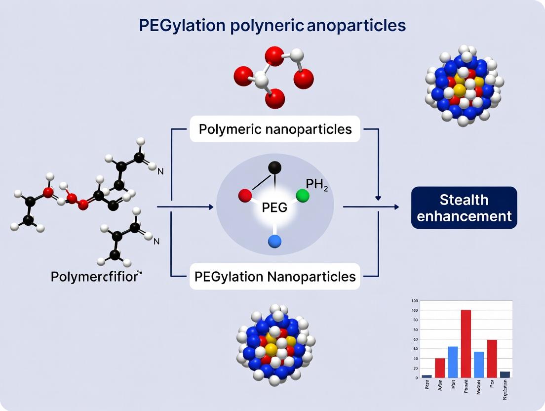
Achieving Stealth Drug Delivery: A Comprehensive Guide to PEGylation of Polymeric Nanoparticles
This article provides a detailed, current overview of PEGylation strategies for polymeric nanoparticles, focusing on creating an effective stealth effect to evade immune clearance.
Achieving Stealth Drug Delivery: A Comprehensive Guide to PEGylation of Polymeric Nanoparticles
Abstract
This article provides a detailed, current overview of PEGylation strategies for polymeric nanoparticles, focusing on creating an effective stealth effect to evade immune clearance. It explores the foundational principles of the 'stealth' concept and Protein Corona formation, details current synthesis and characterization methodologies, and presents troubleshooting for common challenges like the Accelerated Blood Clearance (ABC) phenomenon. The guide further validates approaches through comparative analysis of PEG architectures and alternative stealth polymers, culminating in a synthesis of best practices for designing long-circulating nanocarriers for targeted therapeutic delivery.
The Science of Stealth: Understanding PEGylation and Immune Evasion in Nanomedicine
Within the broader thesis on PEGylation of polymeric nanoparticles (NPs) for stealth effect research, this document defines the "stealth effect" as the engineered ability of NPs to evade the host's immune surveillance, primarily by minimizing opsonization. This effect directly translates to prolonged systemic circulation half-life, a critical parameter for enhancing drug delivery efficacy. The application of poly(ethylene glycol) (PEG) chains to NP surfaces remains the gold standard for conferring stealth properties, primarily through the formation of a hydrophilic, steric barrier.
Core Mechanisms: From Opsonization to Evasion
Key Concepts and Quantitative Benchmarks
The stealth effect is quantifiable through key pharmacokinetic and immunological parameters. The following table summarizes critical benchmarks for PEGylated vs. non-PEGylated polymeric NPs.
Table 1: Quantitative Impact of PEGylation on Stealth Properties
| Parameter | Non-PEGylated NPs | PEGylated NPs (Optimal) | Measurement Technique |
|---|---|---|---|
| Circulation Half-life (t1/2) | Minutes to few hours | 10 - 30+ hours | Pharmacokinetic analysis (blood sampling) |
| Protein Corona Formation | High-density, dysopsonin-rich | Low-density, dysopsonin-rich | SDS-PAGE, LC-MS/MS, DLS |
| Macrophage Uptake (in vitro) | High (>70% fluorescence) | Low (<20% fluorescence) | Flow cytometry (J774, RAW 264.7 cells) |
| Complement Activation (C3a, SC5b-9) | Significant increase | Minimal increase | ELISA-based complement assay |
| Zeta Potential Shift in Serum | Large shift (e.g., -10mV to -25mV) | Minimal shift (e.g., -5mV to -7mV) | Dynamic Light Scattering (DLS) |
| Liver/Spleen Accumulation (%ID/g) | High (e.g., 60-80% ID/g liver) | Reduced (e.g., 20-40% ID/g liver) | Biodistribution study (IV injection, tissue homogenization) |
Signaling Pathways in Immune Recognition
The primary pathway leading to NP clearance is the opsonin-mediated phagocytosis. PEGylation interrupts this cascade.
Diagram Title: Opsonization and Phagocytosis Signaling Cascade
Application Notes & Protocols
Protocol: Synthesis of PEGylated PLGA Nanoparticles (Single Emulsion-Solvent Evaporation)
Objective: To prepare reproducible, stealth-effect PLGA-PEG NPs.
Materials (The Scientist's Toolkit): Table 2: Key Research Reagent Solutions
| Item | Function & Specification |
|---|---|
| PLGA-PEG Copolymer (e.g., PLGA(50:50)-b-PEG(5k)) | Core polymer providing biodegradability and conjugated stealth PEG shell. |
| Dichloromethane (DMC), HPLC grade | Organic solvent for polymer and hydrophobic drug dissolution. |
| Polyvinyl Alcohol (PVA), 2% w/v | Emulsifier/stabilizer for forming primary oil-in-water emulsion. |
| Phosphate Buffered Saline (PBS), 1X, pH 7.4 | Aqueous medium for emulsion and final NP washing/resuspension. |
| Amicon Ultra Centrifugal Filters (100 kDa MWCO) | For purification and buffer exchange via diafiltration. |
| Lyophilizer with trehalose or sucrose (5% w/v) | For long-term NP storage while maintaining colloidal stability. |
| Dynamic Light Scattering (DLS)/Zetasizer | For measuring hydrodynamic diameter, PDI, and zeta potential. |
Procedure:
- Organic Phase: Dissolve 100 mg PLGA-PEG copolymer (and optional hydrophobic drug) in 5 mL DCM.
- Aqueous Phase: Prepare 50 mL of 2% PVA solution in ultrapure water.
- Emulsification: Add the organic phase dropwise to the aqueous phase while probe-sonicating (70% amplitude, 2 min on ice).
- Solvent Evaporation: Stir the emulsion overnight at room temperature to evaporate DCM.
- Purification: Centrifuge the NP suspension at 15,000 x g for 30 min, discard supernatant, and resuspend in PBS. Repeat 2x. Alternatively, use centrifugal filters (100 kDa MWCO) for 3 wash cycles.
- Characterization: Dilute purified NPs in PBS. Use DLS for size/PDI. Measure zeta potential in 1 mM KCl.
- Storage: Add cryoprotectant (5% trehalose), freeze at -80°C, and lyophilize for 48h.
Protocol: In Vitro Serum Protein Binding Assay
Objective: To quantify and qualify the "protein corona" formed on NPs, indicating opsonization potential.
Workflow Diagram:
Diagram Title: Serum Protein Binding Assay Workflow
Procedure:
- Incubate 1 mL of NP suspension (1 mg/mL in PBS) with 1 mL of 100% fetal bovine serum (FBS) at 37°C for 1 hour with gentle rotation.
- Underlay the mixture with a 500 μL cushion of 40% sucrose in PBS. Ultracentrifuge at 100,000 x g for 1 hour at 4°C.
- Carefully aspirate the supernatant and sucrose cushion. Gently wash the pellet (hard corona-NP complex) with 1 mL PBS. Repeat centrifugation and washing twice.
- Resuspend the final pellet in 100 μL 1X LDS sample buffer for SDS-PAGE or in PBS for DLS.
- For SDS-PAGE, heat samples at 95°C for 5 min, load 20 μL per well, and run gel. Stain with Coomassie Blue or silver stain.
- For DLS analysis, dilute the resuspended complex 1:10 in PBS and measure hydrodynamic diameter and zeta potential, comparing to pristine NPs.
Protocol: In Vivo Circulation Half-life Determination
Objective: To quantify the pharmacokinetic enhancement conferred by the stealth effect.
Procedure:
- NP Labeling: Label NPs with a near-infrared (NIR) dye (e.g., DiR) or a radioisotope (e.g., ¹¹¹In) during formulation.
- Animal Dosing: Administer a bolus IV injection (dose: ~5 mg NPs/kg) via the tail vein to groups of mice (n=5 per time point).
- Blood Sampling: At predetermined time points (e.g., 5 min, 30 min, 2h, 6h, 12h, 24h, 48h), collect ~20 μL of blood from the retro-orbital plexus into heparinized tubes.
- Quantification:
- For NIR dyes: Lyse 10 μL blood in 1% Triton X-100. Measure fluorescence using a plate reader (ex/em specific to dye). Create a standard curve with known NP concentrations in whole blood lysate.
- For radioisotopes: Measure radioactivity in whole blood samples using a gamma counter.
- Pharmacokinetic Analysis: Plot blood concentration (% of injected dose per mL) vs. time. Use non-compartmental analysis (e.g., with PK Solver) to calculate: Terminal half-life (t1/2), Area Under the Curve (AUC), and Clearance (CL).
Within the broader thesis on PEGylation of polymeric nanoparticles (PNPs) for stealth effect research, understanding the protein corona is paramount. Upon intravenous administration, nanoparticles (NPs) are instantly coated by a dynamic layer of biomolecules, primarily proteins, forming the "protein corona." This corona dictates the NP's biological identity, overriding its synthetic surface properties and critically impacting its pharmacokinetics, biodistribution, cellular uptake, and toxicity. PEGylation—the covalent attachment of poly(ethylene glycol) (PEG) chains—aims to mask the NP surface to minimize opsonin adsorption, thereby imparting a "stealth" character and prolonging systemic circulation.
Application Notes
Note 1: Corona Formation Dynamics & Composition
The corona evolves through a Vroman effect: initially, abundant, high-mobility proteins (e.g., albumin) adsorb, but are gradually displaced by proteins with higher affinity (e.g., immunoglobulins, apolipoproteins, complement factors). The "hard corona" consists of tightly bound proteins, while the "soft corona" is a loosely associated, rapidly exchanging layer. Composition is influenced by NP properties (size, charge, hydrophobicity) and biological fluid (plasma vs. serum, species, disease state).
Note 2: Impact on Cellular Fate and Pharmacokinetics
The corona mediates biological interactions. A corona rich in opsonins (e.g., IgG, C3b) promotes recognition by mononuclear phagocyte system (MPS) cells, leading to rapid clearance. Conversely, dysopsonins (e.g., albumin, apolipoprotein A-I) can promote stealth. Corona composition directly influences cellular internalization pathways (e.g., clathrin-mediated endocytosis vs. caveolae-mediated uptake) and subsequent intracellular trafficking.
Note 3: The Masking Role of PEG
PEG chains create a hydrophilic, steric barrier that reduces protein adsorption through:
- Steric Repulsion: The hydrated, flexible PEG brush layer presents a physical barrier to approaching proteins.
- Reduced Hydrophobic Interactions: PEG shields the hydrophobic core of polymeric NPs.
- Entropic Effects: Compression of mobile PEG chains upon protein approach is thermodynamically unfavorable. Dense PEG brush conformation is more effective than mushroom conformation. However, recent evidence of anti-PEG antibodies necessitates investigation into next-generation stealth polymers.
Table 1: Key Plasma Proteins in the Corona and Their Impact on NP Fate
| Protein | Approx. Concentration in Plasma (mg/mL) | Typical Affinity for NPs | Primary Consequence for NP Fate |
|---|---|---|---|
| Human Serum Albumin (HSA) | 35-50 | Low-Moderate | Can promote stealth (dysopsonin); abundant in initial corona. |
| Immunoglobulin G (IgG) | ~10 | Moderate-High | Promotes opsonization; MPS recognition via Fc receptors. |
| Fibrinogen | 2-4 | High | Strong opsonin; activates phagocytes. |
| Apolipoprotein A-I (ApoA-I) | 1.0-1.5 | Moderate | May promote targeting to hepatocytes; potential stealth effect. |
| Apolipoprotein E (ApoE) | 0.03-0.06 | High | Can mediate brain targeting via LDL receptor interaction. |
| Complement C3 | 1.0-1.4 | High | Activates complement cascade; leads to opsonization (C3b) and inflammation. |
Table 2: Effect of PEG Density/MW on Corona Formation and Clearance Half-life (Example Data from PLGA-PEG NPs)
| PEG Molecular Weight (kDa) | PEG Chain Density (chains/nm²) | Relative Protein Adsorption (%)* | Clearance Half-life (in mice, min)* |
|---|---|---|---|
| 2 | 0.2 | 100 (Baseline) | ~30 |
| 5 | 0.2 | 75 | ~120 |
| 5 | 0.5 | 40 | ~360 |
| 10 | 0.5 | 25 | >480 |
*Representative synthesized data based on literature trends. Actual values vary with NP core and administration specifics.
Experimental Protocols
Protocol 1: Isolation and Characterization of the Hard Protein Corona
Objective: To isolate the hard corona from PEGylated and non-PEGylated PNPs after incubation in human plasma and identify its composition via LC-MS/MS. Materials: See "Scientist's Toolkit" below. Procedure:
- NP Incubation: Incubate 1 mg of PNPs (e.g., PLGA-PEG) with 1 mL of 100% human plasma (diluted in PBS if necessary) at 37°C for 1 hour under gentle rotation.
- Corona Isolation: Ultracentrifuge the NP-protein complex at 100,000 x g for 1 hour at 4°C. Carefully discard the supernatant.
- Hard Corona Washing: Resuspend the pellet in 1 mL of cold PBS. Repeat ultracentrifugation (100,000 x g, 45 min, 4°C). Perform this wash step three times total to remove loosely bound (soft corona) proteins.
- Protein Elution: Resuspend the final pellet in 100 µL of 2x Laemmli buffer with 5% β-mercaptoethanol. Heat at 95°C for 10 minutes to denature and elute proteins from the NP surface.
- Analysis:
- SDS-PAGE: Load eluate onto a 4-20% gradient gel. Stain with Coomassie or silver stain to visualize protein bands.
- LC-MS/MS (Proteomics): Digest proteins in-gel or in-solution with trypsin. Analyze peptides by LC-MS/MS. Use database search (e.g., against Swiss-Prot Human) for protein identification and label-free quantification to compare corona profiles.
Protocol 2: Quantifying Cellular Uptake Impacted by Corona
Objective: To compare cellular internalization of corona-coated vs. bare PEGylated PNPs in macrophages (e.g., RAW 264.7). Procedure:
- NP Labeling & Corona Formation: Use fluorescently labeled PNPs (e.g., Dy647 or Cy5). Prepare two sets:
- Set A (Corona-coated): Incubate NPs with 50% plasma for 30 min at 37°C, then wash 2x with PBS via centrifugation.
- Set B (Bare/Naked NPs): NPs in PBS only.
- Cell Culture: Seed RAW 264.7 cells in 24-well plates at 2.5 x 10^5 cells/well. Culture overnight.
- Uptake Experiment: Treat cells with both NP sets (equivalent fluorescent dose) in serum-free media. Incubate for 2 hours at 37°C, 5% CO₂.
- Flow Cytometry Analysis: Wash cells 3x with cold PBS, trypsinize, and resuspend in PBS with 1% FBS. Analyze using a flow cytometer (FL4 channel for Cy5). Measure median fluorescence intensity (MFI) of 10,000 single-cell events. Compare MFI of Set A vs. Set B. Include controls: Cells only (autofluorescence) and NPs incubated with cells at 4°C (to measure surface binding vs. internalization).
Visualization: Diagrams & Pathways
Diagram Title: Formation of the Hard Protein Corona on Nanoparticles.
Diagram Title: PEG's Masking Role vs. Non-PEGylated NP Fate.
The Scientist's Toolkit: Key Research Reagent Solutions
| Item | Function/Description |
|---|---|
| Poly(D,L-lactide-co-glycolide)-PEG (PLGA-PEG) | The canonical block copolymer for forming PEGylated NP core. PLGA provides biodegradable core, PEG confers stealth shell. |
| Human Plasma (Citrated or EDTA) | The most physiologically relevant fluid for in vitro corona formation studies. Must be handled ethically and with appropriate biosafety. |
| Size-Exclusion Chromatography (SEC) Columns | For gentle separation of NP-corona complexes from unbound proteins, an alternative to ultracentrifugation. |
| Trypsin, Sequencing Grade | Protease for digesting corona proteins into peptides for mass spectrometric identification and quantification. |
| Label-Free Quantification Software (e.g., MaxQuant, Proteome Discoverer) | Software platforms to process LC-MS/MS data, identifying proteins and comparing their abundance across different NP samples. |
| RAW 264.7 Cell Line | A murine macrophage cell line widely used as an in vitro model for studying NP uptake by the MPS. |
| Fluorescent Dye (e.g., Cy5-NHS, DIR) | Used to covalently or physically label PNPs for tracking in cellular uptake or biodistribution studies. |
| Density Gradient Medium (e.g., Sucrose, Iodixanol) | Used in gradient ultracentrifugation for highly pure isolation of NP-corona complexes. |
Application Notes
Within the research on PEGylation of polymeric nanoparticles for stealth effect, PEG's physicochemical properties confer critical biocompatibility advantages. These notes detail the relationship between PEG properties and their biological impact.
Property-Driven Stealth Performance
PEG's high chain mobility and hydrophilicity create a dense, hydrating layer at the nanoparticle surface. This layer sterically hinders opsonin adsorption and reduces interfacial free energy, minimizing recognition by the mononuclear phagocyte system (MPS). The effectiveness correlates directly with PEG surface density and chain length.
Pharmacokinetics and Biodistribution Modulation
PEGylation significantly alters the pharmacokinetic profile of nanoparticles. It decreases clearance rates, increases circulation half-life, and promotes enhanced permeability and retention (EPR) effect-mediated tumor targeting. The stealth effect is quantifiable through changes in key pharmacokinetic parameters.
Table 1: Key Physicochemical Properties of Common PEGs for Nanoparticle Stealth Coating
| PEG Property / Type | PEG 2kDa | PEG 5kDa | PEG 10kDa | PEG 20kDa | Impact on Stealth Effect |
|---|---|---|---|---|---|
| Hydrodynamic Radius (nm) | ~3.5 | ~6.0 | ~9.5 | ~15.0 | Longer chains enhance steric barrier. |
| Cloud Point (°C) | >100 | >100 | >100 | >100 | High solubility across physiological temps. |
| Surface Density for Optimum Effect (chains/nm²) | 0.5-1.0 | 0.2-0.5 | 0.1-0.3 | 0.05-0.15 | Lower density required for longer chains. |
| Reduction in Protein Adsorption (%)* | 50-70% | 70-85% | 85-95% | >95% | Correlates with stealth efficacy. |
| Typical Half-life Increase (vs. non-PEGylated) | 2-4x | 5-10x | 10-20x | 20-50x | Directly impacts therapeutic window. |
*Measured for model nanoparticles in 10% FBS.
Table 2: Biocompatibility Advantages Linked to PEG Properties
| PEG Property | Biocompatibility Advantage | Mechanism | Supporting Data / Metric |
|---|---|---|---|
| Hydrophilicity & Hydrogen Bonding | Reduced immune recognition | Forms hydration shell, minimizes opsonin binding | >90% reduction in macrophage uptake in vitro. |
| Chain Flexibility & Conformational Entropy | Steric repulsion of proteins | Dynamic "mushroom-to-brush" transition creates energy barrier | 10-100 fold decrease in plasma clearance in murine models. |
| Chemical Stability (Ether Linkage) | Low toxicity, non-biodegradable in short term | Resists metabolic breakdown, inert in biological milieu | LD50 > 20g/kg in rodents; safe for chronic administration. |
| Low Immunogenicity | Low incidence of anti-PEG antibodies (initial exposure) | Lacks common antigenic motifs | <1% of naive population has pre-existing anti-PEG IgM. |
Experimental Protocols
Protocol 1: Assessing PEG Surface Density on Polymeric Nanoparticles (Colorimetric Iodine Assay)
Objective: Quantify the number of PEG chains per unit area on nanoparticle surface. Materials: See "The Scientist's Toolkit" below. Procedure:
- Nanoparticle Synthesis & Purification: Prepare PEGylated PLGA nanoparticles via nanoprecipitation. Purify by three cycles of centrifugation at 21,000 x g for 30 minutes and resuspension in deionized water.
- Standard Curve Preparation: Prepare a series of pure PEG (same molecular weight as coating) solutions in water (0-100 µg/mL).
- Iodine Reagent Preparation: Mix 1.3 g iodine and 2.5 g potassium iodide in 50 mL water. Dilute 1:10 with water before use.
- Assay Execution: a. Dispense 500 µL of nanoparticle suspension (or PEG standard) into a 1.5 mL microcentrifuge tube. b. Add 500 µL of diluted iodine reagent, vortex immediately. c. Incubate at room temperature for 15 minutes, protected from light. d. Centrifuge nanoparticle samples at 21,000 x g for 10 minutes to pellet nanoparticles. e. Transfer 200 µL of supernatant (or standard solution) to a 96-well plate. f. Measure absorbance at 535 nm using a microplate reader.
- Calculation: Determine PEG concentration in supernatant from standard curve. Calculate surface density using nanoparticle concentration (from particle sizing) and surface area (calculated from mean diameter).
Protocol 2: In Vitro Protein Adsorption (Opsonization) Assay
Objective: Measure the stealth effect by quantifying protein corona formation. Procedure:
- Incubation with Plasma: Incubate 1 mL of PEGylated nanoparticle suspension (1 mg/mL) with 9 mL of 50% human plasma in PBS (v/v) at 37°C for 1 hour with gentle rotation.
- Isolation of Protein Corona: Separate nanoparticles via ultracentrifugation at 100,000 x g for 1 hour at 4°C. Wash pellet gently with cold PBS twice.
- Protein Elution & Quantification: Resuspend nanoparticle pellet in 100 µL of 2% SDS solution. Heat at 95°C for 10 minutes to elute adsorbed proteins. Quantify using a microBCA assay.
- Analysis: Normalize adsorbed protein mass to nanoparticle surface area. Compare PEGylated vs. non-PEGylated controls.
Visualizations
PEG Mediated Stealth Effect Pathway
Stealth Efficacy Validation Workflow
The Scientist's Toolkit: Key Research Reagent Solutions
Table 3: Essential Materials for PEGylation and Stealth Effect Evaluation
| Item | Function/Application | Key Considerations |
|---|---|---|
| Methoxy-PEG-NHS Ester (various MW) | Chemically grafts PEG terminal to amine groups on nanoparticle surface. | Higher MW (5k-20k Da) often provides longer circulation. Store desiccated at -20°C. |
| PLGA (50:50, acid terminated) | Core biodegradable polymer for nanoparticle formulation. | Acid end groups allow for PEG conjugation via carbodiimide chemistry. |
| Dialysis Membranes (MWCO 50kDa) | Purifies nanoparticles, removes unreacted PEG and solvents. | MWCO should be 3-5x smaller than PEG molecular weight used. |
| Iodine-Potassium Iodide (I₂/KI) Solution | Colorimetric quantification of PEG surface density. | Prepare fresh; light sensitive. |
| Pre-cleaned Ultracentrifuge Tubes | For isolating nanoparticles with protein corona. | Polycarbonate tubes recommended for minimal protein binding. |
| MicroBCA Protein Assay Kit | Quantifies total protein adsorbed onto nanoparticle surface. | More sensitive than Bradford assay for dilute, detergent-containing samples. |
| Dynamic Light Scattering (DLS) Instrument | Measures nanoparticle hydrodynamic diameter and PDI. | Key for confirming PEG brush layer (size increase post-PEGylation). |
| Near-IR Fluorescent Dye (e.g., Cy7.5 NHS ester) | Labels nanoparticles for in vivo biodistribution imaging. | Conjugate to polymer pre-nanoparticle formation for encapsulation. |
Historical Context and Evolution of PEGylation in Drug Delivery Systems
Application Notes
Historical Development
PEGylation, the covalent attachment of poly(ethylene glycol) (PEG) chains to molecules and particulates, has evolved from a concept in the 1970s to a cornerstone of modern drug delivery. Initial work by Frank Davis and colleagues in 1977, modifying proteins with PEG, demonstrated reduced immunogenicity and prolonged circulation. This principle was later extended to liposomes in the early 1990s, culminating in the 1995 FDA approval of Doxil, the first PEGylated nanomedicine (liposomal doxorubicin). The field's evolution has been driven by the need to overcome biological barriers, primarily the mononuclear phagocyte system (MPS), to achieve the "stealth effect."
The Stealth Effect: Mechanism and Quantitative Impact
The primary thesis for PEGylation of polymeric nanoparticles is to confer a "stealth" character, minimizing opsonization and subsequent clearance by the MPS. This is achieved through:
- Steric Stabilization: A dense, hydrophilic PEG corona creates a physical and energetic barrier that impedes the adsorption of opsonin proteins.
- Reduced Surface Charge: PEGylation masks the surface charge of the core nanoparticle, decreasing electrostatic interactions with blood components.
- Increased Hydrophilicity: The highly hydrated PEG layer reduces hydrophobic interactions with cellular components.
Table 1: Quantitative Impact of PEGylation on Nanoparticle Pharmacokinetics
| Nanoparticle Core | PEG Chain Length (kDa) / Density | Circulation Half-life (Non-PEGylated) | Circulation Half-life (PEGylated) | Key Model | Source |
|---|---|---|---|---|---|
| PLGA Nanoparticle | 5 kDa, dense brush | ~1-2 hours | ~12-24 hours | Murine | Current Literature |
| Poly(alkyl cyanoacrylate) | 2 kDa, medium density | < 0.5 hours | ~6-8 hours | Murine | Early 2000s Studies |
| Polyplex (PEI/DNA) | 20 kDa, low density | Minutes | ~45-60 minutes | Murine | Gene Therapy Studies |
| Liposome (DSPC/Chol) | 2 kDa PEG-DSPE (5 mol%) | ~2 hours (Classical liposome) | ~55 hours (Doxil-like) | Human (Clinical) | Approved Product Data |
Evolution of PEGylation Chemistry and Architectures
The chemistry has evolved from simple amine coupling (e.g., with PEG-succinimidyl succinate) to more controlled, site-specific conjugations (e.g., with maleimide, DBCO, or click chemistry). For polymeric nanoparticles, PEG is typically incorporated as a block copolymer (e.g., PLGA-PEG) or grafted onto the surface post-formulation. The recognition of anti-PEG antibodies (APAs) and the "accelerated blood clearance" (ABC) phenomenon has driven the development of alternatives like polysarcosine, PEG alternatives, and releasable PEG coatings.
Table 2: Evolution of PEGylation Strategies for Polymeric Nanoparticles
| Era | Dominant Strategy | Key Advantage | Primary Limitation |
|---|---|---|---|
| 1990s-2000s | Physical Adsorption / Simple Grafting | Simplicity | PEG shedding, poor stability |
| 2000s-2010s | Block Copolymer (e.g., PLGA-PEG) Self-assembly | Stable, dense corona | Fixed PEG density/length, batch variability |
| 2010s-Present | Post-Particle Formation "Click" Grafting | Tunable density, site-specificity | Multi-step synthesis, reagent cost |
| Present-Future | Releasable PEG (e.g., pH-/enzyme-sensitive linkers) | Aims to mitigate ABC effect | Increased formulation complexity |
Experimental Protocols
Protocol: Synthesis of PLGA-PEG Diblock Copolymer Nanoparticles via Nano-precipitation
Objective: To prepare stealth polymeric nanoparticles with a core-shell structure, where the PEG block forms the stealth corona. Materials: See The Scientist's Toolkit below. Procedure:
- Dissolve 50 mg of PLGA-PEG diblock copolymer (e.g., 15 kDa PLGA-5 kDa PEG) and 5 mg of a model drug (e.g., coumarin-6 for tracking) in 5 mL of acetone (organic phase).
- Prepare 20 mL of an aqueous receiving phase (typically 0.5% w/v PVA or water) in a beaker under magnetic stirring at 600 rpm.
- Using a syringe pump, add the organic phase dropwise (rate: 1 mL/min) into the aqueous phase under constant stirring.
- Allow stirring to continue for 3-4 hours at room temperature to ensure complete evaporation of the organic solvent.
- Transfer the nanoparticle suspension to centrifugal filter units (MWCO 100 kDa) and centrifuge at 4,000 x g for 10 minutes. Wash three times with deionized water to remove excess stabilizer and unencapsulated drug.
- Re-suspend the purified nanoparticles in 5 mL of PBS or water. Characterize for size (PDI) by DLS, surface charge by zeta potential, and morphology by TEM.
Protocol: Assessing Stealth Effect viaIn VitroProtein Adsorption (Opsonization) Assay
Objective: To quantify the reduction in plasma protein adsorption on PEGylated vs. non-PEGylated nanoparticles. Procedure:
- Prepare fluorescently labelled nanoparticles (e.g., using Cy5-labelled polymer) of both PEGylated (PLGA-PEG) and non-PEGylated (PLGA) formulations at identical concentrations (e.g., 1 mg/mL in PBS).
- Incubate 500 µL of each nanoparticle suspension with 500 µL of 100% fetal bovine serum (FBS) or mouse/rat plasma at 37°C for 1 hour with gentle agitation.
- Separate the nanoparticles from unbound proteins by ultracentrifugation (e.g., 100,000 x g, 45 min, 4°C) or size-exclusion chromatography (e.g., using Sepharose CL-4B columns).
- Wash the pellet gently 3 times with cold PBS. Re-suspend the protein-corona-coated nanoparticles in 100 µL of 2X Laemmli buffer.
- Heat samples at 95°C for 5 min. Load 20 µL onto an SDS-PAGE gel (4-20% gradient). Run the gel and visualize total protein using a sensitive stain like Silver Stain or SYPRO Ruby.
- Quantify band intensities using densitometry software. The PEGylated sample should show a significant reduction in total adsorbed protein bands.
Protocol: Evaluating Pharmacokinetics and Stealth EffectIn Vivo
Objective: To compare the blood circulation half-life of PEGylated vs. non-PEGylated nanoparticles. Procedure:
- Prepare DiR or ICG near-infrared fluorescent dye-loaded PLGA and PLGA-PEG nanoparticles. Purify and concentrate to 5 mg/mL in sterile PBS.
- Divide mice (e.g., Balb/c, n=5 per group) into two groups: Group A (non-PEGylated NP) and Group B (PEGylated NP).
- Inject each mouse via the tail vein with a dose of 100 µL (10 mg/kg nanoparticle equivalent).
- At predetermined time points (e.g., 5 min, 30 min, 2 h, 8 h, 24 h, 48 h), collect ~20 µL of blood from the retro-orbital plexus into heparinized capillaries.
- Lyse each blood sample in 200 µL of 1% Triton X-100 in PBS. Measure the fluorescence intensity (Ex/Em for DiR: 748/780 nm) using a plate reader.
- Generate a standard curve of fluorescence vs. known nanoparticle concentrations in blood lysate. Plot nanoparticle concentration in blood (% of injected dose) vs. time. Calculate pharmacokinetic parameters (half-life, AUC) using non-compartmental analysis.
Diagrams
The Scientist's Toolkit
Table 3: Essential Reagents for PEGylation and Stealth Nanoparticle Research
| Reagent / Material | Function & Relevance in Stealth Research |
|---|---|
| PLGA-PEG Diblock Copolymer (e.g., PLGA(15k)-PEG(5k)) | The foundational material for forming stealth nanoparticles via self-assembly. The PEG block length and ratio determine corona density and stealth efficacy. |
| mPEG-NHS Ester (Methoxy-PEG-Succinimidyl Ester) | For post-synthesis "grafting-to" PEGylation of amine-bearing nanoparticle surfaces. A standard for covalent PEG attachment. |
| DSPE-PEG (e.g., DSPE-PEG(2000)) | A lipid-PEG conjugate used to PEGylate liposomes or to impart stealth properties to hybrid lipid-polymer nanoparticles. |
| Polyvinyl Alcohol (PVA) | A common stabilizer/emulsifier in nanoparticle formulation (e.g., emulsion-solvent evaporation). Affects initial surface properties prior to PEGylation. |
| Size-Exclusion Chromatography (SEC) Columns (e.g., Sepharose CL-4B) | Critical for purifying nanoparticles from unreacted PEG, free drug, or serum proteins after in vitro opsonization assays. |
| Dynamic Light Scattering (DLS) / Zeta Potential Analyzer | Essential instrument for characterizing nanoparticle hydrodynamic diameter, polydispersity (PDI), and surface charge (zeta potential) before/after PEGylation. |
| Near-Infrared (NIR) Fluorescent Dye (e.g., DiR, Cy7) | For in vivo tracking of nanoparticle biodistribution and pharmacokinetics without significant tissue interference. |
| Densitometry Software (e.g., Image Lab, ImageJ) | For quantifying protein adsorption from SDS-PAGE gels in opsonization studies, providing a semi-quantitative measure of stealth effect. |
Application Notes
Within the broader thesis on PEGylation of polymeric nanoparticles (PNPs) for stealth effect research, quantifying stealth performance is paramount. Effective PEGylation reduces opsonization and recognition by the mononuclear phagocyte system (MPS), thereby improving pharmacokinetics. The critical triad of metrics to evaluate this includes:
- Circulation Half-life (t₁/₂): The primary indicator of stealth, measured via blood pharmacokinetic (PK) studies. Longer t₁/₂ correlates with effective MPS evasion.
- Biodistribution: The quantitative measurement of nanoparticle accumulation in target (e.g., tumor) and off-target organs (especially liver and spleen), defining the stealth profile and potential for passive targeting via the Enhanced Permeability and Retention (EPR) effect.
- Targeting Efficacy: For actively targeted stealth PNPs, this measures the specific accumulation and retention at the disease site, often expressed as a targeting ratio (Target Organ : Non-Target Organ).
These metrics are interdependent. Optimal stealth (prolonged circulation) is a prerequisite for effective biodistribution and targeting.
Data Presentation
Table 1: Comparison of Key Metrics for Non-PEGylated vs. PEGylated Polymeric Nanoparticles (Representative Data)
| Nanoparticle Formulation | Polymeric Core | PEG Density (Chain Length & Surface Coverage) | Circulation Half-life (t₁/₂, h) | Tumor Accumulation (%ID/g)* | Liver Uptake (%ID/g)* | Tumor-to-Liver Ratio |
|---|---|---|---|---|---|---|
| Plain PLGA NP | PLGA | None | 0.5 - 2 | ~2.5 | ~25 | 0.10 |
| PEG-PLGA NP (Low Density) | PLGA | 2kDa, ~5% coverage | 4 - 8 | ~5.0 | ~15 | 0.33 |
| PEG-PLGA NP (High Density) | PLGA | 5kDa, ~20% coverage | 12 - 24 | ~8.5 | ~8 | 1.06 |
| PEG-PLGA NP (Targeted) | PLGA | 5kDa, ~15% coverage + Ligand | 10 - 18 | ~15.0 | ~10 | 1.50 |
*%ID/g: Percentage of Injected Dose per gram of tissue at 24h post-injection. Data is a synthesis of current literature values for murine models.
Table 2: Essential Assays and Their Outputs for Stealth Evaluation
| Metric | Primary Assay(s) | Key Readout Parameters | Instrumentation |
|---|---|---|---|
| Circulation Time | Pharmacokinetic (PK) Study | AUC (Area Under Curve), t₁/₂ (half-life), Clearance (CL) | HPLC, Fluorescence Spectrometer, Gamma Counter (for radiolabels) |
| Biodistribution | Ex vivo Organ Analysis | %ID/g in blood, liver, spleen, kidney, lung, tumor | Near-Infrared (NIR) Imaging System, Gamma Counter, ICP-MS (for inorganic NPs) |
| Stealth Profile | Protein Corona Analysis | Protein composition & abundance on NP surface | SDS-PAGE, LC-MS/MS |
| Targeting Efficacy | Competitive Blocking Studies, In vivo Imaging | Specific vs. non-specific uptake, Targeting Index (Target/Non-target) | In vivo Imaging System (IVIS), CT, PET |
Experimental Protocols
Protocol 1: Measuring Circulation Half-Life via Blood Pharmacokinetics
Objective: To determine the plasma concentration-time profile and calculate pharmacokinetic parameters of dye/radiolabel-loaded PEGylated PNPs.
Materials: See "The Scientist's Toolkit" below. Method:
- NP Administration: Inject a known dose (e.g., 100 µL of 5 mg/mL NPs) of sterile-filtered PNPs intravenously (IV) via the tail vein in mice (n=5 per time point).
- Blood Collection: At pre-determined time points (e.g., 5 min, 30 min, 2h, 8h, 24h, 48h), collect ~20 µL of blood from the retro-orbital plexus or tail nick into heparinized tubes.
- Plasma Separation: Centrifuge blood at 5000 rpm for 10 min at 4°C. Collect the plasma supernatant.
- Quantification:
- For fluorescent NPs: Lyse 10 µL plasma with 1% Triton X-100. Measure fluorescence intensity (FI) using a plate reader. Compare to a standard curve of NPs in plasma.
- For radiolabeled NPs: Measure radioactivity in 10 µL plasma using a gamma counter.
- Data Analysis: Express plasma concentration as % of Injected Dose (%ID) per mL. Fit data using non-compartmental analysis (e.g., with PK Solver) to calculate t₁/₂, AUC, and CL.
Protocol 2: QuantitativeEx VivoBiodistribution Study
Objective: To quantify the accumulation of PNPs in major organs and tumors.
Method:
- Dosing & Sacrifice: Administer NPs as in Protocol 1. At terminal time points (e.g., 24h and 72h), euthanize animals (n=5 per group).
- Organ Harvest: Systematically harvest organs of interest (blood, heart, lungs, liver, spleen, kidneys, tumor). Weigh each organ precisely.
- Tissue Processing: Homogenize each whole organ (or a representative ~100 mg portion for large organs) in PBS (1:4 w/v) using a tissue homogenizer.
- NP Quantification:
- For NIR-labeled NPs: For each homogenate, measure fluorescence intensity. Use a standard curve of NPs in homogenates from control organs to convert FI to %ID/g.
- Calculation: %ID/g = (Amount in organ / Weight of organ) / Total Injected Dose * 100.
- Imaging Validation: Optional: Before homogenization, image excised organs using an NIR imaging system to corroborate quantitative data.
Visualization: Diagrams and Pathways
Title: Workflow for Key Stealth & Targeting Metrics
Title: Causal Pathway of PEGylated NP Stealth Effect
The Scientist's Toolkit: Research Reagent Solutions
| Item | Function & Relevance |
|---|---|
| mPEG-PLGA Copolymer | The building block for stealth NP formulation. The mPEG chain length (2k-5k Da) and copolymer ratio determine PEG density and stealth properties. |
| Cyanine Dye (Cy5.5, DiR) | Near-infrared (NIR) fluorophores for labeling NPs. Enables sensitive in vivo imaging and ex vivo quantification with minimal tissue autofluorescence. |
| ³H or ¹²⁵I Radiolabels | Provides the gold-standard for absolute, quantitative biodistribution and PK studies without optical interference. |
| Size-Exclusion Chromatography (SEC) Columns | For purification of NPs from unencapsulated dye/unreacted ligand and for analyzing serum protein corona composition. |
| Polycarbonate Membranes (100-200 nm) | Used for extruding NP suspensions to achieve a uniform, monodisperse size distribution, critical for reproducible PK/BD. |
| Plasma/Serum from Model Species | For in vitro protein corona studies and for creating standard curves in biological matrices for accurate quantification. |
| Target-Specific Ligand (e.g., cRGD, Folate, Antibody) | Conjugated to PEG termini to confer active targeting, enabling evaluation of targeting efficacy beyond passive stealth. |
| Non-Compartmental Analysis Software (PK Solver, Phoenix WinNonlin) | Essential for calculating pharmacokinetic parameters (t₁/₂, AUC, MRT) from blood concentration-time data. |
Synthesizing Stealth Nanoparticles: Techniques for PEG Conjugation and Characterization
In the context of a thesis on polymeric nanoparticle PEGylation for stealth effect research, the selection of a grafting strategy is paramount. PEGylation, the covalent or non-covalent attachment of poly(ethylene glycol) (PEG) chains, is a critical process to confer a "stealth" character to nanoparticles, reducing opsonization and prolonging systemic circulation. The two principal chemical strategies are "grafting-to" and "grafting-from." This application note details the protocols, comparative data, and strategic considerations for these approaches, providing a practical guide for researchers and drug development professionals.
Strategic Comparison & Core Principles
Grafting-To: Pre-synthesized, end-functionalized PEG chains are reacted with complementary functional groups on the surface of pre-formed nanoparticles. This is a convergent approach.
Grafting-From: PEG chains are polymerized directly from initiator sites immobilized on the nanoparticle surface. This is a divergent approach.
Thesis Context Relevance: The choice impacts final nanoparticle architecture, PEG grafting density, chain conformation ("mushroom" vs. "brush" regime), and ultimately, the in vivo stealth performance. High-density brush conformations, often more achievable via grafting-from, are frequently targeted for optimal stealth effects.
Quantitative Data Comparison
Table 1: Comparative Analysis of Grafting-To vs. Grafting-From Strategies
| Parameter | Grafting-To Approach | Grafting-From Approach | Impact on Stealth Properties |
|---|---|---|---|
| Typical Grafting Density | Low to Moderate (0.1 - 0.3 chains/nm²) | High (≥ 0.5 chains/nm²) | Higher density promotes brush regime, enhancing steric repulsion. |
| PEG Chain Length Control | Excellent (Pre-characterized PEG) | Moderate (Influenced by polymerization kinetics) | Defined length is critical for reproducible stealth layer thickness. |
| Reaction Conditions | Milder (Often in aqueous buffer, room temp) | Harsher (May require anhydrous conditions, catalysts, heat) | Harsh conditions may destabilize pre-formed nanoparticle cores. |
| Synthetic Complexity | Lower (One-step coupling) | Higher (Requires initiator attachment & controlled polymerization) | Complexity affects reproducibility and scalability. |
| Purification Post-Grafting | Simple (Remove unreacted PEG) | Complex (Remove monomer, catalyst, homopolymer) | Purity is essential for accurate biological evaluation. |
| Typical Coupling Chemistry | NHS-Ester, Maleimide, Click Chemistry (CuAAC, SPAAC) | ATRP, RAFT, Ring-Opening Polymerization | Chemistry choice dictates functional group tolerance. |
Table 2: Representative Experimental Outcomes from Recent Literature
| Nanoparticle Core | Grafting Method | PEG Mn (kDa) | Grafting Density (chains/nm²) | Resulting Circulation Half-life (vs. Bare NP) | Key Reference (Type) |
|---|---|---|---|---|---|
| PLGA | Grafting-To (NHS-PEG) | 5 | 0.15 | ~2x increase | 2023, J. Control. Release |
| PCL | Grafting-From (ATRP) | 2 | 0.62 | ~8x increase | 2024, Biomacromolecules |
| Polystyrene | Grafting-To (Click) | 10 | 0.25 | ~3x increase | 2023, Langmuir |
| Poly(acrylate) | Grafting-From (RAFT) | 3 | 0.85 | ~10x increase | 2024, ACS Nano |
Detailed Experimental Protocols
Protocol 4.1: Grafting-To via NHS-Ester Coupling to Amine-Functionalized NPs
Aim: To attach methoxy-PEG-carboxylate (mPEG-COOH) to polymeric nanoparticles bearing surface amine groups.
Materials: See "The Scientist's Toolkit" (Section 6).
Procedure:
- NP Activation: Dissolve 50 mg of amine-functionalized NPs (e.g., PLGA-NH₂) in 5 mL of anhydrous DMSO or phosphate buffer (0.1 M, pH 7.4).
- PEG Activation: Separately, dissolve 100 mg of mPEG-COOH (5 kDa) and 30 mg of N-Hydroxysuccinimide (NHS) in 2 mL of anhydrous DMSO. Add 40 mg of N-(3-Dimethylaminopropyl)-N′-ethylcarbodiimide (EDC). Stir for 20 minutes at room temperature to form the NHS-ester.
- Conjugation: Add the activated PEG solution dropwise to the stirring NP suspension. Adjust pH to 7.4-8.0 if necessary. React for 12-16 hours at 4°C under gentle stirring.
- Purification: Transfer the reaction mixture to a pre-washed dialysis membrane (MWCO 50 kDa). Dialyze against distilled water (4 L, changed 5 times over 48 hours) to remove unreacted PEG, NHS, and EDC byproducts.
- Characterization: Recover NPs by lyophilization. Determine grafting density via ¹H NMR in D₂O or by colorimetric assay for residual surface amines.
Protocol 4.2: Grafting-From via Surface-Initiated ATRP of PEG Methacrylate
Aim: To grow PEG brushes from initiator-decorated nanoparticles using Atom Transfer Radical Polymerization (ATRP).
Materials: See "The Scientist's Toolkit" (Section 6).
Procedure: Part A: Nanoparticle Initiator Functionalization
- Synthesize or purchase NPs with surface hydroxyl groups (e.g., PCL-OH).
- In a Schlenk flask under N₂, disperse 100 mg of NPs in 10 mL of dry THF. Add 0.2 mL of triethylamine.
- Using a syringe, add 0.15 mL of 2-bromoisobutyryl bromide dropwise at 0°C. Allow to warm to room temperature and react for 12 hours.
- Centrifuge and wash NPs sequentially with THF, methanol, and acetone to remove excess reagent. Dry under vacuum.
Part B: Surface-Initiated ATRP
- In a Schlenk tube, add 50 mg of initiator-functionalized NPs, 1.0 g of poly(ethylene glycol) methacrylate (PEGMA, Mn 500 g/mol), 5 mg of CuBr, and 10 mg of bipyridine ligand.
- Seal and cycle with N₂/vacuum three times. Under N₂, inject 5 mL of degassed anisole.
- Place the flask in an oil bath at 60°C and stir for 4 hours.
- Stop polymerization by exposing to air and diluting with THF.
- Purification: Centrifuge NPs and wash thoroughly with THF and water to remove copper catalyst and any homopolymer. Pass through a chelating resin column to remove trace metal ions.
- Characterization: Analyze by GPC (after cleaving brushes) and XPS to confirm PEG grafting and determine brush thickness via DLS or ellipsometry.
Diagrams & Visualizations
Title: Grafting-To vs. Grafting-From Conceptual Workflow
Title: PEG Conformation vs. Grafting Density
The Scientist's Toolkit
Table 3: Essential Research Reagents & Materials
| Item | Function in PEGylation | Typical Example/Specification |
|---|---|---|
| Functionalized PEG | The graft polymer for "grafting-to." | mPEG-NHS (MW: 2k, 5k, 10k Da), Maleimide-PEG-NHS, Azide-PEG-Alkyne. |
| PEG Monomers | Building blocks for "grafting-from." | Poly(ethylene glycol) methacrylate (PEGMA), Oligo(ethylene glycol) methyl ether methacrylate (OEGMA). |
| Coupling Agents | Activates carboxylates for conjugation. | EDC (1-Ethyl-3-(3-dimethylaminopropyl)carbodiimide) with NHS or Sulfo-NHS. |
| Polymerization Catalyst | Drives controlled radical polymerization. | CuBr for ATRP; AIBN for conventional radical; organocatalysts for ROP. |
| Ligands & Chain Transfer Agents | Controls polymerization in "grafting-from." | PMDETA, bipyridine (for ATRP); CPDB (for RAFT). |
| Functionalized Nanoparticles | The substrate for grafting. | PLGA-COOH, PLGA-NH₂; Polystyrene with surface initiators (e.g., -Br for ATRP). |
| Purification Systems | Removes unreacted reagents, catalysts. | Dialysis membranes (MWCO appropriate), Size Exclusion Chromatography, Centrifugal filters. |
| Characterization Buffers | For analysis and stability testing. | Phosphate Buffered Saline (PBS, pH 7.4), HEPES buffer. |
Within the broader research on PEGylation of polymeric nanoparticles (NPs) for enhanced stealth properties, surface functionalization is the critical step that enables precise, covalent attachment of polyethylene glycol (PEG) chains. This process mitigates opsonization and rapid clearance by the mononuclear phagocyte system (MPS), thereby prolonging systemic circulation. The choice of coupling chemistry is dictated by the functional groups present on the nanoparticle polymer (e.g., PLGA, PLA, PCL) and the terminal group of the PEG derivative. N-hydroxysuccinimide (NHS), maleimide, and click chemistries represent the most robust and widely adopted strategies.
- NHS Ester Chemistry: Reacts efficiently with primary amines (-NH₂) under mild aqueous conditions (pH 7-9) to form stable amide bonds. Ideal for coupling amine-terminal PEG (e.g., mPEG-NH₂) to carboxylated nanoparticle surfaces activated with EDC/NHS.
- Maleimide Chemistry: Offers highly selective, rapid conjugation to sulfhydryl groups (-SH) at physiological pH (6.5-7.5), forming stable thioether bonds. This is the standard for "PEGylation via thiol" strategies, crucial for attaching PEG to thiolated surfaces or for conjugating thiol-bearing ligands to maleimide-PEGylated NPs.
- Click Chemistry (Copper-Catalyzed Azide-Alkyne Cycloaddition, CuAAC): Provides exceptional specificity and bioorthogonality, linking azide (-N₃) and alkyne (-C≡CH) groups to form a 1,2,3-triazole ring. Particularly valuable for multi-step functionalization where other reactive groups must remain inert, enabling precise "stealth + targeting" nanoparticle engineering.
Table 1: Key Parameters of Common Coupling Chemistries for PEGylation
| Parameter | NHS Ester-Amine | Maleimide-Thiol | CuAAC Click (Azide-Alkyne) |
|---|---|---|---|
| Reactive Pair | NHS ester & primary amine | Maleimide & sulfhydryl (thiol) | Azide & terminal alkyne |
| Optimal pH | 7.2 - 9.0 (amine deprotonated) | 6.5 - 7.5 (avoids thiol deprotonation & hydrolysis) | 7.0 - 8.0 (with Cu(I) catalyst) |
| Reaction Time | 15 min - 2 hours | 30 min - 1 hour | 30 min - 2 hours |
| Coupling Efficiency | ~70-90% (can vary with amine accessibility) | >90% (highly specific) | >95% (near-quantitative) |
| Bond Formed | Amide | Thioether | 1,2,3-Triazole |
| Key Advantage | Fast, simple, widely used. | Extremely selective, fast, stable product. | Highly specific, bioorthogonal, works in complex matrices. |
| Key Limitation | NHS esters hydrolyze in water; non-specific if other nucleophiles present. | Maleimide can hydrolyze at high pH; potential for thiol exchange in vivo. | Requires cytotoxic Cu(I) catalyst (can use strained alkynes for Cu-free). |
Detailed Experimental Protocols
Protocol 3.1: PEGylation of PLGA-NPs via NHS/EDC Chemistry (Amine-PEG Coupling) Objective: Attach mPEG-NH₂ (5 kDa) to carboxylate-terminated PLGA nanoparticles. Materials: Carboxyl-PLGA NPs (1 mg/mL in MES buffer, pH 6.0), mPEG-NH₂ (5 kDa), EDC hydrochloride, NHS, Zeba Spin Desalting Columns (7K MWCO).
- Activation: To 1 mL of NP suspension, add EDC (final 5 mM) and NHS (final 10 mM). React for 15 min at RT with gentle mixing.
- Purification: Immediately purify the activated NPs using a pre-equilibrated (pH 6.0 MES buffer) desalting column to remove excess EDC/NHS. Collect eluate.
- Conjugation: Add mPEG-NH₂ to the eluted NPs at a 10:1 molar excess (PEG:estimated NP surface COOH). Incubate for 2 hours at RT with mixing.
- Quenching & Final Purification: Add glycine (final 10 mM) to quench unreacted esters for 15 min. Purify PEGylated NPs via dialysis (100 kDa MWCO) against PBS for 24h. Characterize by DLS and ζ-potential.
Protocol 3.2: Functionalization of PEGylated NPs via Maleimide-Thiol Chemistry (Ligand Attachment) Objective: Conjugate a thiolated targeting ligand (e.g., RGD-SH) to maleimide-PEG-PLGA nanoparticles. Materials: Mal-PEG-PLGA NPs (1 mg/mL in PBS, pH 7.2), RGD-SH peptide, TCEP hydrochloride, EDTA.
- Ligand Preparation: Reduce any disulfide bonds in the RGD-SH ligand by incubating with TCEP (10x molar excess) for 1h at RT. Purify using a micro-spin desalting column into degassed PBS (pH 7.2) with 1 mM EDTA.
- Conjugation: Add the freshly reduced RGD-SH ligand to the NP suspension at a 50:1 molar excess (ligand:estimated maleimide). Incubate for 1 hour at 4°C, protected from light, with gentle agitation.
- Quenching: Quench the reaction by adding a 1000x molar excess of L-cysteine (relative to ligand) for 15 min to block unreacted maleimides.
- Purification: Purify functionalized NPs by ultracentrifugation (100,000 x g, 45 min). Resuspend in formulation buffer. Confirm conjugation via HPLC analysis of supernatant.
Protocol 3.3: Copper-Catalyzed Azide-Alkyne Cycloaddition (CuAAC) on Polymeric NPs Objective: Perform "click" conjugation of an alkyne-modified fluorescent dye to azide-functionalized PEG-PLGA NPs. Materials: N₃-PEG-PLGA NPs (1 mg/mL in PBS/5% DMSO), Alkyne-fluorophore (e.g., DBCO-Cy5), CuSO₄, THPTA ligand, Sodium ascorbate.
- Catalyst Premix: Prepare a fresh catalyst mixture: 10 μL of CuSO₄ (10 mM in water), 20 μL of THPTA ligand (50 mM in water), and 70 μL PBS. Mix and incubate 5 min to form the active Cu(I)-THPTA complex.
- Reaction Assembly: To 1 mL of NP suspension, add the catalyst premix (final Cu: 100 μM, THPTA: 500 μM). Add alkyne-fluorophore (final 200 μM). Initiate the reaction by adding sodium ascorbate (final 1 mM, from fresh stock).
- Incubation: React for 1-2 hours at RT with gentle mixing, protected from light.
- Purification: Remove copper catalyst and excess dye by size-exclusion chromatography (e.g., Sephadex G-25) or extensive dialysis against EDTA-containing buffer followed by PBS. Verify labeling via fluorescence spectrometry.
Visualization of Experimental Workflows
Diagram 1: NHS-PEGylation workflow.
Diagram 2: Maleimide-thiol conjugation.
Diagram 3: CuAAC click reaction workflow.
The Scientist's Toolkit: Research Reagent Solutions
Table 2: Essential Materials for Surface Functionalization
| Item | Function & Relevance |
|---|---|
| EDC (1-Ethyl-3-(3-dimethylaminopropyl)carbodiimide) | Zero-length crosslinker; activates carboxyl groups for reaction with amines in NHS chemistry. |
| Sulfo-NHS (N-hydroxysulfosuccinimide) | Water-soluble analog of NHS; enhances EDC-mediated coupling efficiency and stability in aqueous buffers. |
| Maleimide-PEG-NHS | Heterobifunctional crosslinker; enables sequential conjugation: NHS end to amine on NP, maleimide end to thiolated ligand. |
| TCEP (Tris(2-carboxyethyl)phosphine) | Reducing agent; cleaves disulfide bonds to generate free thiols for maleimide conjugation without side reactions. |
| THPTA (Tris(3-hydroxypropyltriazolylmethyl)amine) | Chelating ligand for CuAAC; binds Cu(I), stabilizing it in aqueous solution and reducing cytotoxicity/ side reactions. |
| DBCO-PEG-NHS (Dibenzocyclooctyne) | Bioorthogonal, copper-free click chemistry reagent; reacts with azides via strain-promoted alkyne-azide cycloaddition (SPAAC). |
| Zeba Spin Desalting Columns | Rapid (<2 min) buffer exchange and removal of small molecule reagents (EDC, TCEP, free dye) from NP suspensions. |
| Dialysis Membranes (MWCO 100kDa) | Standard method for final purification of PEGylated NPs from reaction mixtures and transfer into storage/formulation buffers. |
Application Notes
The efficacy of PEGylated polymeric nanoparticles (NPs) in achieving a "stealth" effect—evading the mononuclear phagocyte system (MIPS) and prolonging systemic circulation—is not a singular function of PEG's presence but is critically dependent on three inter-related parameters: PEG surface density, PEG molecular weight (MW), and resulting PEG chain conformation. Optimization of this "PEG corona" is paramount for successful drug delivery.
- PEG Density: A minimum surface density is required to form a dense, brush-like conformation that effectively shields the NP core. Below a critical density, PEG chains adopt a "mushroom" conformation, which offers insufficient protection against protein adsorption (opsonization).
- PEG Molecular Weight: Higher MW PEG chains extend further from the surface, enhancing steric repulsion. However, the benefit plateaus and must be balanced against increased viscosity, potential immunogenicity, and reduced drug loading capacity.
- Chain Conformation: The operative parameter is the interplay of density and MW, often described by the Flory radius and the reduced tethering density (Σ). A brush conformation (high Σ) is optimal for stealth.
Recent data consolidates the quantitative relationships between these parameters and key biological outcomes:
Table 1: Impact of PEG Parameters on Nanoparticle Performance
| Parameter | Optimal Range | Effect on Hydrodynamic Size | Effect on Plasma Circulation Half-life (t₁/₂) | Key Reference Findings |
|---|---|---|---|---|
| PEG Density | > 0.2 chains/nm² for brush | Linear increase with density | Sharp increase up to ~0.2 chains/nm², then plateau | PLGA-PEG NPs with density >0.2 chains/nm² showed >90% reduction in macrophage uptake in vitro. |
| PEG MW (for PLGA NPs) | 2 kDa - 5 kDa | ~5-15 nm increase per 2 kDa MW | 2 kDa: t₁/₂ ~4-6h; 5 kDa: t₁/₂ ~12-24h (mouse model) | Circulation time peaks at 5 kDa; 10 kDa showed no significant further benefit in recent murine studies. |
| Conformation (Σ) | Σ > 1 (Brush regime) | N/A (conformational state) | Brush regime: 5-10x longer t₁/₂ vs. mushroom | NPs in brush regime reduced fibrinogen adsorption by >80% compared to mushroom in SPR studies. |
Experimental Protocols
Protocol 1: Synthesis of PLGA-PEG Diblock Copolymers with Varied PEG MW Objective: To synthesize a series of NPs with varying PEG MW while keeping density constant. Materials: PLGA-COOH (various MWs), mPEG-NH₂ (1kDa, 2kDa, 5kDa), N,N'-Dicyclohexylcarbodiimide (DCC), N-Hydroxysuccinimide (NHS), Dimethylformamide (DMF), Dialysis tubing (MWCO 3.5 kDa). Procedure:
- Activate PLGA-COOH (0.1 mmol) by reaction with DCC (0.12 mmol) and NHS (0.12 mmol) in anhydrous DMF (5 mL) for 4h at 0°C, then 12h at RT.
- Filter to remove dicyclohexylurea precipitate.
- Add the activated PLGA solution dropwise to a DMF solution of mPEG-NH₂ (0.11 mmol). Stir for 48h under nitrogen.
- Precipitate the crude PLGA-PEG copolymer in cold diethyl ether, collect by centrifugation.
- Purify by dialysis (DMF for 24h, then water for 48h) to remove unreacted PEG. Lyophilize to obtain white solid.
- Confirm conjugation and MW via ¹H NMR in CDCl₃ (characteristic PEG -OCH₂ peak at ~3.6 ppm, PLGA -CH peaks at ~1.6, 4.8, 5.2 ppm).
Protocol 2: Nanoparticle Fabrication & Surface PEG Density Quantification Objective: To prepare NPs from synthesized copolymers and quantify surface PEG density. Materials: PLGA-PEG copolymer, PLGA-COOH, Fluorescamine, Sodium phosphate buffer (0.1M, pH 8.0). Procedure (Nanoprecipitation):
- Dissolve the PLGA-PEG copolymer (or blend with PLGA-COOH for density variation) in acetone (10 mg/mL).
- Inject 2 mL of the organic solution rapidly into 8 mL of stirring Milli-Q water.
- Evaporate acetone under reduced pressure. Filter NP suspension through a 0.8 μm filter.
- PEG Density Quantification (Fluorescamine Assay): a. Prepare a series of mPEG-NH₂ standards (0-50 μg/mL) in borate buffer (pH 8.5). b. Mix 500 μL of NP suspension (lyophilized and reconstituted at known concentration) or standard with 500 μL of fluorescamine solution in acetone (0.3 mg/mL). Vortex immediately for 30s. c. Measure fluorescence (λex=390 nm, λem=475 nm). d. Calculate surface PEG density: Determine moles of PEG/ mg NP from standard curve. Calculate NP surface area from DLS-measured radius (r). Density = (Moles PEG * Avogadro's #) / (Surface Area of NPs).
Protocol 3: In Vitro Protein Adsorption & Macrophage Uptake Assay Objective: To correlate PEG parameters with stealth performance. Materials: FITC-labeled NPs, Fetal Bovine Serum (FBS), RAW 264.7 macrophage cell line, Flow cytometry buffer. Procedure (Protein Adsorption - SDS-PAGE):
- Incubate 1 mL of NP suspension (1 mg/mL) with 1 mL of 50% FBS in PBS for 1h at 37°C.
- Centrifuge at 21,000 x g for 30 min to pellet protein-coated NPs.
- Wash pellet 3x with PBS. Resuspend in 50 μL SDS-PAGE loading buffer, boil for 5 min.
- Run on a 10% polyacrylamide gel, stain with Coomassie Blue. Analyze band intensity. Procedure (Macrophage Uptake - Flow Cytometry):
- Seed RAW 264.7 cells in 24-well plates (2x10⁵ cells/well). Incubate overnight.
- Add FITC-labeled NPs (100 μg/mL final concentration) to cells. Incubate for 2h.
- Wash cells 3x with cold PBS, detach, and resuspend in flow buffer containing propidium iodide.
- Analyze using flow cytometry. Gate on viable cells, measure mean fluorescence intensity (MFI) of FITC channel.
Visualizations
Diagram 1: PEG Density & Conformation Impact on Stealth
Diagram 2: Workflow for Optimizing PEGylated NPs
The Scientist's Toolkit
Table 2: Essential Research Reagents and Materials
| Item | Function/Explanation |
|---|---|
| PLGA-COOH (various MWs) | Core nanoparticle polymer; carboxyl end-group allows covalent conjugation to PEG-NH₂. |
| mPEG-NH₂ (varied MWs: 1k, 2k, 5k) | Methoxy-PEG-amine; provides the stealth layer. MW variation is key for parameter optimization. |
| DCC (N,N'-Dicyclohexylcarbodiimide) | Carbodiimide crosslinker; activates PLGA carboxyl groups for amide bond formation with PEG-NH₂. |
| NHS (N-Hydroxysuccinimide) | Enhances stability of the activated ester intermediate, improving conjugation efficiency. |
| Fluorescamine | Fluorogenic reagent that reacts with primary amines (PEG-NH₂ terminus) to quantify surface PEG density. |
| Dialysis Tubing (MWCO 3.5-14 kDa) | Critical for purifying synthesized copolymers and removing organic solvents from NP suspensions. |
| RAW 264.7 Cell Line | A murine macrophage cell line; standard in vitro model for assessing NP uptake by the MPS. |
| Dynamic Light Scattering (DLS) Instrument | For measuring hydrodynamic diameter, polydispersity index (PDI), and zeta potential of NPs. |
Application Notes
Within the thesis context of PEGylation for stealth effect in polymeric nanoparticles, advanced PEG architectures are critical for optimizing pharmacokinetics, minimizing opsonization, and achieving targeted biodistribution. These architectures address the limitations of linear PEG grafting.
Brush-like PEG Layers: Dense, surface-tethered PEG chains create a steric and hydration barrier that is highly effective at reducing protein adsorption and macrophage uptake. The "brush" regime (high grafting density) is superior to the "mushroom" regime for stealth properties.
PEG-Polymer Block Copolymers: These are fundamental building blocks for self-assembled nanocarriers (e.g., polymeric micelles). PEG forms the stealth corona, while the hydrophobic block (e.g., PLA, PLGA, PCL) forms the drug-encapsulating core. The copolymer's molecular weight and block ratio dictate critical micelle concentration (CMC), size, and stability.
PEG-Lipid Conjugates: Primarily used for post-insertion into liposomal membranes or as stabilizers for solid lipid nanoparticles (SLNs). The lipid anchor (e.g., DSPE) integrates into hydrophobic domains, presenting the PEG chain outward to confer stealth functionality to lipid-based systems.
The comparative efficacy of these architectures is summarized in Table 1.
Table 1: Comparative Analysis of Advanced PEG Architectures for Stealth Nanoparticles
| Architecture | Common Synthesis Method | Key Advantage for Stealth | Typical Hydrodynamic Size (nm) | Protein Adsorption Reduction (vs. non-PEG) | Primary Application |
|---|---|---|---|---|---|
| Brush-like PEG Layer | "Grafting-to" of multi-arm PEG or surface-initiated polymerization | High grafting density maximizes steric repulsion | Core NP + 5-15 nm PEG layer | 85-95% | Coating of pre-formed polymeric NPs (PLGA, PLA) |
| PEG-Polymer Diblock | Ring-opening polymerization (ROP) or RAFT | Forms stable, defined core-shell structures; tunable CMC | 20-100 nm (micelle) | 75-90% | Self-assembled micelles for hydrophobic drugs |
| PEG-Lipid Conjugate | Chemical conjugation of PEG to phospholipid (e.g., DSPE) | Excellent for lipid membrane integration; simple post-insertion | Liposome/NP + 5-10 nm PEG layer | 80-92% | Stealth liposomes, SLNs, hybrid lipid-polymer NPs |
Experimental Protocols
Protocol 1: Formulation of Brush-like PEG-coated PLGA Nanoparticles via "Grafting-To"
Objective: To coat pre-formed PLGA nanoparticles with a dense brush of 4-arm PEG-amine for enhanced stealth properties.
Materials: PLGA nanoparticles (100 nm, carboxylic acid terminal), 4-arm PEG-amine (10 kDa), MES buffer (0.1 M, pH 5.5), EDC (1-ethyl-3-(3-dimethylaminopropyl)carbodiimide), NHS (N-hydroxysuccinimide), PBS (pH 7.4), Purification columns (Sephadex G-25) or centrifugal filters (100 kDa MWCO).
Procedure:
- Activation of PLGA NPs: Resuspend 10 mg of PLGA NPs in 2 mL of MES buffer. Add EDC (2 mg) and NHS (1.2 mg). React for 20 minutes at room temperature with gentle agitation.
- PEG Grafting: Add 40 mg of 4-arm PEG-amine to the activated NP solution. Allow the reaction to proceed for 4 hours at room temperature.
- Purification: Quench the reaction by adding 100 µL of 1M glycine. Purify the PEG-coated NPs via size-exclusion chromatography (Sephadex G-25 column equilibrated with PBS) or by 3 cycles of centrifugation/filtration (100 kDa MWCO filter) with PBS.
- Characterization: Determine grafting density by measuring the decrease in free PEG in the supernatant via iodine complex assay. Confirm coating by an increase in hydrodynamic diameter (DLS) and a shift in zeta potential towards neutral.
Protocol 2: Preparation of PEG-PLGA Diblock Copolymer Micelles via Nanoprecipitation
Objective: To fabricate stealth polymeric micelles from PEG(5k)-PLGA(15k) diblock copolymer for drug delivery.
Materials: PEG-PLGA diblock copolymer (5k-15k), Acetone (HPLC grade), PBS (pH 7.4), Drug of interest (e.g., Paclitaxel), Dialysis tubing (MWCO 3.5 kDa) or Tangential Flow Filtration (TFF) system.
Procedure:
- Organic Phase Preparation: Dissolve 50 mg of PEG-PLGA copolymer (and 5 mg of drug for loaded micelles) in 5 mL of acetone.
- Nanoprecipitation: Using a syringe pump, add the organic phase dropwise (1 mL/min) into 20 mL of rapidly stirring PBS.
- Organic Solvent Removal: Stir the suspension open to air for 2 hours to allow for acetone evaporation. Alternatively, place the suspension in dialysis tubing against PBS for 4 hours.
- Purification & Concentration: Concentrate and wash the micelle solution using TFF or centrifugal filtration (100 kDa MWCO) to remove unencapsulated drug and solvent traces.
- Characterization: Measure size and PDI by DLS. Determine CMC using pyrene fluorescence assay. Analyze drug loading via HPLC.
Protocol 3: Post-Insertion of PEG-Lipid Conjugates into Liposomal Membranes
Objective: To confer stealth properties to pre-formed liposomes by incorporating DSPE-PEG(2000).
Materials: Pre-formed liposomes (e.g., DOPC/Cholesterol, 100 nm), DSPE-PEG(2000) powder, PBS (pH 7.4), Water bath or heating block.
Procedure:
- PEG-Lipid Solution: Prepare a solution of DSPE-PEG(2000) in PBS at 5 mg/mL. Gently warm to 60°C until fully dispersed.
- Liposome Preparation: Warm the pre-formed liposome suspension to 60°C.
- Insertion: Slowly add the warm DSPE-PEG solution to the warm liposome suspension under gentle stirring to achieve a final concentration of 5 mol% PEG-lipid relative to total phospholipid.
- Incubation: Maintain the mixture at 60°C for 45-60 minutes with occasional gentle mixing.
- Cooling & Characterization: Allow the stealth liposomes to cool slowly to room temperature. Characterize final size (DLS) and confirm surface modification via a slight change in zeta potential. Assess stability in serum-containing media.
The Scientist's Toolkit
Table 2: Key Research Reagent Solutions for Advanced PEGylation Studies
| Item | Function/Description |
|---|---|
| Multi-arm PEG Derivatives (e.g., 4-arm PEG-NH₂) | Provides multiple attachment points for high-density "brush" formation on NP surfaces. |
| Heterobifunctional PEG (e.g., HO-PEG-NHS, Mal-PEG-NHS) | Enables controlled, directional conjugation of PEG to specific functional groups on polymers or lipids. |
| Diblock Copolymers (e.g., PEG-PLGA, PEG-PCL) | The foundational material for self-assembled, core-shell stealth nanocarriers (micelles). |
| PEG-Lipid Conjugates (e.g., DSPE-PEG(2000)) | Industry-standard reagent for imparting stealth properties to lipid-based nanoparticles (Liposomes, LNPs). |
| EDC/NHS Coupling Kit | Standard carbodiimide chemistry for activating carboxyl groups to conjugate PEG-amines. |
| Size-Exclusion Chromatography (SEC) Columns | Essential for purifying PEG-conjugated nanoparticles from unreacted polymers and small molecules. |
| Pyrene | Fluorescent probe used in the standard protocol to determine the Critical Micelle Concentration (CMC) of block copolymers. |
Visualizations
Title: Brush-like PEG Layer Prevents Opsonization and Uptake
Title: Self-Assembly of PEG-Polymer into Stealth Micelles
Title: PEG-Lipid Post-Insertion into Liposomes
Title: Thesis Context of Advanced PEG Architectures
Within the context of PEGylation for stealth effect research in polymeric nanoparticles (PNPs), comprehensive characterization is critical. This article details the application, protocols, and data interpretation for five essential tools: Dynamic Light Scattering (DLS), Zeta Potential, Nuclear Magnetic Resonance (NMR), X-ray Photoelectron Spectroscopy (XPS), and In Vitro Serum Stability Assays. These techniques collectively validate PEGylation success, surface properties, and the conferred "stealth" functionality.
Application Notes & Protocols
Dynamic Light Scattering (DLS) for Hydrodynamic Size and PDI
Application: Measures the hydrodynamic diameter (Dh) and polydispersity index (PDI) of PEGylated PNPs. Confirms successful PEGylation (expected slight increase in Dh) and assesses batch uniformity, which is crucial for predictable biodistribution.
Protocol:
- Sample Preparation: Dilute the PEGylated PNP suspension in the same buffer used for synthesis (e.g., 1x PBS, 10 mM HEPES) to achieve a recommended particle concentration of 0.1-1 mg/mL. Filter the diluent through a 0.1 µm or 0.22 µm syringe filter prior to use.
- Instrument Setup: Equilibrate the DLS instrument (e.g., Malvern Zetasizer) at 25°C for 10 minutes. Set measurement angle to 173° (backscatter).
- Measurement: Load 1 mL of sample into a clean, disposable polystyrene cuvette. Insert into the instrument. Set number of runs to 3-5 measurements per sample, with an automatic duration for each run.
- Data Analysis: Use the instrument software to obtain the intensity-weighted mean Dh (Z-average) and the PDI via the cumulants analysis method. Report as mean ± standard deviation from at least three independent samples.
Table 1: Typical DLS Data for PEGylation Assessment
| Nanoparticle Type | Hydrodynamic Diameter (nm) | Polydispersity Index (PDI) | Interpretation |
|---|---|---|---|
| Unmodified PNP | 105.2 ± 3.5 | 0.12 ± 0.03 | Baseline core size |
| PEGylated PNP (5kDa) | 128.7 ± 4.1 | 0.15 ± 0.02 | Successful PEG coating, minimal aggregation |
| PEGylated PNP (10kDa) | 145.6 ± 5.3 | 0.14 ± 0.03 | Larger corona, size increase correlates with PEG MW |
Title: DLS Workflow for Nanoparticle Size Analysis
Zeta Potential for Surface Charge Analysis
Application: Determines the effective surface charge (electrokinetic potential) of nanoparticles in suspension. Successful PEGylation of charged polymeric cores often leads to a reduction in absolute zeta potential magnitude and a shift towards neutral values (e.g., -30 mV to -10 mV), indicating shielding and predicting reduced non-specific interactions.
Protocol:
- Sample Preparation: Dilute PEGylated PNPs in 1 mM KCl or 10 mM NaCl (low ionic strength) to a conductivity < 1 mS/cm. Use filtered (0.22 µm) diluent. Final concentration ~0.1 mg/mL.
- Cell Loading: Rinse a clear disposable zeta cell (e.g., DTS1070) with filtered diluent. Load 800 µL of sample using a pipette, ensuring no air bubbles.
- Instrument Settings: Set temperature to 25°C. Input material refractive index and dispersant viscosity (water parameters as default). Set number of runs to >15.
- Measurement: Insert cell. Use automatic voltage selection for the applied field. The software will perform phase analysis light scattering (PALS).
- Analysis: Report the zeta potential as the mean ± standard deviation (in mV) from the electrophoretic mobility using the Smoluchowski model.
Table 2: Zeta Potential Changes with PEGylation
| Nanoparticle Type | Zeta Potential (mV) | Observation |
|---|---|---|
| Unmodified PNP (PLGA-COOH) | -42.5 ± 2.1 | Highly negative surface |
| PEGylated PNP (5kDa) | -16.8 ± 1.7 | Charge shielding evident |
| PEGylated PNP (10kDa) | -8.3 ± 1.2 | Near-neutral surface achieved |
Nuclear Magnetic Resonance (NMR) for Chemical Confirmation
Application: ¹H NMR confirms covalent PEG conjugation and quantifies grafting density. The appearance of characteristic PEG peak (e.g., -OCH₂CH₂- at ~3.6 ppm) and the shift/disappearance of polymer end-group peaks provide direct chemical evidence.
Protocol:
- Sample Preparation: Lyophilize ~5 mg of purified PEGylated PNPs. Re-dissolve the dried sample in 0.75 mL of deuterated solvent (e.g., CDCl₃, D₂O, or d⁶-DMSO depending on polymer solubility).
- Acquisition: Transfer to a 5 mm NMR tube. Acquire ¹H NMR spectrum on a spectrometer (e.g., 400 MHz). Use standard parameters: 64 scans, spectral width 12 ppm, relaxation delay 2 seconds.
- Data Processing: Reference the spectrum to the solvent peak. Integrate the characteristic PEG peak and a unique core polymer peak.
- Grafting Density Calculation: Calculate using the ratio of integrated peak areas, known molecular weights, and nanoparticle concentration (determined separately).
Research Reagent Solutions for NMR:
| Reagent/Solution | Function |
|---|---|
| Deuterated Chloroform (CDCl₃) | NMR solvent for hydrophobic polymers. Provides lock signal. |
| Deuterium Oxide (D₂O) | NMR solvent for water-soluble/PEGylated systems. |
| Tetramethylsilane (TMS) | Internal chemical shift reference standard (0 ppm). |
| Purified PEG-NH₂/COOH | Functionalized PEG reagent for covalent conjugation. |
Title: NMR Confirmation of PEGylation Chemistry
X-ray Photoelectron Spectroscopy (XPS) for Surface Composition
Application: Quantifies elemental composition of the nanoparticle's outermost surface (~10 nm). A successful PEG layer is indicated by a significant increase in the atomic % of oxygen (O) and the ether carbon (C-O) component in the C1s high-resolution spectrum.
Protocol:
- Sample Preparation: Deposit a concentrated suspension of PEGylated PNPs onto a clean silicon wafer or indium foil. Allow to air-dry completely under a laminar hood to form a thin film.
- Instrument Setup: Load sample into ultra-high vacuum chamber. Use monochromatic Al Kα X-ray source (1486.6 eV).
- Acquisition: First, acquire a wide/survey scan (0-1100 eV binding energy) to identify elements. Then, perform high-resolution scans for relevant core levels: C1s, O1s, N1s (if present). Pass energy: 20-50 eV for high-res.
- Data Analysis: Use software (e.g., CasaXPS) for background subtraction (Shirley/Tougaard) and peak fitting. Deconvolute C1s peak into components: C-C/C-H (~284.8 eV), C-O (~286.5 eV), O-C=O (~289 eV).
Table 3: XPS Surface Elemental Analysis
| Sample | Atomic % C | Atomic % O | C-O / C-C Ratio | Key Finding |
|---|---|---|---|---|
| PLGA Core | 72.1 | 27.9 | 0.38 | Dominant C-C from polymer backbone |
| PEGylated PNP | 65.4 | 34.6 | 1.25 | Significant increase in O% and C-O bond |
In Vitro Serum Stability Assay
Application: Evaluates the "stealth" efficacy of PEGylation by monitoring nanoparticle size and aggregation in biologically relevant media (e.g., 10-50% FBS) over time. Stable Dh indicates resistance to protein opsonization.
Protocol:
- Reagent Preparation: Prepare 50% (v/v) Fetal Bovine Serum (FBS) in DPBS. Filter through a 0.22 µm filter.
- Incubation: Mix PEGylated PNP suspension with an equal volume of 50% FBS to achieve a final 25% FBS concentration. Inculate at 37°C under gentle agitation.
- Sampling: At predetermined time points (0, 1, 2, 4, 8, 24 h), aliquot 50 µL from the mixture. Dilute immediately with 950 µL of pre-warmed DPBS to minimize further protein interaction.
- Analysis: Measure the Dh and PDI of each diluted sample via DLS as per Protocol 1. Compare to a control of nanoparticles in plain buffer.
- Data Interpretation: An increase in Dh > 20% from baseline or a significant rise in PDI indicates aggregation due to insufficient serum stability.
Table 4: Serum Stability Assay Results Over 24h
| Time (h) | Unmodified PNP Dh (nm) / PDI | PEGylated (10kDa) PNP Dh (nm) / PDI |
|---|---|---|
| 0 | 105 / 0.12 | 146 / 0.14 |
| 2 | 185 / 0.35 | 151 / 0.16 |
| 8 | Aggregated (>1000 nm) | 155 / 0.18 |
| 24 | Aggregated | 162 / 0.21 |
Title: Logic of PEGylated Nanoparticle Serum Stability
Overcoming PEGylation Challenges: The ABC Phenomenon and Optimization Strategies
Within the broader thesis on PEGylation of polymeric nanoparticles for stealth effect research, the Accelerated Blood Clearance (ABC) phenomenon presents a critical paradox. While polyethylene glycol (PEG) coatings are employed to confer "stealth" properties and prolong systemic circulation, repeated administration of PEGylated nanocarriers can trigger an unexpected immune-mediated clearance, drastically reducing their half-life upon subsequent doses. This application note details the mechanisms, experimental protocols for study, and clinical implications of the ABC effect.
Mechanisms of the ABC Phenomenon
The ABC phenomenon is a biphasic, T-cell independent immune response. The primary mechanisms involve anti-PEG IgM production and subsequent complement activation.
Key Signaling and Cellular Events
Upon first injection, PEGylated nanoparticles are recognized by the innate immune system, particularly in the spleen. This triggers a T-cell independent B-cell response (likely involving B-1 B-cells or marginal zone B-cells), leading to the production of anti-PEG IgM antibodies. Upon a second, subsequent injection, these pre-formed anti-PEG IgMs rapidly opsonize the nanoparticles, leading to complement activation (primarily via the classical pathway) and swift clearance by Kupffer cells in the liver.
Diagram 1: ABC Phenomenon Mechanism
Factors Influencing ABC Induction
The magnitude of the ABC effect is influenced by multiple formulation and dosing parameters. Key quantitative relationships are summarized below.
Table 1: Factors Influencing the ABC Phenomenon
| Factor | Effect on ABC Magnitude | Typical Experimental Range / Observation | Key Reference Insights |
|---|---|---|---|
| PEG Density & Conformation | High density & brush conformation reduces initial ABC. | >5 mol% PEG for brush; <2 mol% for mushroom. | Dense brush sterically shields particle core, reducing IgM epitope diversity. |
| PEG Molecular Weight | Higher MW (>2000 Da) induces stronger ABC. | 2000 Da vs 5000 Da: ABC stronger with 5kDa. | Longer PEG chains are more immunogenic. |
| Dosing Interval | Peak effect at 5-7 days post-initial dose. | Interval: 1 day (weak), 7 days (strong), 21 days (weak). | Time required for IgM production and decay. |
| Nanoparticle Core | Lipid-based (e.g., liposomes) induce stronger ABC than polymeric. | Poly(D,L-lactide-co-glycolide) (PLGA) shows attenuated ABC vs. liposomes. | Core composition affects splenic trafficking and B-cell interaction. |
| Dose | High first dose (>5 mg/kg) can attenuate ABC. | Low dose (0.001-0.1 mg/kg): strong ABC. High dose (>5 mg/kg): weak ABC. | Possible B-cell tolerance or exhaustion at high antigen load. |
Experimental Protocols for ABC Evaluation
Protocol: In Vivo Pharmacokinetic (PK) Study of ABC
Objective: To quantify the accelerated clearance of a second dose of PEGylated nanoparticles. Materials: See "Scientist's Toolkit" below. Procedure:
- Animal Groups: Randomize rodents (typically rats or mice) into at least two groups: a "Primed" group and a "Naïve" control group (n=5-6).
- Priming Dose (Day 0): Administer the PEGylated nanoparticle formulation intravenously to the Primed group at a defined dose (e.g., 1 mg/kg). Administer PBS or non-PEGylated particles to the Naïve group.
- Challenging Dose (Day 7): On day 7, administer a second, identical dose of PEGylated nanoparticles to all animals. This dose should be traceable (e.g., radiolabeled, fluorescently labeled).
- Blood Sampling: Collect blood samples (e.g., 10-20 µL from tail vein) at multiple time points post-injection (e.g., 2 min, 30 min, 2h, 8h, 24h).
- Sample Analysis: Quantify nanoparticle concentration in plasma using appropriate methods (gamma counter for radiolabels, fluorescence plate reader).
- PK Analysis: Calculate pharmacokinetic parameters: AUC (area under the curve), clearance (CL), and terminal half-life (t1/2). Compare Primed vs. Naïve groups.
Diagram 2: In Vivo ABC PK Study Workflow
Protocol: Detection of Anti-PEG IgM Antibodies
Objective: To measure anti-PEG IgM levels in serum following the priming dose. Procedure (ELISA-based):
- Coating: Coat a 96-well plate with 100 µL/well of a PEG-conjugated carrier protein (e.g., PEG-BSA, 10 µg/mL in carbonate buffer). Incubate overnight at 4°C.
- Washing & Blocking: Wash plate 3x with PBS containing 0.05% Tween-20 (PBST). Block with 200 µL/well of 1% BSA in PBS for 2h at 37°C.
- Serum Incubation: Dilute test sera (from primed animals) in blocking buffer. Add 100 µL/well in duplicate. Include a negative control (naïve serum) and a blank (buffer). Incubate 2h at 37°C.
- Detection Antibody: Wash plate. Add 100 µL/well of HRP-conjugated anti-rodent IgM antibody (diluted per manufacturer's instructions). Incubate 1h at 37°C.
- Substrate & Stop: Wash plate. Add 100 µL/well of TMB substrate. Incubate for 15-30 min in the dark. Stop reaction with 50 µL/well of 2M H2SO4.
- Analysis: Read absorbance at 450 nm. Report titers as the reciprocal of the highest serum dilution giving an absorbance >2x the negative control.
The Scientist's Toolkit: Research Reagent Solutions
Table 2: Essential Materials for ABC Phenomenon Research
| Item | Function in ABC Research | Example/Notes |
|---|---|---|
| PEGylated Liposomes (e.g., Doxil mimic) | Standard inducer of ABC; model PEGylated nanocarrier. | Composed of HSPC, cholesterol, and PEG-DSPE. Can be loaded with a fluorescent dye (e.g., DiD) or radiolabel (³H-CHE). |
| PEGylated Polymeric Nanoparticles | Core-dependent ABC study; thesis-relevant stealth NPs. | PLGA-PEG or PCL-PEG block copolymers. Allows study of how polymer chemistry affects immunogenicity. |
| Anti-PEG IgM ELISA Kit | Quantifies anti-PEG antibody titers in serum. | Commercial kits (e.g., from Academia Sinica spin-offs) or custom plates coated with PEG-BSA. |
| ³H-Cholesteryl Hexadecyl Ether (³H-CHE) | A non-exchangeable, non-metabolized radiolabel for in vivo PK tracking. | Incorporated into lipid bilayer for liposomal tracking. Enables precise blood clearance measurements via scintillation counting. |
| Near-IR Fluorescent Dyes (DiR, DiD) | Non-radioactive tracer for in vivo imaging and ex vivo quantification. | Allows real-time imaging of biodistribution and blood clearance using IVIS systems. |
| HRP-conjugated Anti-Rodent IgM | Key reagent for detecting anti-PEG IgM in ELISA. | Species-specific secondary antibody (e.g., anti-mouse IgM, anti-rat IgM). |
| Complement Depletion Agents (e.g., Cobra Venom Factor) | Tool to validate complement's role in ABC. | Pre-treatment depletes complement, and attenuation of ABC confirms pathway involvement. |
Clinical Implications and Mitigation Strategies
The ABC phenomenon poses a significant challenge for chronic therapies requiring repeated dosing of PEGylated therapeutics (e.g., PEGylated liposomal doxorubicin, enzyme therapies, mRNA-LNPs).
- Impact: Reduced therapeutic efficacy of subsequent doses, potential altered biodistribution leading to increased liver toxicity, and risk of hypersensitivity reactions from complement activation.
- Current Mitigation Approaches:
- Dosing Schedule Optimization: Extending the interval between doses (>3 weeks) may reduce ABC.
- Alternative Polymers: Research into non-PEG stealth coatings (e.g., poly(2-oxazoline)s, poly(glycerol), zwitterionic polymers) as part of the broader stealth effect thesis.
- PEG Variants: Using lower immunogenicity PEG alternatives (e.g., cleavable PEG, branched PEG, or altering terminal groups).
- Pre-treatment Regimens: Exploring the use of immunosuppressants or complement inhibitors in specific clinical scenarios.
Table 3: Clinical Candidates Affected by ABC and Mitigation Status
| Therapeutic Class | Example | Reported ABC in Clinics? | Mitigation Strategy in Development |
|---|---|---|---|
| PEGylated Liposomal Chemotherapy | PEGylated liposomal doxorubicin | Yes, observed in some patients | Dose interval management; next-gen non-PEG stealth lipids. |
| PEGylated Protein/Enzyme | Pegloticase (for gout) | Anti-PEG antibodies linked to loss of efficacy | Immunosuppression co-therapy (methotrexate). |
| PEGylated Nucleic Acid Nanoparticles | mRNA-LNP vaccines (COVID-19) | Anti-PEG antibodies prevalent, but clinical impact on boosters debated | Investigation into alternative ionizable lipids & polymers. |
The efficacy of PEGylated polymeric nanoparticles (NPs) for drug delivery hinges on the "stealth" effect conferred by poly(ethylene glycol) (PEG) chains, which reduce opsonization and extend systemic circulation. However, a significant challenge has emerged: the widespread prevalence of pre-existing anti-PEG antibodies (Abs) in up to 72% of the population and the robust induction of PEG-specific Abs upon repeated administration. This anti-PEG immunity accelerates blood clearance (ABC phenomenon), reduces therapeutic efficacy, and can cause severe hypersensitivity reactions. This document, framed within a broader thesis on stealth polymer nanotechnology, details application notes and protocols for characterizing and mitigating anti-PEG immunity.
Table 1: Reported Prevalence of Pre-existing Anti-PEG Antibodies in Human Sera
| Population / Cohort | IgM Prevalence | IgG Prevalence | Assay Method | Citation (Year) |
|---|---|---|---|---|
| Healthy Donors (US) | 25-40% | ~0.3-2% | ELISA | Yang et al. (2022) |
| Healthy Donors (China) | 44.5% | 22.8% | ELISA & SERS | Liu et al. (2023) |
| Patients pre-mRNA COVID-19 vaccine | 32-42% (IgM) | 2-3% (IgG) | Chemiluminescent Immunoassay | Kiai et al. (2023) |
| Meta-analysis | Up to 72% (Any Ab) | Up to 23% (IgG) | Various | Kozma et al. (2022) |
Table 2: Impact of Anti-PEG Antibodies on Pharmacokinetics of PEGylated NPs
| Nanoparticle Formulation | Anti-PEG Ab Status | Half-Life Reduction | Clearance Increase | Model |
|---|---|---|---|---|
| PEG-PLGA NP (200 nm) | PEG-IgM Positive | ~85% (vs. naive) | >10-fold | Mouse (ABC) |
| PEGylated Liposome | Induced (2nd dose) | ~70% | ~8-fold | Rat |
| PEG-Polyester Micelle | Pre-existing IgG | ~60% | ~5-fold | Mouse |
Core Strategies & Experimental Protocols
Strategy A: Engineering Alternative Polymer Coronal Architectures
Objective: Synthesize and evaluate PEG-alternative polymers for nanoparticle stealth coating.
Protocol 3.1.1: Synthesis of Poly(phosphoester)-Based Stealth Nanoparticles
- Materials: ε-Caprolactone, 2-Ethoxy-2-oxo-1,3,2λ⁵-dioxaphospholane, Stannous octoate, Methoxy-PEG-OH (5kDa), Diethyl ether.
- Procedure:
- Random Copolymer Synthesis: In a flame-dried Schlenk flask, combine ε-caprolactone (2.28 g, 20 mmol) and 2-ethoxy-2-oxo-1,3,2λ⁵-dioxaphospholane (0.68 g, 4 mmol). Add stannous octoate catalyst (0.1 wt%).
- Purge with argon and immerse in an oil bath at 110°C for 24h with stirring.
- Termination with mPEG: Cool to 70°C. Add methoxy-PEG-OH (0.5 g, 0.1 mmol). React for an additional 6h.
- Purification: Precipitate the final block copolymer (PEG-b-P(CL-ran-EP)) into cold diethyl ether twice. Dry under vacuum.
- Nanoparticle Formation: Dissolve copolymer (50 mg) in acetone (5 mL). Add dropwise to stirring DI water (20 mL). Evaporate acetone under reduced pressure. Filter through a 0.22 µm filter.
- Characterization: Use DLS for size/PDI, NMR for composition, and assess protein adsorption via quartz crystal microbalance (QCM).
Strategy B: Minimizing Immunogenic PEG Presentation
Objective: Utilize low molecular weight (MW) PEG and dense brush architecture to reduce antigenic epitope availability.
Protocol 3.2.1: Grafting Density Optimization via "Grafting-To" Approach
- Materials: Poly(lactic acid) (PLA) nanoparticle core, NHS-activated mPEG (2 kDa, 5 kDa), Dichloromethane (DCM), Borate buffer (pH 8.5).
- Procedure:
- NP Core Synthesis: Prepare PLA NPs via nanoprecipitation. Isolate and lyophilize with 1% sucrose as cryoprotectant.
- Surface Aminolysis: Re-disperse PLA NPs (100 mg) in hexylamine/DCM solution (1% v/v, 10 mL). React for 5 min with vortexing. Centrifuge and wash 3x with DCM to generate surface amine groups.
- PEG Grafting: Disperse amine-functionalized NPs in borate buffer (10 mL). Add NHS-mPEG at varying molar excesses (10x, 50x, 100x relative to estimated surface amines). React for 4h at RT.
- Purification: Centrifuge and wash 3x with DI water to remove unreacted PEG.
- Characterization: Calculate grafting density (chains/nm²) via TNBSA assay for residual amines and TGA for organic content. Evaluate immunogenicity via in vitro anti-PEG IgM binding ELISA (see Protocol 3.3.1).
Strategy C: Preclinical Assessment of Anti-PEG Immune Response
Objective: Quantify anti-PEG antibody levels and evaluate the ABC phenomenon in vivo.
Protocol 3.3.1: In Vitro ELISA for Detection of Anti-PEG IgM/IgG
- Materials: 96-well ELISA plates coated with PEG-BSA (20 µg/mL), Test serum/plasma, HRP-conjugated Goat anti-mouse/human IgM (µ-chain) or IgG (Fc-specific), TMB Substrate, Stop Solution (1M H₂SO₄).
- Procedure:
- Coating: Coat plates with 100 µL/well PEG-BSA in PBS overnight at 4°C.
- Blocking: Wash 3x with PBST (0.05% Tween-20). Block with 200 µL/well 1% BSA in PBST for 1h at 37°C.
- Primary Ab Incubation: Wash 3x. Add serial dilutions of test serum (e.g., 1:50 to 1:6400 in dilution buffer). Incubate 2h at 37°C. Include a negative (naive serum) and positive control (commercial anti-PEG Ab).
- Secondary Ab Incubation: Wash 5x. Add 100 µL/well HRP-conjugated secondary Ab (1:5000 dilution). Incubate 1h at 37°C.
- Detection: Wash 7x. Add 100 µL TMB substrate. Incubate 10-15 min in the dark. Stop with 50 µL 1M H₂SO₄.
- Reading: Measure absorbance at 450 nm immediately. Titers are defined as the highest dilution giving an absorbance >2.1x the negative control.
Protocol 3.3.2: In Vivo Accelerated Blood Clearance (ABC) Assay in Mice
- Materials: C57BL/6 mice (6-8 weeks), Test PEGylated NP (DiR-labeled), Control non-PEGylated NP, IVIS Imaging System or blood sampling materials.
- Procedure:
- Priming Dose: Administer a low "priming" dose (e.g., 0.1 µmol phospholipid for liposomes) of the test PEGylated NP intravenously to mice (n=5/group). Inject control group with PBS.
- Wait Period: Allow 7-14 days for anti-PEG Ab response to develop.
- Challenging Dose: Administer a second, fluorescently (DiR) labeled dose of the same NP formulation.
- Pharmacokinetic Monitoring: At time points (5 min, 1h, 4h, 12h, 24h) post-injection, image mice using IVIS or collect retro-orbital blood samples (20 µL). For blood, lyse cells and measure fluorescence intensity.
- Analysis: Plot % of injected dose remaining in blood vs. time. Compare AUC and half-life between primed and control groups. A significant reduction indicates ABC phenomenon.
Visualization: Pathways & Workflows
Title: Anti-PEG Immune Response Leading to ABC
Title: Immunogenicity Assessment Workflow for Stealth NPs
The Scientist's Toolkit
Table 3: Essential Research Reagents & Materials
| Item | Function/Benefit | Example/Note |
|---|---|---|
| Functionalized PEGs | Enable covalent grafting to NP surface via controlled chemistry. | NHS-PEG, Maleimide-PEG, DBCO-PEG for click chemistry. |
| PEG Alternatives | Provide stealth properties while avoiding anti-PEG immunity. | Poly(phosphoesters), Poly(2-oxazoline)s (e.g., PMeOx), Poly(sarcosine). |
| PEG-BSA Conjugate | Essential coating antigen for anti-PEG ELISA assays. | Commercial or synthesized via NHS chemistry; validates PEG as epitope. |
| Anti-PEG Antibody Standards | Positive controls for immunoassays; allow for quantification. | Mouse/Human anti-PEG IgM/IgG monoclonal antibodies. |
| Fluorescent Lipophilic Dyes (DiR, DiD) | For in vivo and in vitro tracking of nanoparticle pharmacokinetics. | DiR is near-infrared, ideal for deep-tissue IVIS imaging in mice. |
| Dynamic Light Scattering (DLS) Instrument | Critical for measuring nanoparticle hydrodynamic diameter, PDI, and zeta potential. | Key metrics for batch consistency and predicting in vivo behavior. |
| Quartz Crystal Microbalance with Dissipation (QCM-D) | Label-free, real-time measurement of protein adsorption (opsonization) onto NP surfaces. | Directly assesses "stealth" capability. |
| IVIS Imaging System | Non-invasive, longitudinal monitoring of fluorescently labeled NPs in live animals. | Enables full in vivo PK/ABC studies with fewer animals. |
Polyethylene glycol (PEG) conjugation (PEGylation) to polymeric nanoparticles (PNPs) is a cornerstone strategy for achieving a "stealth" effect, prolonging systemic circulation by reducing opsonization and subsequent clearance by the mononuclear phagocyte system (MPS). However, a critical trade-off exists: excessive PEG density or chain length can sterically hinder the binding of surface-conjugated targeting ligands (e.g., antibodies, peptides) to their intended receptors, diminishing active targeting efficacy. This application note details protocols and data analysis for optimizing PEG surface coverage on model polymeric nanoparticles to balance stealth properties with preserved targeting functionality, framed within a broader thesis on stealth-effect engineering.
Table 1: Impact of PEG Surface Density on Nanoparticle Properties
| PEG Density (chains/nm²) | Hydrodynamic Diameter (nm) PDI | Zeta Potential (mV) | % Serum Protein Adsorption (vs. non-PEG) | Cellular Uptake in Macrophages (% of non-PEG control) | Target Cell Binding Efficiency (% of theoretical max) |
|---|---|---|---|---|---|
| 0.00 | 152.3 ± 2.1 (0.12) | -12.5 ± 1.8 | 100.0 ± 5.2 | 100.0 ± 8.1 | 5.2 ± 1.1 |
| 0.15 | 155.7 ± 3.4 (0.09) | -10.2 ± 2.1 | 68.4 ± 4.7 | 45.3 ± 6.2 | 58.7 ± 7.3 |
| 0.30 | 159.2 ± 1.9 (0.08) | -8.5 ± 1.5 | 25.6 ± 3.1 | 18.9 ± 3.5 | 92.4 ± 5.8 |
| 0.45 | 162.8 ± 2.5 (0.07) | -5.8 ± 0.9 | 12.3 ± 2.4 | 9.5 ± 2.1 | 85.6 ± 6.9 |
| 0.60 | 168.5 ± 3.7 (0.10) | -3.1 ± 0.7 | 8.9 ± 1.8 | 7.2 ± 1.8 | 62.3 ± 8.4 |
| 0.75 | 175.1 ± 4.2 (0.11) | -1.5 ± 0.5 | 7.5 ± 1.5 | 6.5 ± 1.5 | 41.5 ± 7.1 |
Data represents mean ± SD (n=3). PDI: Polydispersity Index. Target: anti-HER2 scFv ligand.
Table 2: Effect of PEG Chain Length (at Fixed Density of 0.30 chains/nm²)
| PEG Mw (Da) | Hydrodynamic Diameter (nm) | % Serum Protein Adsorption | Macrophage Uptake (% control) | Target Cell Binding Efficiency (%) |
|---|---|---|---|---|
| 2000 | 156.4 ± 2.2 | 38.5 ± 3.8 | 32.1 ± 4.2 | 95.1 ± 4.3 |
| 5000 | 159.2 ± 1.9 | 25.6 ± 3.1 | 18.9 ± 3.5 | 92.4 ± 5.8 |
| 10000 | 165.7 ± 2.8 | 15.2 ± 2.6 | 11.2 ± 2.8 | 80.7 ± 6.5 |
| 20000 | 178.9 ± 3.5 | 10.8 ± 2.1 | 8.5 ± 2.0 | 55.9 ± 9.2 |
Experimental Protocols
Protocol 3.1: Synthesis of PLGA-PEG-Targeting Ligand Nanoparticles with Tunable PEG Density
Objective: To prepare a library of Poly(lactic-co-glycolic acid) (PLGA) nanoparticles with a surface functionalized with a mixture of methoxy-PEG-PLGA and maleimide-PEG-PLGA copolymers, allowing post-formation conjugation of a targeting ligand (e.g., thiolated scFv) at controlled PEG densities.
Materials:
- PLGA (50:50, 24kDa)
- PLGA-mPEG (5kDa PLGA, 2-20kDa mPEG) and PLGA-PEG-Maleimide (5kDa PLGA, 2-20kDa PEG) co-polymers.
- Dichloromethane (DCM), analytical grade.
- Polyvinyl alcohol (PVA, 87-90% hydrolyzed, 30-70 kDa).
- Thiolated targeting ligand (e.g., anti-HER2 scFv with engineered C-terminal cysteine).
- Zeba Spin Desalting Columns, 7K MWCO.
- Dynamic Light Scattering (DLS)/Zetasizer.
- Probe sonicator.
Procedure:
- Formulation Planning: Calculate the total mass of polymer required for 10 mg of nanoparticles. Based on the target PEG surface density (e.g., 0.30 chains/nm²), calculate the molar ratio of PLGA-mPEG and PLGA-PEG-Mal to unmodified PLGA. Use the known copolymer molecular weight and nanoparticle target size (~150 nm) to estimate total surface area and thus PEG chain number.
- Oil Phase Preparation: Dissolve the calculated amounts of PLGA, PLGA-mPEG, and PLGA-PEG-Mal in 1 mL of DCM to form a clear solution.
- Aqueous Phase Preparation: Dissolve 2% (w/v) PVA in 4 mL of deionized water.
- Emulsification: Pour the oil phase into the aqueous phase under vigorous vortexing for 30 seconds. Immediately emulsify the mixture using a probe sonicator (70% amplitude, 30 seconds on/10 seconds off, 2 minutes total) on ice.
- Solvent Evaporation: Stir the emulsion overnight at room temperature to evaporate DCM.
- Washing & Collection: Centrifuge the nanoparticle suspension at 15,000 x g for 20 minutes at 4°C. Discard the supernatant and resuspend the pellet in 5 mL of 1x PBS (pH 7.4). Repeat twice.
- Ligand Conjugation: Resuspend the final nanoparticle pellet (with exposed maleimide groups) in 2 mL of PBS. Add a 1.5x molar excess (relative to estimated maleimide groups) of thiolated ligand. React for 2 hours at room temperature with gentle shaking.
- Purification: Pass the reaction mixture through a Zeba spin column pre-equilibrated with PBS to remove unreacted ligand. Characterize the final product by DLS for size, PDI, and zeta potential.
Protocol 3.2: In Vitro Opsonization and Macrophage Uptake Assay
Objective: To quantitatively assess the stealth effect of PEGylated nanoparticles by measuring their uptake by RAW 264.7 murine macrophages in the presence of serum opsonins.
Materials:
- RAW 264.7 macrophage cell line.
- Complete DMEM media, Fetal Bovine Serum (FBS).
- Fluorescently labelled nanoparticles (e.g., loaded with DiD or Cy5).
- Flow cytometry buffer (PBS + 1% BSA).
- Flow cytometer.
- Microplate reader (optional for fluorescence quantification).
Procedure:
- Cell Seeding: Seed RAW 264.7 cells in a 24-well plate at 1x10^5 cells/well in complete DMEM. Incubate for 24 hours at 37°C, 5% CO2.
- Opsonization: Incubate fluorescent nanoparticles (50 µg/mL) in 50% FBS/DMEM for 1 hour at 37°C under gentle rotation.
- Uptake Experiment: Replace cell media with the opsonized nanoparticle suspension (diluted to 10% FBS final). Incubate cells with nanoparticles for 2 hours.
- Washing: Aspirate media, wash cells three times with ice-cold PBS to remove non-internalized particles.
- Analysis (Flow Cytometry): Detach cells using trypsin, quench with serum-containing media, centrifuge, and resuspend in flow cytometry buffer. Analyze at least 10,000 events per sample using a flow cytometer. Gate on live cells and measure median fluorescence intensity (MFI) in the appropriate channel. Express uptake as a percentage relative to non-PEGylated control nanoparticles.
- Analysis (Fluorescence): Alternatively, lyse washed cells with 1% Triton X-100 and measure fluorescence intensity with a microplate reader. Generate a standard curve with known amounts of nanoparticles to quantify cell-associated mass.
Protocol 3.3: Targeted Cell Binding and Internalization Assay
Objective: To evaluate the targeting efficiency of ligand-conjugated nanoparticles on receptor-positive cells, ensuring that PEG stealth does not compromise specific binding.
Materials:
- Receptor-positive (e.g., SK-BR-3 for HER2) and receptor-negative (e.g., MCF-7) cell lines.
- Fluorescently labelled targeted and non-targeted nanoparticles.
- Flow cytometer or confocal microscope.
- Trypan Blue (0.4%) for fluorescence quenching of surface-bound particles.
Procedure:
- Cell Preparation: Seed receptor-positive and negative cells in 24-well plates (1x10^5 cells/well) 24 hours prior.
- Binding on Ice: Chill cells and nanoparticle suspensions on ice. Replace media with ice-cold serum-free media containing fluorescent nanoparticles (20 µg/mL). Incubate on a rocking platform at 4°C for 1 hour to allow binding but inhibit internalization.
- Surface vs. Total Binding: For total association (surface + internalized), proceed to step 4. To measure specific surface binding only, add 0.4% Trypan Blue (equal volume to media) to quench extracellular fluorescence after washing, then analyze immediately.
- Washing: Wash cells three times with ice-cold PBS.
- Analysis: Analyze by flow cytometry (as in Protocol 3.2) or fix cells for confocal microscopy. Specific binding is calculated as: (MFI Positive Cell Line with Targeted NP) - (MFI Positive Cell Line with Non-Targeted NP). Normalize to the maximum value obtained in the library to determine efficiency.
Visualizations
Title: PEG Density Trade-off: Stealth vs. Targeting
Title: Workflow for Optimizing PEG Coverage
The Scientist's Toolkit: Research Reagent Solutions
Table 3: Essential Materials for PEGylation Optimization Studies
| Item | Function/Benefit | Example Product/Chemical |
|---|---|---|
| PLGA-mPEG & PLGA-PEG-Maleimide Copolymers | Provide controlled, covalent anchoring of PEG chains and functional groups for ligand conjugation. Enables precise tuning of surface chemistry. | LACTEL Custom PEG Copolymers; Sigma-Aldrich Polymeric Encapsulants. |
| Thiolated Targeting Ligands | Enable site-specific conjugation via maleimide-thiol "click" chemistry, preserving ligand activity. | Cysteine-engineered scFvs or peptides; commercial thiolation kits (Traut's Reagent). |
| Zeba Spin Desalting Columns | Rapid, efficient removal of unreacted small molecules (ligands, dyes) from nanoparticle suspensions with minimal sample loss. | Thermo Fisher Scientific, 7K-40K MWCO. |
| Dynamic Light Scattering (DLS) Instrument | Critical for measuring hydrodynamic diameter, polydispersity index (PDI), and zeta potential of nanoparticles in suspension. | Malvern Panalytical Zetasizer; Brookhaven Instruments. |
| Fluorescent Lipophilic Tracers (DiD, DiI) | Incorporate into nanoparticle polymer matrix for robust, stable fluorescent labeling for cellular uptake and binding assays. | Invitrogen CellTrace dyes; DiIC18(5) (DiD). |
| Specific Cell Lines (Positive & Negative Control) | Essential for validating targeting specificity. Requires well-characterized receptor expression profiles. | SK-BR-3 (HER2+), MCF-7 (HER2 low); purchased from ATCC. |
| Standardized Fetal Bovine Serum (FBS) | Source of consistent opsonin proteins for in vitro opsonization experiments. Batch selection is critical for reproducibility. | Characterized FBS, heat-inactivated. |
Within the context of stealth effect research for polymeric nanoparticles (NPs), PEGylation—the covalent attachment or physical adsorption of poly(ethylene glycol) (PEG) chains—is a cornerstone strategy to impart colloidal stability, reduce opsonization, and prolong systemic circulation. However, achieving consistent, effective, and stable stealth coatings presents significant technical challenges. This Application Note details three critical pitfalls: incomplete surface coverage leading to immune recognition, instability of the PEG layer during storage, and variability between production batches, which collectively hinder translational success.
Pitfall 1: Incomplete Coating
Quantitative Analysis
Table 1: Impact of PEGylation Density on Key Physicochemical and Biological Parameters
| PEG Density (chains/nm²) | Hydrodynamic Size (nm) | ζ-Potential (mV) | Protein Adsorption (% Reduction vs. Non-PEGylated) | Macrophage Uptake (% Reduction vs. Non-PEGylated) |
|---|---|---|---|---|
| 0.0 | 150 ± 5 | -25 ± 3 | 0% | 0% |
| 0.2 | 155 ± 8 | -18 ± 4 | 40% | 35% |
| 0.5 | 160 ± 6 | -10 ± 2 | 75% | 78% |
| 0.8 (Optimal) | 165 ± 4 | -5 ± 1 | 92% | 95% |
| 1.2 | 170 ± 7 | -3 ± 1 | 90% | 93% |
Protocol: Quantifying PEG Coating Density via H-NMR
Objective: To determine the number of PEG chains per nanoparticle.
- Sample Preparation: Precisely weigh 10 mg of freeze-dried PEGylated NPs. Dissolve in 0.7 mL of deuterated solvent (e.g., D₂O or deuterated chloroform, depending on polymer core).
- NMR Acquisition: Acquire a ¹H NMR spectrum at 25°C using a high-field spectrometer (e.g., 500 MHz). Key signals: PEG backbone (-O-CH₂-CH₂-) at ~3.6 ppm and characteristic core polymer signal (e.g., PLA -CH₃ at ~1.5 ppm).
- Calculation: Integrate the area under the PEG peak (APEG) and the core polymer reference peak (ACore). Use the known number of protons contributing to each peak (nPEG, nCore) and the mass of polymer core (mcore) and PEG (mPEG) in the sample.
- Formula: PEG Density = (APEG / nPEG) / (ACore / nCore) * (MWcore / MWPEG) * (1 / (N_A * Surface Area per NP))
- Surface area is calculated from DLS-measured radius, assuming spherical geometry.
Pitfall 2: Storage Instability
Quantitative Analysis
Table 2: Stability Profile of PEGylated PLGA Nanoparticles Under Different Storage Conditions
| Storage Condition | Time Point | Size Change (PDI) | ζ-Potential Change | % PEG Desorbed/ Degraded | Bioactivity Retention (Stealth Assay) |
|---|---|---|---|---|---|
| 4°C, Aqueous Suspension | 1 month | +8 nm (0.12→0.18) | +4 mV | 15% | 85% |
| 25°C, Aqueous Suspension | 1 month | +25 nm (0.12→0.35) | +8 mV | 45% | 40% |
| -80°C, Lyophilized (+5% Trehalose) | 6 months | +2 nm (0.12→0.13) | +1 mV | <5% | 98% |
Protocol: Accelerated Stability Testing for PEG Layer Integrity
Objective: To assess the shelf-life of the PEG stealth coating.
- Stress Conditions: Aliquot identical NP suspensions (1 mg/mL in PBS). Subject them to: a) 4°C, b) 25°C/60% RH, c) 40°C/75% RH (ICH Q1A guideline). Include lyophilized samples with cryoprotectant.
- Time Points: Analyze at t=0, 1, 2, 4, 8, and 12 weeks.
- Key Metrics: Monitor hydrodynamic diameter and PDI (DLS), ζ-potential (ELS), and critical stealth metric via serum protein adsorption assay:
- Incubate NPs with 50% FBS at 37°C for 1h.
- Centrifuge at high speed to pellet protein-coated NPs.
- Use a BCA or Bradford assay on the supernatant to determine the depletion of protein, quantifying adsorption.
- A significant increase in adsorption over storage time indicates PEG layer failure.
Pitfall 3: Batch-to-Batch Variability
Quantitative Analysis
Table 3: Inter-Batch Consistency Analysis for a PEG-PLGA Nanoparticle Formulation
| Batch No. | PEG MW (kDa) | NP Size (nm) | PDI | ζ-Potential (mV) | PEG Density (chains/nm²) | Encapsulation Efficiency (%) |
|---|---|---|---|---|---|---|
| B01 | 5.0 | 152 ± 3 | 0.09 | -6 ± 1 | 0.78 | 88.5 |
| B02 | 5.0 | 168 ± 7 | 0.21 | -12 ± 3 | 0.52 | 76.2 |
| B03 | 5.2 | 155 ± 5 | 0.11 | -5 ± 2 | 0.81 | 90.1 |
| Target ± SD | 5.0 ± 0.2 | 155 ± 5 | <0.1 | -5 ± 2 | 0.8 ± 0.1 | >85% |
Protocol: Standardized Preparation for Reduced Variability (Single Emulsion)
Objective: To produce consistent batches of PEGylated NPs via a controlled process.
- Polymer Solution: Dissolve 50 mg of PLGA-PEG copolymer (and core polymer if blend) + drug in 2 mL of organic solvent (e.g., ethyl acetate) with precise vortexing and timed sonication (30s).
- Emulsification: Inject the organic phase into 4 mL of 2% (w/v) PVA aqueous solution using a syringe pump at a fixed rate (1 mL/min). Emulsify immediately using a probe sonicator at a fixed power (e.g., 40 W) for exactly 60 seconds on ice.
- Solvent Evaporation: Stir the emulsion at 400 rpm under reduced pressure (using a rotary evaporator) at 25°C for 3 hours.
- Purification: Centrifuge at 21,000 x g for 20 min at 4°C. Wash pellet with Milli-Q water. Repeat 3x. Critical: Record exact centrifugation time/temperature.
- Characterization: Follow SOPs for DLS, ELS, and ¹H NMR as described above on a sample from the final suspension before proceeding.
The Scientist's Toolkit
Table 4: Essential Research Reagent Solutions for Robust PEGylation Studies
| Item | Function & Rationale |
|---|---|
| Functionalized PEG (e.g., mPEG-NHS, HO-PEG-COOH) | Enables controlled covalent conjugation to amine/carboxyl groups on NP surface, improving coating stability versus physical adsorption. |
| PLGA-PEG Diblock Copolymer | The gold-standard material for forming stealth NPs with an intrinsic, entangled PEG brush layer via nano-precipitation or emulsion. |
| Size-Exclusion Chromatography (SEC) Columns | For precise purification of PEGylated NPs from free, unreacted PEG polymers, a crucial step for accurate density quantification. |
| Deuterated Solvents (D₂O, CDCl₃) | Required for ¹H NMR analysis to quantify PEG grafting density and confirm conjugation chemistry. |
| Polyvinyl Alcohol (PVA) | Common surfactant/stabilizer in emulsion-based NP preparation; batch variability of PVA itself is a major source of inter-batch NP variability. |
| Cryoprotectants (Trehalose, Sucrose) | Essential for lyophilization to maintain NP size and PEG layer integrity during long-term storage by preventing aggregation and ice crystal damage. |
| Dynamic Light Scattering (DLS) with Zeta Potential Module | Core instrument for measuring hydrodynamic size, PDI (polydispersity index), and surface charge (ζ-potential), the first indicators of coating quality. |
| BCA Protein Assay Kit | Colorimetric method to quantify protein adsorption from serum, the definitive functional assay for "stealth" efficacy. |
Visualizations
Title: Consequences of Incomplete PEG Coating Workflow
Title: Mechanisms of PEG Layer Storage Instability
Title: Sources and Impact of Batch-to-Batch Variability
Within the broader thesis on optimizing polymeric nanoparticle PEGylation for stealth effect research, the stability of the polyethylene glycol (PEG) corona is paramount. Premature desorption of PEG chains from the nanoparticle surface compromises the stealth effect, leading to accelerated blood clearance, reduced circulation times, and potential off-target effects. This application note focuses on robust bridging chemistries that form covalent, stable anchors between PEG derivatives and common polymeric nanoparticle matrices (e.g., PLGA, PLA, PCL).
Key Strategies & Comparative Data
Effective anchoring involves a "bridge" — a functional group on the nanoparticle surface reacting with a complementary group on the PEG terminus. The choice of chemistry depends on the nanoparticle polymer and the desired coupling environment (in-situ during formulation vs. post-particle formation grafting).
Table 1: Comparison of Common PEG Anchoring Chemistries
| Chemistry Type | Nanoparticle Surface Group | PEG Terminal Group | Reaction Conditions | Bond Formed | Stability (Key Advantage) | Potential Drawback |
|---|---|---|---|---|---|---|
| NHS Ester-Amine | Primary amine (-NH₂) | N-hydroxysuccinimide ester (NHS) | pH 7-9, aqueous/organic buffer, 2-4 hrs, RT | Amide (C-N) | High hydrolytic stability; fast kinetics. | NHS ester susceptible to hydrolysis pre-reaction. |
| Maleimide-Thiol | Thiol (-SH) | Maleimide | pH 6.5-7.5, degassed buffer, 1-2 hrs, RT | Thioether (C-S-C) | Highly specific, rapid under mild conditions. | Maleimide can hydrolyze; thiols may oxidize. |
| Click Chemistry (CuAAC) | Alkyne | Azide | Cu(I) catalyst, ambient temp, 1-24 hrs | 1,2,3-Triazole | Extremely selective, high yield, bioorthogonal. | Cytotoxic copper catalyst requires removal. |
| Click Chemistry (SPAAC) | Cyclooctyne (e.g., DBCO) | Azide | No catalyst, pH 7-8, 4-12 hrs, RT | Triazoline | Catalyst-free, excellent for sensitive systems. | Slower kinetics; cyclooctyne reagents are costly. |
| Epoxide-Amine/Thiol | Amine or Thiol | Epoxide | pH 9-11 (amine) or pH 7.5-8.5 (thiol), 6-24 hrs, 25-40°C | Secondary amine or thioether | Stable bond; epoxides are relatively stable. | Slower reaction; may require elevated pH/temp. |
Table 2: Impact of Anchoring Chemistry on Nanoparticle Properties (Representative Data)
| Formulation (PLGA Core) | PEG Anchor Type | PEG Density (chains/nm²) | In Vitro Protein Adsorption (% Reduction vs. Bare NP) | In Vivo t₁/₂ (Hours, Murine Model) | % PEG Remaining After 48h in Serum |
|---|---|---|---|---|---|
| PLGA-PEG (NHS) | Amide | 0.35 | 87% | 12.4 ± 1.8 | 95 ± 3 |
| PLGA-PEG (Maleimide) | Thioether | 0.41 | 91% | 14.1 ± 2.1 | 98 ± 2 |
| PLGA-PEG (CuAAC) | Triazole | 0.38 | 89% | 13.5 ± 2.0 | 97 ± 2 |
| PLGA-PEG (Physical Adsorption) | N/A | ~0.30 | 65% | 3.2 ± 0.7 | 22 ± 8 |
Detailed Experimental Protocols
Protocol 3.1: In-Situ Conjugation via NHS Ester Chemistry during PLGA NP Formulation (Single Emulsion)
Objective: To synthesize PEGylated PLGA nanoparticles with stable amide bond anchors using carbodiimide chemistry in-situ.
Materials:
- PLGA-COOH (e.g., Resomer RG 503H)
- mPEG-NH₂ (MW 2000-5000 Da)
- N-(3-Dimethylaminopropyl)-N'-ethylcarbodiimide (EDC)
- N-Hydroxysuccinimide (NHS)
- Dichloromethane (DCM), anhydrous
- Polyvinyl alcohol (PVA) solution (1% w/v)
- Ultrapure Water
Procedure:
- Dissolve 100 mg PLGA-COOH and 10 mg mPEG-NH₂ in 5 mL anhydrous DCM.
- In a separate vial, dissolve 15 mg EDC and 9 mg NHS in 1 mL DCM. Add this activator solution to the polymer/PEG solution. Stir magnetically for 2 hours at room temperature under nitrogen to pre-activate PLGA-COOH and allow coupling to mPEG-NH₂.
- Add this organic phase to 20 mL of 1% PVA aqueous solution. Emulsify using a probe sonicator (70% amplitude, 2 min on ice).
- Pour the emulsion into 50 mL of 0.1% PVA solution under gentle stirring. Stir overnight to evaporate DCM.
- Collect nanoparticles by ultracentrifugation (20,000 g, 30 min, 4°C). Wash three times with ultrapure water to remove PVA, unreacted reagents, and free PEG.
- Resuspend the final nanoparticle pellet in buffer or lyophilize with a cryoprotectant.
Protocol 3.2: Post-Formulation "Grafting-To" via Maleimide-Thiol Click Chemistry
Objective: To conjugate thiolated PEG onto pre-formed, maleimide-functionalized polymeric nanoparticles.
Materials:
- PLGA-NP-Mal (pre-formed nanoparticles with surface maleimide groups)
- mPEG-SH (Thiol-terminated PEG, MW 2000 Da)
- Phosphate Buffered Saline (PBS), pH 7.0, degassed
- Tris(2-carboxyethyl)phosphine (TCEP) (optional, for reducing disulfides)
- PD-10 Desalting Columns
Procedure:
- PEG-SH Preparation: Reduce mPEG-SH with 5x molar excess TCEP in degassed PBS, pH 7.0, for 1 hour at RT. Purify via PD-10 column equilibrated with degassed PBS. Confirm thiol concentration via Ellman's assay.
- Conjugation: Add a 1.5x molar excess (relative to estimated surface Mal groups) of purified mPEG-SH to a suspension of PLGA-NP-Mal (5-10 mg/mL in degassed PBS, pH 7.0).
- React for 4 hours at room temperature under gentle rotation in an inert atmosphere (N₂ or Ar).
- Quenching: Add a 10x molar excess of L-cysteine (relative to initial Mal) to the reaction mixture. Stir for 30 min to quench any unreacted maleimide groups.
- Purify the conjugated nanoparticles by centrifugation/washing or size exclusion chromatography. Characterize for size, zeta potential, and PEG density.
Visualization: Reaction Pathways & Workflow
Diagram 1: General Workflow for Stable PEG Anchoring
Diagram 2: Key Chemical Reaction Mechanisms for PEG Anchoring
The Scientist's Toolkit: Essential Reagents & Materials
Table 3: Research Reagent Solutions for Stable PEG Anchoring
| Item | Function & Rationale | Key Considerations |
|---|---|---|
| Functionalized Polymers (e.g., PLGA-COOH, PLGA-NH₂, PLGA-Mal) | Provides the reactive "anchor point" on the nanoparticle core. Choice dictates available coupling chemistry. | Batch-to-batch variability in end-group concentration must be quantified (e.g., titration, NMR). |
| Heterobifunctional PEGs (e.g., mPEG-NHS, mPEG-Mal, HO-PEG-COOH, NH₂-PEG-NH₂) | The stealth agent. One end couples to the NP, the other (usually methoxy) provides the brush corona. | Poly-dispersity index (PDI) and degree of substitution are critical for reproducibility. |
| Coupling Agents (EDC, NHS, DCC) | Activates carboxyl groups for efficient amide bond formation with amines. | EDC is water-sensitive; reactions in anhydrous organic solvents often yield higher conjugation efficiency. |
| Click Chemistry Reagents (Azide-PEG, DBCO-PEG, CuBr/ligand systems) | Enables highly specific, bioorthogonal conjugation, often with fast kinetics. | Catalyst toxicity (CuAAC) requires rigorous purification. SPAAC is catalyst-free but slower. |
| Thiol Reducing Agents (TCEP, DTT) | Reduces disulfide bonds in thiolated PEGs to ensure high reactive -SH concentration. | TCEP is preferred for its stability at neutral pH and lack of odorous byproducts. |
| Degassed Buffers (PBS, HEPES, pH 6.5-7.5) | Provides optimal, oxygen-minimized environment for thiol and maleimide stability during reaction. | Use nitrogen/argon sparging and sealed vessels to prevent thiol oxidation. |
| Purification Tools (Size Exclusion Columns, Centrifugal Filters, Dialysis Membranes) | Removes unreacted PEG, coupling agents, catalysts, and byproducts to obtain pure conjugates. | Choice depends on nanoparticle size and stability; SEC (e.g., Sepharose CL-4B) is gentle and effective. |
Beyond Standard PEG: Evaluating Alternative Stealth Polymers and Clinical Translation
This document, framed within a thesis on PEGylation for stealth effect in polymeric nanoparticles, provides application notes and detailed protocols for the synthesis, characterization, and in vitro evaluation of leading PEG-alternative stealth coatings.
Application Notes & Quantitative Comparison
The drive to circumvent limitations of PEG—such as accelerated blood clearance (ABC) and non-biodegradability—has spurred research into alternative stealth polymers. The following table summarizes key performance metrics of these alternatives in preclinical nanoparticle (NP) formulations.
Table 1: Comparative Performance Metrics of PEG Alternatives in Nanoparticle Stealth Coatings
| Polymer Class | Example Polymer | Hydrodynamic Layer Thickness (nm)⁽¹⁾ | Protein Adsorption Reduction vs. Bare NP (%)⁽²⁾ | In Vivo Circulation Half-life (h)⁽³⁾ | Key Advantage | Primary Challenge |
|---|---|---|---|---|---|---|
| Poly(2-oxazoline)s | Poly(2-methyl-2-oxazoline) (PMeOx) | 5-10 | 85-92 | 8-15 (Rodent) | Low immunogenicity, structural tunability | Scalable, reproducible synthesis |
| Poly(glycerol) | Linear polyglycerol (LPG) | 8-15 | 88-95 | 10-18 (Rodent) | High hydrophilicity, multifunctionality | Potential oxidative degradation |
| Zwitterionic Polymers | Poly(carboxybetaine methacrylate) (pCBMA) | 3-8 | 90-98 | 12-24 (Rodent) | Ultra-low fouling via hydration | Complex conjugation chemistry |
| Polysaccharides | Hyaluronic acid (HA) | 10-25 | 70-85 | 4-9 (Rodent) | Natural, biodegradable, targeting potential | Batch variability, enzymatic degradation |
⁽¹⁾ As measured by dynamic light scattering (DLS) or atomic force microscopy (AFM). ⁽²⁾ Measured via fluorescence assay or quartz crystal microbalance (QCM) using human serum. ⁽³⁾ Highly model-dependent; values represent ranges for well-optimized, ~100 nm particles in murine models.
Detailed Experimental Protocols
Protocol 2.1: Synthesis of PMeOx-b-PLA Diblock Copolymer for Nanoparticle Formulation
Objective: To synthesize a poly(2-methyl-2-oxazoline)-block-polylactide (PMeOx-b-PLA) copolymer via a two-step cationic ring-opening polymerization (CROP) and ring-opening polymerization (ROP) for use in nanoprecipitation.
Materials (Research Reagent Solutions Toolkit):
- Methyl triflate (MeOTf): Cationic initiator for CROP of 2-methyl-2-oxazoline.
- 2-Methyl-2-oxazoline (MeOx): Monomer, distilled over CaH₂ under argon prior to use.
- Acetonitrile (anhydrous): Solvent for CROP reaction.
- D,L-Lactide: Monomer for ROP, recrystallized from ethyl acetate.
- Tin(II) 2-ethylhexanoate (Sn(Oct)₂): Catalyst for ROP.
- Termination solution: Saturated aqueous K₂CO₃.
- Dialysis membrane (MWCO 3.5 kDa): For polymer purification.
Procedure:
- Synthesis of PMeOx Macroinitiator: In a flame-dried Schlenk flask under argon, add anhydrous acetonitrile (50 mL) and MeOTf (0.5 mmol). Cool to 0°C. Slowly add distilled MeOx (50 mmol) via syringe. Stir at 70°C for 18 hours.
- Termination & Isolation: Cool the reaction to room temperature. Add saturated K₂CO₃ solution (5 mL) to terminate the reaction. Stir for 2 hours. Filter the solution to remove salts. Concentrate the filtrate by rotary evaporation and precipitate the polymer into cold diethyl ether. Recover by filtration and dry in vacuo.
- Chain Extension to form PMeOx-b-PLA: Transfer the dry PMeOx (10 mmol -OH end groups) to a dried flask with recrystallized D,L-lactide (100 mmol). Add dry toluene (30 mL) and Sn(Oct)₂ (0.1 mmol). Perform three freeze-pump-thaw cycles. React under argon at 110°C for 24 hours.
- Purification: Cool the mixture, dissolve in dichloromethane, and precipitate into a 10:1 mixture of cold methanol and water. Filter and dry the resulting block copolymer under vacuum until constant weight. Confirm structure via ¹H-NMR and GPC.
Protocol 2.2: Preparation and Characterization of Zwitterionic pSBMA-Coated PLGA Nanoparticles
Objective: To fabricate poly(lactic-co-glycolic acid) (PLGA) nanoparticles grafted with poly(sulfobetaine methacrylate) (pSBMA) via a carbodiimide coupling and in situ free radical polymerization.
Materials (Research Reagent Solutions Toolkit):
- PLGA-COOH (50:50, MW 24kDa): Core nanoparticle polymer with carboxylic acid termini.
- Cystamine dihydrochloride: Provides disulfide-linked amine for initiator attachment.
- 1-Ethyl-3-(3-dimethylaminopropyl)carbodiimide (EDC) / N-Hydroxysuccinimide (NHS): Activates PLGA carboxyl groups for amide coupling.
- Sulfobetaine methacrylate (SBMA) monomer: Purified by recrystallization from acetone/ethanol.
- Ammonium persulfate (APS) & Tetramethylethylenediamine (TMEDA): Redox initiator pair for aqueous polymerization.
- Phosphate Buffered Saline (PBS, 0.01 M, pH 7.4): Reaction and purification medium.
Procedure:
- Surface Priming: Prepare PLGA NPs via standard emulsion-solvent evaporation. Resuspend 50 mg of NPs in 10 mL MES buffer (0.1 M, pH 6.0). Add EDC (20 mM) and NHS (10 mM) and activate for 15 minutes. Add cystamine dihydrochloride (50 mM). React for 4 hours at RT. Purify cystamine-decorated NPs via three centrifugation/wash cycles (15,000 x g, 20 min) with deionized water.
- Surface-Initiated Polymerization: Redisperse functionalized NPs in 10 mL of degassed PBS. Add SBMA monomer (final concentration 0.5 M). Purge with N₂ for 20 min. Quickly add APS and TMEDA (final conc. 1 mM each). React under N₂ atmosphere for 2 hours at 30°C with gentle stirring.
- Purification & Characterization: Purify pSBMA-grafted NPs by exhaustive dialysis (MWCO 300 kDa) against water for 48h. Characterize final product:
- Size & Zeta Potential: By DLS in PBS and 10 mM NaCl, respectively.
- Stealth Performance: Incubate NPs (1 mg/mL) with 50% FBS at 37°C for 1h. Centrifuge, wash, and use a Micro BCA assay to quantify adsorbed protein on the pellet.
Visualizations
PEG vs. Alternative Stealth Coating Pathways
Workflow for Grafting pSBMA onto PLGA Nanoparticles
This application note, framed within a thesis on PEGylation of polymeric nanoparticles (PNPs) for stealth effect research, provides a structured protocol for the comparative evaluation of alternative stealth coatings. As concerns over PEG immunogenicity and non-biodegradability grow, researchers require standardized methods to assess next-generation polymers like polysarcosine (pSar), poly(2-oxazoline)s (POx), poly(glycerol) (PG), and zwitterionic polymers.
Table 1: Key Characteristics of Prominent Stealth Coatings
| Coating Material | Mw Range (kDa) | Hydrophilicity (Contact Angle, °) | Protein Adsorption (% Reduction vs. bare PNP) | In Vivo Circulation Half-life (t1/2, h) | Biodegradable? | Reported Immunogenicity Concerns |
|---|---|---|---|---|---|---|
| PEG (Control) | 2 - 20 | ~30 | 85-95% | 12-24 | No | Anti-PEG antibodies; CLEAR effect |
| Polysarcosine (pSar) | 5 - 30 | ~35 | 80-90% | 10-20 | Yes (amide hydrolysis) | Minimal to none observed |
| Poly(2-methyl-2-oxazoline) (PMeOx) | 5 - 50 | ~40 | 75-88% | 8-18 | Limited | Very low; rare non-specific responses |
| Poly(glycerol) (PG) | 3 - 25 | ~25 | 88-95% | 15-22 | Yes (ether cleavage) | Low; some complement activation at high Mw |
| Zwitterionic (e.g., PCB) | 2 - 15 | <20 | >95% | 6-14 | Varies | Low; dependent on charge balance |
Table 2: Quantitative ELISA Results for Anti-Coating Antibodies (Mean ± SD)
| Coating | IgM (OD450nm) Day 7 | IgG (OD450nm) Day 28 | IgM (OD450nm) Day 28 | IgG (OD450nm) Day 28 |
|---|---|---|---|---|
| PEG (5kDa) | 0.85 ± 0.12 | 0.45 ± 0.08 | 0.62 ± 0.10 | 1.58 ± 0.25 |
| pSar (10 kDa) | 0.22 ± 0.05 | 0.15 ± 0.03 | 0.18 ± 0.04 | 0.31 ± 0.07 |
| PMEtOx (8 kDa) | 0.28 ± 0.06 | 0.20 ± 0.04 | 0.25 ± 0.05 | 0.40 ± 0.09 |
| PG (6 kDa) | 0.30 ± 0.07 | 0.18 ± 0.04 | 0.22 ± 0.05 | 0.35 ± 0.08 |
| Bare PNP | 0.95 ± 0.15 | 0.75 ± 0.12 | 0.82 ± 0.13 | 1.20 ± 0.20 |
Experimental Protocols
Protocol 1: Synthesis & Conjugation of Stealth Coatings to PLGA Nanoparticles Objective: To synthesize PNPs with controlled, dense surface grafting of different stealth polymers. Materials: PLGA (50:50), PVA, mPEG-NH₂, pSar-NH₂, PMeOx-NHS, PG-OH, Carbodiimide (EDC), NHS. Procedure:
- PNP Formation: Prepare PLGA nanoparticles via standard single-emulsion solvent evaporation. Dissolve 100 mg PLGA in 3 mL DCM. Emulsify in 10 mL 2% PVA using a probe sonicator (70 W, 60 s). Pour into 50 mL 0.3% PVA, stir overnight for solvent evaporation.
- Surface Functionalization (Carboxyl Activation): Wash particles 3x via centrifugation (15,000 x g, 20 min). Resuspend pellet in 5 mL MES buffer (pH 6.0). Add 10 mg EDC and 15 mg NHS, react for 30 min with gentle stirring.
- Coating Conjugation: Wash activated PNPs 2x. Split suspension into 5 equal aliquots. To each, add a 5x molar excess (relative to surface COOH groups) of the respective amine- or hydroxyl-terminated stealth polymer (e.g., mPEG-NH₂). React for 4 h at RT.
- Purification: Wash coated PNPs 3x with deionized water. Resuspend in PBS, lyophilize, and store at 4°C. Validation: Determine grafting density via TNBS assay (for residual amines) or ¹H NMR of dissolved particles.
Protocol 2: In Vitro Protein Corona & Macrophage Uptake Assay Objective: Quantify stealth efficacy via protein adsorption and cellular uptake. Materials: Coated PNPs (fluorescently labeled with Cy5), DMEM + 10% FBS, RAW 264.7 cells, flow cytometer. Procedure:
- Protein Corona Formation: Incubate 1 mg/mL of each PNP type in DMEM + 10% FBS at 37°C for 1 h. Centrifuge (21,000 x g, 30 min) to pellet coronated particles.
- Protein Quantification: Analyze supernatant via BCA assay. Calculate adsorbed protein mass by difference from control.
- Cellular Uptake: Seed RAW 264.7 cells in 24-well plates (2x10⁵ cells/well). Add Cy5-labeled PNPs (100 µg/mL). Incubate for 3 h.
- Analysis: Wash, trypsinize, and resuspend cells in PBS. Analyze median fluorescence intensity (MFI) via flow cytometry. Express results as % of uptake relative to bare PNPs.
Protocol 3: In Vivo Immunogenicity Assessment (Anti-Polymer IgG/IgM) Objective: Measure humoral immune response against stealth coatings. Materials: C57BL/6 mice (n=5/group), coated PNPs (1 mg/mL in PBS), ELISA plates, anti-mouse IgM/IgG-HRP, TMB substrate. Procedure:
- Immunization: Inject mice intravenously with 100 µL (100 µg) of each PNP type on Day 0 and Day 14. Control group receives PBS.
- Serum Collection: Collect blood via retro-orbital bleed on Days 7, 14, 21, and 28. Isolate serum by centrifugation.
- ELISA:
- Coat high-binding ELISA plates with 2 µg/mL of the respective free polymer (e.g., mPEG-COOH) overnight at 4°C.
- Block with 3% BSA for 2 h.
- Add serially diluted serum samples (1:50 to 1:5000) for 2 h.
- Add HRP-conjugated anti-mouse IgM or IgG (1:5000) for 1 h.
- Develop with TMB, stop with 1M H₂SO₄, read absorbance at 450 nm.
- Analysis: Report endpoint titers or OD values at a standard dilution (e.g., 1:200).
Diagrams
Title: Stealth Coatings Prevent Clearance
Title: Coating Conjugation Workflow
The Scientist's Toolkit: Research Reagent Solutions
| Item | Function in Experiment | Key Consideration |
|---|---|---|
| PLGA (50:50, acid-terminated) | Core biodegradable polymer nanoparticle; provides carboxyl groups for conjugation. | Lot-to-lot consistency in molecular weight and end-group ratio is critical. |
| mPEG-NH₂ (various Mw) | Standard stealth coating control for PEGylation. Enables EDC coupling. | Ensure low polydispersity (Đ < 1.05) and verify amine functionality. |
| pSar-NH₂ (NCA-derived) | Biodegradable poly(amino acid) alternative. Mimics PEG but enzymatically cleavable. | Requires rigorous purification to remove trace monomers. |
| Poly(2-oxazoline) NHS Ester | Enables controlled, dense grafting of POx stealth layer. | Reactivity depends on chain end fidelity; check by ¹H NMR. |
| Carbodiimide (EDC) & NHS | Activates surface carboxyl groups on PNPs for amide bond formation with polymer amines. | Fresh solutions required; reaction pH must be 6.0 for optimal efficiency. |
| Fluorescent Dye (e.g., Cy5-NHS) | Labels PNPs for quantitative cellular uptake and biodistribution studies. | Conjugate dye before stealth coating to avoid altering surface properties. |
| Anti-Polymer Antibody (IgM/IgG) ELISA Kit | Quantifies immunogenicity by detecting anti-coating antibodies in serum. | Requires plate-coating antigen (e.g., pure polymer) matching the tested coating. |
| Dynamic Light Scattering (DLS) System | Measures hydrodynamic diameter, PDI, and zeta potential of coated PNPs. | Always measure in relevant physiological buffer (e.g., PBS) for accurate stealth assessment. |
This application note details experimental protocols for validating the stealth performance of PEGylated polymeric nanoparticles (PNPs) through in vivo pharmacokinetic/pharmacodynamic (PK/PD) studies. The work is situated within a broader thesis investigating the structure-function relationship of PEG (polyethylene glycol) density, chain length, and conformation on the surface of PNPs. The primary objective is to correlate specific PEGylation parameters with in vivo performance metrics, thereby providing a quantitative framework for designing long-circulating, stealth drug delivery systems that evade the mononuclear phagocyte system (MPS).
Key PK/PD Parameters for Stealth Assessment
The stealth performance of PEGylated PNPs is quantified by measuring the following parameters in an appropriate animal model (typically rodents).
Table 1: Core PK/PD Parameters for Stealth Nanoparticle Evaluation
| Parameter | Abbreviation | Definition | Significance for Stealth Performance |
|---|---|---|---|
| Area Under the Curve | AUC0-∞ | Total systemic exposure over time. | Higher AUC indicates prolonged circulation and reduced clearance by MPS. |
| Elimination Half-Life | t1/2,β | Time for plasma concentration to reduce by half in elimination phase. | Longer t1/2 directly reflects enhanced stealth properties. |
| Clearance | CL | Volume of plasma cleared of nanoparticles per unit time. | Lower CL indicates reduced recognition and uptake by phagocytic cells. |
| Volume of Distribution | Vd | Apparent volume into which nanoparticles distribute. | Vd influenced by avoidance of MPS organs and extravasation potential. |
| Mean Residence Time | MRT | Average time nanoparticles remain in circulation. | Complementary to t1/2; longer MRT suggests effective stealth coating. |
| Target Tissue Accumulation | - | % Injected Dose per Gram of tissue (%ID/g) in target vs. MPS organs. | High target (e.g., tumor) and low MPS (liver, spleen) uptake demonstrates successful active/passive targeting. |
Experimental Protocols
Protocol: In Vivo Pharmacokinetic Study in Rodents
Objective: To determine the plasma concentration-time profile and PK parameters of PEGylated PNPs compared to non-PEGylated controls.
Materials:
- Test Articles: PEGylated PNP (varying PEG MW/density) and non-PEGylated PNP control, fluorescently or radio-labeled.
- Animals: Healthy male/female Sprague-Dawley rats or BALB/c mice (n=6 per group).
- Equipment: IVIS Spectrum Imaging System or Gamma Counter, heparinized microcentrifuge tubes, nanofluidic HPLC system (for drug-loaded NPs).
Procedure:
- NP Administration: Inject nanoparticles intravenously via tail vein at a standardized dose (e.g., 5 mg/kg NP or equivalent fluorescence/radioactivity).
- Blood Sampling: Collect blood samples (e.g., ~50 µL for mice) at predetermined time points (e.g., 2 min, 15 min, 30 min, 1h, 2h, 4h, 8h, 12h, 24h, 48h) into heparinized tubes.
- Plasma Separation: Centrifuge blood at 5,000 rpm for 10 min at 4°C. Collect plasma supernatant.
- Quantification:
- For fluorescent NPs: Measure fluorescence in plasma (using a plate reader) against a standard curve of known NP concentrations.
- For radiolabeled NPs: Measure radioactivity in a gamma counter.
- For drug-loaded NPs: Process plasma via protein precipitation or solid-phase extraction and analyze drug content via HPLC-MS/MS.
- Data Analysis: Fit plasma concentration vs. time data using non-compartmental analysis (NCA) software (e.g., Phoenix WinNonlin) to calculate AUC, t1/2, CL, Vd, and MRT.
Protocol: Biodistribution Study
Objective: To quantify nanoparticle accumulation in major organs, particularly MPS organs (liver, spleen) and target tissue (e.g., tumor).
Materials: Same as 3.1, plus dissection tools.
Procedure:
- NP Administration & Termination: Administer NPs as in 3.1. At terminal time points (e.g., 4h and 24h), euthanize animals (n=4 per time point per group).
- Organ Harvest: Excise organs of interest: heart, lungs, liver, spleen, kidneys, and target tissue (e.g., tumor). Weigh each organ.
- Tissue Homogenization: Homogenize organs in PBS or lysis buffer.
- Quantification: Quantify NP signal (fluorescence, radioactivity, or drug content) in homogenates as in Step 3.1.4.
- Data Expression: Calculate %ID/g for each organ. Compare PEGylated vs. non-PEGylated NP profiles.
Protocol: Ex Vivo Macrophage Uptake Assay
Objective: To provide a mechanistic link between PK results and cellular-level stealth performance.
Materials: Primary murine peritoneal macrophages or RAW 264.7 cell line, flow cytometer, fluorescently labeled NPs.
Procedure:
- Cell Seeding: Seed macrophages in 24-well plates.
- NP Incubation: Incubate cells with fluorescent PEGylated or non-PEGylated NPs (at equivalent concentration) for 2-4 hours.
- Washing & Trypsinization: Wash cells thoroughly with PBS, trypsinize, and resuspend in FACS buffer.
- Flow Cytometry: Analyze cell-associated fluorescence using flow cytometry. Report results as Mean Fluorescence Intensity (MFI). Lower MFI indicates reduced macrophage uptake and better stealth performance.
Visualizations
Title: Workflow for Validating Nanoparticle Stealth Performance
Title: Mechanism of PEG-Mediated Stealth Effect
The Scientist's Toolkit: Research Reagent Solutions
Table 2: Essential Materials for In Vivo PK/PD Stealth Studies
| Item | Function & Relevance |
|---|---|
| mPEG-PLGA or mPEG-PLGA-COOH | The block copolymer for forming the PEGylated nanoparticle core. PEG block provides stealth; PLGA biodegradable core encapsulates payloads. |
| Cyanine Dyes (Cy5.5, Cy7) | Near-infrared fluorescent labels for NP tracking. Enable sensitive in vivo imaging and ex vivo quantification in tissues with low autofluorescence. |
| Zetasizer Nano ZS | Instrument for measuring hydrodynamic diameter (size), polydispersity index (PDI), and zeta potential. Critical for NP characterization pre-injection. |
| Phoenix WinNonlin Software | Industry-standard software for non-compartmental PK analysis. Calculates AUC, t1/2, CL, MRT from concentration-time data. |
| Heparin-Coated Microcentrifuge Tubes | Prevents blood clotting during serial sampling, ensuring accurate plasma volume and analyte measurement. |
| IVIS Spectrum In Vivo Imaging System | Allows real-time, non-invasive longitudinal imaging of fluorescently labeled NP distribution in live animals. |
| Luminex/xMAP Assay Kits | For multiplex cytokine analysis. Assess potential immune reactions (e.g., complement activation) to different PEGylated formulations. |
Application Notes
Within the broader thesis on PEGylation for stealth effect research, these notes provide a detailed examination of clinically approved agents and those under investigation. The focus is on how poly(ethylene glycol) (PEG) surface conjugation to polymeric nanoparticles (NPs) confers a "stealth" property by reducing opsonization and mononuclear phagocyte system (MPS) uptake, thereby prolonging systemic circulation and enhancing tumor accumulation via the Enhanced Permeability and Retention (EPR) effect.
Clinically Approved PEGylated Polymeric Nanomedicines
The following table summarizes key quantitative data for approved therapeutics.
Table 1: Clinically Approved PEGylated Polymeric Nanomedicines
| Product Name (Generic) | Polymer Core | PEG Conjugation Method | Approved Indication(s) | Key Pharmacokinetic Improvement (vs. non-PEGylated) | Typical Dose & Regimen |
|---|---|---|---|---|---|
| Oncaspar (Pegaspargase) | L-asparaginase enzyme | Succinimidyl carbonate PEG (SC-PEG) linkage | Acute Lymphoblastic Leukemia (ALL) | t½: ~5.5 days vs. 1.2 days for native enzyme | 2500 IU/m² IM/IV, every 14 days |
| PEG-PAL (Pegvaliase) | Recombinant Anopheles phenylalanine ammonia-lyase (rAvPAL) | Multiple SC-PEG chains | Phenylketonuria (PKU) | Reduces immunogenicity, increases circulating t½ | 2.5–80 mg SC daily, titrated |
| Adynovate (Pegylated rFVIII) | Recombinant Factor VIII (rFVIII) | PEG linked via cysteine residue | Hemophilia A | t½: ~1.4–1.6x longer than parent rFVIII | Individualized based on weight and bleed control |
| Revcovi (Pegylated rADA) | Recombinant Adenosine Deaminase (rADA) | SC-PEG modification | Adenosine Deaminase Severe Combined Immunodeficiency (ADA-SCID) | Reduces immunogenicity, extends enzyme activity | 0.2 mg/kg IM, twice weekly |
Investigational PEGylated Polymeric Nanomedicines in Clinical Trials
Current research focuses on advanced, multi-functional polymeric NPs.
Table 2: Select Investigational PEGylated Polymeric Nanomedicines in Clinical Trials (Phase I-III)
| Platform/Name | Polymer Core & Structure | Drug Payload/Function | PEG Role & Stealth Metrics | Clinical Trial Phase & Indication | Reported Key Finding |
|---|---|---|---|---|---|
| BIND-014 (Accurin) | PLGA-PEG copolymer nanoparticles (Targeted Polymer Nanoparticle) | Docetaxel | Prolongs circulation (t½ ~20h in humans); enables tumor targeting | Phase II; Non-small cell lung cancer, prostate cancer | Evidence of tumor regression and reduced toxicity vs. conventional docetaxel. |
| CRLX101 (cyclodextrin-based NP) | Cyclodextrin-PEG copolymer | Camptothecin | Provides stealth, t½ ~40h in humans | Phase II; Renal cell carcinoma, ovarian cancer | Demonstrated tumor-selective drug release and reduced systemic exposure. |
| NKTR-102 (PEGylated irinotecan) | 4-arm PEG conjugate (not traditional NP) | Irinotecan (active metabolite SN-38) | Alters biodistribution, extends exposure (t½ ~50 days) | Phase III; Metastatic breast cancer | Improved progression-free survival in some patient subsets. |
| Docetaxel-PNP (Polymeric Nanoparticle) | mPEG-PLGA core-shell | Docetaxel | Stealth coating reduces clearance, increases AUC by ~3x in preclinical models | Phase I; Advanced solid malignancies | Tolerable safety profile with signs of antitumor activity. |
Experimental Protocols
Protocol: Synthesis of PEG-PLGA Nanoparticles via Nanoprecipitation
Objective: To prepare stealth polymeric nanoparticles with a PEGylated corona using a standard nanoprecipitation method. Principle: A hydrophobic polymer (PLGA) and a diblock copolymer (PEG-PLGA) are dissolved in a water-miscible organic solvent. Rapid addition to an aqueous phase under stirring causes nanoprecipitation, forming NPs with a PEG-rich surface.
Materials:
- PLGA (50:50, 10 kDa)
- mPEG-PLGA (5kDa-PEG:10kDa-PLGA)
- Acetone (HPLC grade)
- Double-distilled water (ddH₂O)
- Magnetic stirrer and stir bar
- Sonicator (bath or probe)
- Rotary evaporator or dialysis tubing (MWCO 12-14 kDa)
Procedure:
- Dissolve 50 mg of PLGA and 50 mg of mPEG-PLGA in 5 mL of acetone to form the organic phase. Mix thoroughly.
- Place 20 mL of ddH₂O in a 50 mL beaker. Insert a magnetic stir bar and begin stirring vigorously (800-1000 rpm).
- Using a syringe pump or pipette, add the organic phase dropwise (approx. 1 mL/min) into the vigorously stirring aqueous phase.
- Allow the mixture to stir uncovered for 4-6 hours at room temperature to evaporate the organic solvent.
- Optionally, concentrate the NP suspension using a rotary evaporator (gentle vacuum, 30°C) or dialyze against ddH₂O for 12 hours to remove residual solvent.
- Characterize NP size, polydispersity index (PDI), and zeta potential using dynamic light scattering (DLS).
Protocol: In Vivo Evaluation of Stealth Effect and Pharmacokinetics
Objective: To compare the blood circulation time of PEGylated vs. non-PEGylated polymeric nanoparticles. Principle: NPs are fluorescently labeled, administered intravenously, and blood is serially sampled. Fluorescence intensity quantifies NP concentration in blood over time to calculate pharmacokinetic parameters.
Materials:
- Cy7-labeled PEG-PLGA NPs and non-PEGylated PLGA NPs (from Protocol 2.1)
- Mice (e.g., Balb/c, n=5 per group)
- Heparinized capillary tubes or microtainers
- Fluorescence spectrophotometer or in vivo imaging system (IVIS)
- PBS for dilutions
Procedure:
- Adjust NP suspensions to identical dye concentration (e.g., 1 mg/mL Cy7-equivalent).
- Administer a dose of 100 µL (10 mg/kg NP) via tail vein injection to each mouse.
- Collect blood samples (e.g., ~20 µL) from the retro-orbital plexus or tail nick at predetermined time points (e.g., 5 min, 30 min, 2h, 8h, 24h, 48h).
- Lyse each blood sample in 1 mL of 1% Triton X-100 in PBS. Centrifuge to remove debris.
- Measure the fluorescence intensity (Ex/Em: 750/780 nm for Cy7) of the supernatant.
- Generate a standard curve of fluorescence vs. NP concentration using spiked control blood.
- Plot NP concentration in blood (% of injected dose) vs. time. Calculate pharmacokinetic parameters (AUC, t½, clearance) using non-compartmental analysis.
Diagrams
Stealth Effect of PEGylated Nanoparticles
Workflow for PK/PD Study of PEGylated Nanomedicines
The Scientist's Toolkit
Table 3: Key Research Reagent Solutions for PEGylated Polymeric NP Research
| Item | Function / Application in Stealth Effect Research |
|---|---|
| Diblock Copolymers (e.g., mPEG-PLGA, PEG-PCL) | The fundamental building block. The PEG block forms the stealth corona; the hydrophobic block (PLGA, PCL) forms the core for drug encapsulation. |
| Heterobifunctional PEG Linkers (e.g., MAL-PEG-NHS) | Enables controlled, covalent conjugation of targeting ligands (via thiol) to the NP surface (via amine) after NP formation, preserving stealth properties. |
| Size Exclusion Chromatography (SEC) Columns | For purification of PEGylated polymers or NPs from unreacted precursors, critical for obtaining consistent stealth performance. |
| Dynamic Light Scattering (DLS) / Zeta Potential Analyzer | Essential for characterizing hydrodynamic diameter, polydispersity (PDI), and surface charge (zeta potential) of NPs, key predictors of in vivo behavior. |
| Fluorescent Probes (e.g., DiD, Cy7, DIR) | Hydrophobic or reactive dyes for labeling NP cores or surfaces to enable tracking in pharmacokinetic and biodistribution studies. |
| Pre-formed Protein Corona Assay Kits | Used to incubate NPs with plasma proteins and isolate the hard corona for proteomic analysis, directly measuring stealth efficacy. |
| Macrophage Cell Lines (e.g., RAW 264.7, J774) | For in vitro assessment of stealth properties by quantifying cellular uptake of PEGylated vs. non-PEGylated NPs via flow cytometry. |
| Animal Models (e.g., nude mice, syngeneic models) | Required for definitive in vivo evaluation of prolonged circulation, biodistribution, and EPR-mediated tumor targeting. |
1. Introduction and Thesis Context
The PEGylation of polymeric nanoparticles (PNPs) has been the cornerstone of stealth technology in nanomedicine, providing a steric barrier against opsonization and clearance by the mononuclear phagocyte system (MPS). However, the phenomenon of accelerated blood clearance (ABC) and anti-PEG immunogenicity highlight the limitations of this passive, chemistry-dependent approach. This Application Note frames a novel thesis: the next generation of stealth for PNPs requires a shift from passive physicochemical camouflage to active biological communication, integrating biomimetic surface architectures with the deliberate display of 'Don't-Eat-Me' signaling molecules to achieve robust, future-proof stealth.
2. Core Strategies: Application Notes
2.1 Biomimetic Camouflage (CD47-overexpressing cancer cell membrane-coating) and engineered 'Don't-Eat-Me' ligands (SIRPα-Fc fusion proteins).
- Mechanism: The "Self" signal is reinforced via two synergistic pathways.
- Quantitative Data Summary:
Table 1: In Vivo Circulation Half-Life and Tumor Accumulation of Stealth PNPs
| Nanoparticle Formulation | Circulation Half-life (t1/2, h) | % Injected Dose per Gram Tumor (ID/g) at 24h | Key Signaling Component | Ref. |
|---|---|---|---|---|
| Bare PNP | 0.5 ± 0.2 | 0.8 ± 0.3 | None | [1] |
| PEGylated PNP | 6.5 ± 1.1 | 3.2 ± 0.7 | PEG (Passive) | [1] |
| CD47-Membrane Coated PNP | 12.8 ± 2.3 | 6.5 ± 1.2 | CD47 (Biomimetic) | [2] |
| PEG-PNP + SIRPα-Fc Conjugate | 18.4 ± 3.1 | 8.1 ± 1.5 | SIRPα-Fc (Engineered Ligand) | [3] |
| CD47-Membrane Coated PNP + SIRPα-Fc "Boost" | 28.6 ± 4.5 | 10.7 ± 2.0 | Combined Signal | [Proposed] |
3. Detailed Experimental Protocols
Protocol 3.1: Synthesis of CD47-Enriched Cell Membrane Vesicles (CMVs) for Biomimetic Coating
Objective: To isolate and characterize plasma membrane vesicles from CD47-overexpressing cells (e.g., K562 or engineered HEK293) for coating onto PEGylated PNPs.
Materials: CD47-overexpressing cell line, hypotonic lysis buffer (10 mM Tris, 1 mM EDTA, pH 7.4), sucrose gradient solutions (10%, 30%, 60% w/v in lysis buffer), Dounce homogenizer, ultracentrifuge, extrusion apparatus (200 nm, 100 nm filters), BCA protein assay kit, anti-CD47 antibody for Western blot/flow cytometry.
Procedure:
- Cell Culture & Harvest: Grow cells to 80% confluence. Harvest using a cell scraper (to preserve membrane proteins). Wash 3x with ice-cold PBS.
- Membrane Isolation: Resuspend cell pellet in hypotonic lysis buffer (1 mL per 107 cells) and incubate on ice for 30 min. Homogenize with 30-40 strokes in a Dounce homogenizer (tight pestle). Verify >90% cell lysis via trypan blue.
- Sucrose Gradient Centrifugation: Layer the lysate onto a discontinuous sucrose gradient (60%/30%/10%). Centrifuge at 100,000 x g for 2h at 4°C. Collect the opaque band at the 30%/10% interface (plasma membrane fraction).
- Vesicle Formation & Sizing: Dilute collected fraction 1:5 in PBS. Subject to 3 freeze-thaw cycles (liquid N2/37°C). Sequentially extrude through 200 nm and 100 nm polycarbonate membranes (11 times each).
- Characterization: Determine protein concentration (BCA). Analyze CD47 density via flow cytometry (using latex beads to anchor CMVs) or Western blot. Measure vesicle size and Zeta potential via dynamic light scattering (DLS).
Protocol 3.2: Conjugation of SIRPα-Fc Fusion Protein to PEGylated PNPs
Objective: To covalently conjugate a recombinant SIRPα-Fc protein (high-affinity CD47 ligand) to the terminal end of PEG chains on PNPs via maleimide-thiol chemistry.
Materials: PEGylated PNPs with maleimide-terminated PEG (Mal-PEG-PNP), recombinant SIRPα-Fc protein with reduced cysteine hinge, PD-10 desalting columns, Ellman's reagent, reaction buffer (10 mM HEPES, 1 mM EDTA, pH 6.8-7.2), quenching solution (10 mM L-cysteine).
Procedure:
- Protein Preparation: Reduce SIRPα-Fc in 10 mM DTT for 30 min at room temperature. Purify using a PD-10 column equilibrated with reaction buffer to remove DTT. Confirm free thiols using Ellman's assay.
- Conjugation Reaction: Incubate Mal-PEG-PNPs (1 mg/mL, 10 nmol maleimide) with reduced SIRPα-Fc (20 nmol) in reaction buffer for 4h at 4°C under gentle agitation.
- Quenching & Purification: Add excess L-cysteine (100 nmol) to quench unreacted maleimide groups for 30 min. Purify conjugated PNPs (SIRPα-PEG-PNP) via size-exclusion chromatography or ultracentrifugation.
- Characterization: Use SDS-PAGE (Coomassie staining) to confirm protein conjugation. Quantify the number of SIRPα-Fc per particle via fluorometric assay or ELISA against human Fc. Verify retention of CD47 binding via surface plasmon resonance (SPR) or flow cytometry with CD47-coated beads.
4. Visualization of Signaling Pathways
Diagram Title: Synergistic 'Don't-Eat-Me' Signaling Pathway to Phagocytes
5. The Scientist's Toolkit: Essential Research Reagents
Table 2: Key Reagent Solutions for Advanced Stealth PNP Research
| Reagent / Material | Function / Role | Key Consideration |
|---|---|---|
| Maleimide-PEG-NHS Ester | Functional PEG linker for covalent conjugation to amine-bearing nanoparticles or proteins. Enables site-specific attachment of ligands. | Reactivity: Maleimide reacts with thiols; NHS ester reacts with primary amines. Use fresh, anhydrous DMSO. |
| Recombinant CD47 Protein / SIRPα-Fc Fusion Protein | Critical binding pair for 'Don't-Eat-Me' signaling. Used for surface display, competitive assays, and validation. | Check species specificity (human vs. murine). Verify affinity (KD) via SPR. Monitor aggregation. |
| Cell Membrane Protein Isolation Kit | Standardizes the extraction of plasma membrane fractions for biomimetic coating. Improves reproducibility over homemade buffers. | Assess final vesicle yield and protein profile (markers like Na+/K+ ATPase). |
| Anti-PEG IgM/IgG ELISA Kit | Quantifies anti-PEG antibody titers in serum. Essential for assessing ABC effect and immunogenicity of formulations. | Use pre-injection and post-injection samples for kinetic analysis. |
| Murine Macrophage Cell Lines (e.g., RAW 264.7, J774A.1) | In vitro model for phagocytosis assays (flow cytometry, microscopy). Used to test stealth efficacy. | Differentiate primary macrophages from bone marrow for more physiologically relevant models. |
| Near-Infrared (NIR) Lipophilic Dyes (e.g., DiR, DiD) | Labels nanoparticles or membrane vesicles for sensitive, quantitative in vivo biodistribution and pharmacokinetic imaging. | Ensure dye does not alter surface properties. Purify after labeling to remove free dye. |
Conclusion
PEGylation remains a cornerstone technology for bestowing a stealth effect upon polymeric nanoparticles, significantly advancing their potential for systemic drug delivery. Success hinges on a deep understanding of the interplay between PEG architecture, surface density, and the biological environment, particularly in navigating challenges like the ABC phenomenon. While PEG is highly effective, the exploration of next-generation stealth polymers and biomimetic coatings promises to address its limitations regarding immunogenicity and biodegradability. Future research must focus on developing standardized characterization protocols, designing intelligent responsive PEG layers, and gathering robust long-term clinical safety data. The continued optimization of stealth strategies is critical for realizing the full potential of nanomedicine in creating safe, effective, and targeted therapeutics for a wide range of diseases.